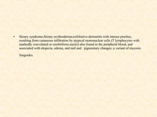
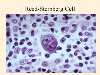
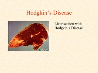
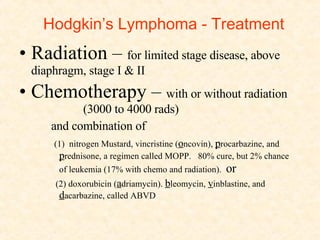
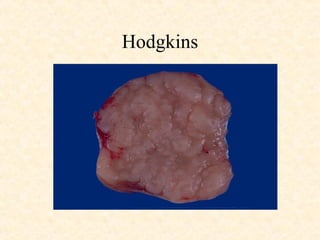
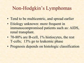
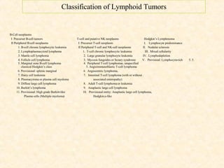
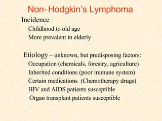
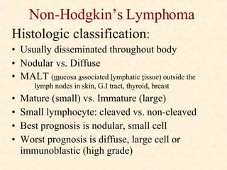
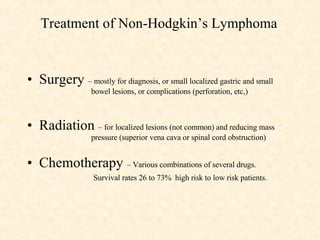
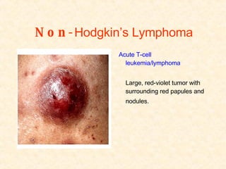
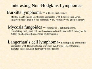
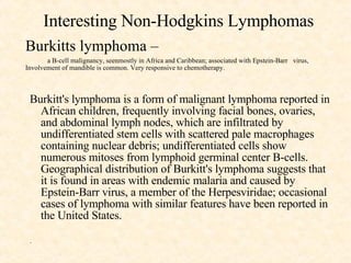
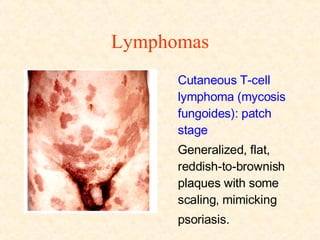
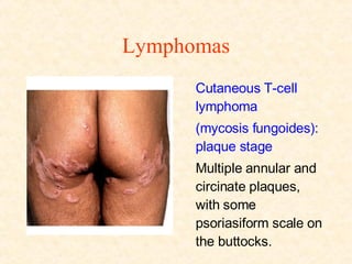
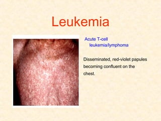
Malignant  Diseases  of  the  Lymphatic  System  and  Soft  Tissues George A. Simpson, M.D., F.A.C.S.
Lymphomas – Sites of Lymphoid tissue Lymph nodes White pulp of spleen Waldeyer’s ring Thymus gland Peyer’s patches  (submucosa of   respiratory and gastrointestinal tracts)
Lymphomas Definition: malignancy of lymphoid tissue Can be T (thymus) or B (bursa) cells T are “killers cells”; B make antibodies B- center of lymphoid follicle; T - around the periphery 2 main types: Hodgkins vs. Non-Hodgkins
Classification of Lymphoid Tumors B-Cell neoplasms I  Precurser B-cell tumors T-cell and putative NK neoplasms Hodgkin’s lymphoma  II Peripheral B-cell neoplasms   I  Precurser T-cell neoplasm   I.  Lymphocyte predominance 1. B-cell chronic lymphocytic leukemia   II Peripheral T-cell and NK-cell neoplasms  II.  Nodular sclerosis 2. Lymphoplasmacytoid lymphoma   1.  T-cell chronic lymphocytic leukemia  III.  Mixed cellularity  3. Mantle cell lymphoma   2.  Large granular lymphocyte leukemia  IV.  Lymphodepletion 4. Follicle cell lymphoma   3.  Mycosis fungoides or sezary syundrome  V.  Provisioal: Lymphocyte-rich  5. 5. 5. Marginal zone B-cell lymphoma   4.  Peripheral T-cell lymphomas, unspecified   classical Hodgkin’sclass  5. Angioimmunoblastic T-cell lymphoma 6. Provisional: splenic marginal   6  Angiocentric lymphoma.. 7. Hairy cell leukemia   7.  Intestinal T-ce;; lymphoma (with or without . 8. Plasmacytoma or plasma cell myeloma   associated enteropathy) 9. Diffuse large cell lymphoma   8.  Adult T-cell lymphoma or leukemia 10. Burkitt’s lymphoma   9.  Anaplastic large cell lymphoma  11. Provisional: High grade Burkitt-like    10.  Provisional entity: Anaplastic large cell lymphoma,   Hodgkin;s-like
Hodgkin’s Lymphoma Age and incidence Young adulthood Older age (Rare before 10 years of age) Approximately 13% of all lymphomas Twin “peaks” 10-20, then after age 50 Unknown etiology, Epstein-Barr virus suspect
Hodgkin’s Lymphoma Classification of Hodgkin’s Lymphoma I  Lymphocyte predominance II  Nodular sclerosis III  Mixed cellularity IV  Lymphocyte depletion V  Provisional entity:  lymphocyte –rich classical Hodgkin’s lymphoma
Symptoms of Hodgkin’s Lymphoma Asymptomatic, painless, discrete rubbery enlarged nodes Usually noticed first in neck  (90% supradiaphragmatic in young  adults, 25% subdiaphragmatic in older adults) Mediastinal adenopathy seen on routine chest x-ray Dry, non-productive cough Fever and night sweats  (Pel-Ebstein cyclical fever rare) Malaise  Weight loss Waxing and waning of lymph node size
Hodgkins - Clinical Presentation Young people, hard lymph node usually in neck or supraclavicular fossa.  Old people, can be anywhere Low grade fever ( Pel-Ebstein ), night sweats, weight loss. Abdominal pain with ETOH. May have immune suppression Diagnosis depends on pathognomonic  Reed-  Sternberg  cell
Hodgkin’s Lymphoma   Diagnosis Biopsy of lymph node.   Reed-Sternberg cells mandatory Staging Ultrasound and CT scan Lymphogram Bone marrow biopsy Laparotomy (rarely used now) Liver biopsy Gallium scan
Staging of Hodgkin’s Lymphoma Stage  Findings I  Involvement of single lymph node region or single extralymphatic organ (Ie) II  2 or more lymph node regions on same side of diaphragm  or localized  involvement of an extralymphatic organ or site (IIe)  III  Lymph node regions on both sides of diaphragm  or localized involvement  of an extralymphatic organ . Spleen (IIIs)  or both (IIIse) IV  Diffuse or disseminated involvement of one or more extralymphatic organs with or without associated node involvement Stage A  Asymptomatic  Stage B  Fever, sweats, weight loss
Hodgkins - Staging Staging determines prognosis:  Laparotomy with tissue biopsies lymphangiogram, CAT scans may preclude laparotomy  Stages:  I - localized to one area  II - Above the diaphragm (or below)  III - Above and below the diaphragm  IV- infiltration of organs.  A is asymptomatic.  B is with symptoms.
Hodgkins Lymphoma - Prognosis Staging and histology determines prognosis:  Lymphocyte –predominant group has best prognosis  nodular-sclerosis group – next best prognosis  then mixed cellularity and lymphocyte depleted 80% of patients cured with adequate treatment Risk of secondary tumor increases in time (perhaps caused by treatment -  Same area as radiation,  different cell type, three year interval ) Risk of leukemia  -3% in 10 years
Normal Lymph Node
Hodgkins Disease with Reed-Sternberg Cells
Reed-Sternberg Cell
Hodgkin’s Disease Liver section with Hodgkin’s Disease
Hodgkin’s Lymphoma - Treatment Radiation –  for limited stage disease, above diaphragm, stage I & II Chemotherapy –  with or without radiation  (3000 to 4000 rads)  and combination of  (1)  nitrogen Mustard, vincristine ( o ncovin),  p rocarbazine, and  p rednisone, a regimen called MOPP.  80% cure, but 2% chance of leukemia (17% with chemo and radiation).  or (2) doxorubicin ( a driamycin).  b leomycin,  v inblastine, and  d acarbazine, called ABVD
Hodgkins
Non-Hodgkin’s Lymphomas Tend to be multicentric, and spread earlier Etiology unknown: more frequent in immunocompromised patients such as: AIDS, renal transplant. 70-80% are B-cell, 1% histiocytes, the rest  T-cells;  13% go to leukemic phase Prognosis depends on histologic classification
Classification of Lymphoid Tumors B-Cell neoplasms I  Precurser B-cell tumors T-cell and putative NK neoplasms Hodgkin’s Lymphmoma  II Peripheral B-cell neoplasms   I  Precurser T-cell neoplasm  I.  Lymphocyte predominance 1. B-cell chronic lymphocytic leukemia   II Peripheral T-cell and NK-cell neoplasms  II.  Nodular sclerosis 2. Lymphoplasmacytoid lymphoma   1.  T-cell chronic lymphocytic leukemia  III.  Mixed cellularity  3. Mantle cell lymphoma   2.  Large granular lymphocyte leukemia  IV.  Lymphodepletion 4. Follicle cell lymphoma   3.  Mycosis fungoides or Sezary syndrome  V.  Provisioal: Lymphocyte-rich  5. 5. 5. Marginal zone B-cell lymphoma   4.  Peripheral T-cell lymphomas, unspecified   classical Hodgkin’s class  5. Angioimmunoblastic T-cell lymphoma 6. Provisional: splenic marginal   6  Angiocentric lymphoma.. 7. Hairy cell leukemia   7.  Intestinal T-cell lymphoma (with or without . 8. Plasmacytoma or plasma cell myeloma   associated enteropathy) 9. Diffuse large cell lymphoma   8.  Adult T-cell lymphoma or leukemia 10. Burkitt’s lymphoma   9.  Anaplastic large cell lymphoma  11. Provisional: High grade Burkitt-like    10.  Provisional entity: Anaplastic large cell lymphoma,   Plasma cells (Multiple myeloma)   Hodgkin;s-like
Non - Hodgkin’s Lymphoma Incidence Childhood to old age More prevalent in elderly  Etiology  – unknown, but predisposing factors: Occupation (chemicals, forestry, agriculture)  Inherited conditions (poor immune system) Certain medications  (Chemotherapy drugs) HIV and AIDS patients susceptible Organ transplant patients susceptible
Non-Hodgkin’s Lymphoma Histologic classification : Usually disseminated throughout body Nodular vs. Diffuse MALT  ( m ucosa  a ssociated  l ymphatic  t issue) outside the  lymph nodes in skin, G.I tract, thyroid, breast Mature (small) vs. Immature (large) Small lymphocyte: cleaved vs. non-cleaved Best prognosis is nodular, small cell Worst prognosis is diffuse, large cell or immunoblastic (high grade)
Evaluation of Non-Hodgkins Lymphoma History and physical examination CBC, liver function tests, chest x-ray, CT scan  of chest, abdomen, and pelvis Bone marrow biopsy Lumbar puncture Staging laparotomy usually not indicated   (disease is disseminated vs. localized disease of Hodgkin’s)
Nodular Lymphoma
Diffuse Lymphoma
High Grade Lymphoma
Non-Hodgkin’s Lymphoma - Treatment Surgery  –  For diagnosis,  (sometimes for Rx of local  focus or for compications- perforation, obstruction) Radiation –   for limited stage disease, above diaphragm, stage I  & II; Superior vena cava obstruction, spinal cord  compression Chemotherapy - with or without radiation (3000 - 4000 rads)  and combination of  (1)  nitrogen  M ustard, vincristine ( O ncovin),  p rocarbazine, and  p rednisone, a regimen called MOPP.  80% cure, but 2% chance of leukemia (17% with chemo and radiation).  or (2) doxorubicin ( a driamycin).  b leomycin,  v inblastine, and  d acarbazine, called ABVD
Non-Hodgkin’s Lymphoma - Prognosis Favorable prognosis Low grade tumors – 5 years even untreated Poor prognosis   high grade tumors  (poor differentiation)  – best response is to  chemotherapy older than 60 yrs. systemic symptoms  bulky disease  extra-nodal disease  G.I or  bone marrow involvement
Multiple Myeloma Definition: malignancy of plasma cells  from B cells-  010-15% of all hematologic cancers Clone of plasma cells produces immunoglobulins. Can be light chain, heavy chain, Kappa or Lambda. Bence-Jones protein in urine Produces bone lysis by OAF -  Osteoclast Activation Factor:
Myeloma
Myeloma of Skull
Myeloma
Multiple myeloma - presentation bone pain pathologic fractures: “moth holes” on X-ray Increased calcium, protein, globulin Problems include: hyperviscosity,  Rouleaux , amyloidosis Survival: 2-3 years.  BMT gives best chance.
Waldenstrom’s Macroglobulinemia Plasmacytoid lymphocyte Produces large immunoglubulin molecules. Main problem is hyperviscosity. Treated with plasmapheresis, sometimes chemotherapy More benign course than myeloma
Classification of Lymphoid Tumors B-Cell neoplasms I  Precurser B-cell tumors T-cell and putative NK neoplasms Hodgkin’s lymphoma  II Peripheral B-cell neoplasms   I  Precurser T-cell neoplasm   I.  Lymphocyte predominance 1. B-cell chronic lymphocytic leukemia   II Peripheral T-cell and NK-cell neoplasms  II.  Nodular sclerosis 2. Lymphoplasmacytoid lymphoma   1.  T-cell chronic lymphocytic leukemia  III.  Mixed cellularity  3. Mantle cell lymphoma   2.  Large granular lymphocyte leukemia  IV.  Lymphodepletion 4. Follicle cell lymphoma   3.  Mycosis fungoides or sezary syundrome  V.  Provisioal: Lymphocyte-rich   Marginal zone B-cell lymphoma   4.  Peripheral T-cell lymphomas, unspecified   classical Hodgkin’sclass  5. Angioimmunoblastic T-cell lymphoma 6. Provisional: splenic marginal   6  Angiocentric lymphoma.. 7. Hairy cell leukemia   7.  Intestinal T-ce;; lymphoma (with or without . 8. Plasmacytoma or plasma cell myeloma   associated enteropathy) 9. Diffuse large cell lymphoma   8.  Adult T-cell lymphoma or leukemia 10. Burkitt’s lymphoma   9.  Anaplastic large cell lymphoma  11. Provisional: High grade Burkitt-like    10.  Provisional entity: Anaplastic large cell lymphoma,   Plasma cells (Multiple myeloma)   Hodgkin;s-like
Treatment of Non-Hodgkin’s Lymphoma   Surgery  – mostly for diagnosis, or small localized gastric and small  bowel lesions, or complications (perforation, etc,) Radiation  – for localized lesions (not common) and reducing mass  pressure (superior vena cava or spinal cord obstruction)  Chemotherapy  – Various combinations of several drugs. Survival rates 26 to 73%  high risk to low risk patients.
Non- Hodgkin’s Lymphoma   Acute T-cell leukemia/lymphoma  Large, red-violet tumor with surrounding red papules and nodules.
Interesting Non-Hodgkins Lymphomas Burkitts lymphoma –  a B-cell malignancy Mostly in Africa and Caribbean; associated with Epstein-Barr virus, Involvement of mandible is common. Very responsive to chemotherapy. Mycosis fungoides –  cutaneous T-cell lymphoma.  Circulating malignant cells with convoluted nuclei are called Sezary cells.  Often misdiagnosed as eczema or dermatosis. Langerhan’s cell lymphoma-  Eosinophilic granuloma, associated with Hand-Schuller-Christian syndrome (Exophthalmus, diabetes insipidus, and destructive bone lesions .
Interesting Non-Hodgkins Lymphomas Burkitts lymphoma –  a B-cell malignancy, seenmostly in Africa and Caribbean; associated with Epstein-Barr  virus, Involvement of mandible is common. Very responsive to chemotherapy. Burkitt's lymphoma is a form of malignant lymphoma reported in African children, frequently involving facial bones, ovaries, and abdominal lymph nodes, which are infiltrated by undifferentiated stem cells with scattered pale macrophages containing nuclear debris; undifferentiated cells show numerous mitoses from lymphoid germinal center B-cells. Geographical distribution of Burkitt's lymphoma suggests that it is found in areas with endemic malaria and caused by Epstein-Barr virus, a member of the Herpesviridae; occasional cases of lymphoma with similar features have been reported in the United States.  .
Lymphomas Cutaneous T-cell lymphoma (mycosis fungoides): patch stage Generalized, flat, reddish-to-brownish plaques with some scaling, mimicking psoriasis.
Lymphomas Cutaneous T-cell lymphoma (mycosis fungoides): plaque stage  Multiple annular and circinate plaques, with some psoriasiform scale on the buttocks.
Leukemia Acute T-cell leukemia/lymphoma  Disseminated, red-violet papules becoming confluent on the chest.
Lymphomas Cutaneous T-cell lymphoma (mycosis fungoides): plaque stage Multiple annular and circinate plaques, some with psoriasi-form scale, some with erosion, indurated upon palpation.
Lymphomas Cutaneous T-cell lymphoma (mycosis fungoides): tumor stage  A fleshy, tomato-like, partially eroded nodule arising from a larger, slightly elevated, psoriasiform plaque with scaling and central atrophy.
Lymphomas   Cutaneous T-cell lymphoma (mycosis fungoides): leonine facies  Diffuse infiltration of the skin has caused a coarsening of the facial features, with thickening of the brows and glabella; the neck and trunk are also involved.
Lymphomas Cutaneous T-cell lymphoma: Sézary syndrome  Sézary syndrome,  Sézary  erythroderma; exfoliative dermatitis with intense pruritus, resulting from cutaneous infiltration by atypical mononuclear cells (T lymphocytes with markedly convoluted or cerebriform nuclei) also found in the peripheral blood, and associated with alopecia, edema, and nail and  pigmentary changes; a variant of mycosis fungoides.   Appearance Generalized erythema, scaling, and thickening of the skin (erythroderma); the palms are hyperkeratotic. Generalized lymphadenopathy was present. The buffy coat contained 45% atypical lymphocytes (Sézary cells).
Lymphomas Cutaneous B-cell lymphoma  Multiple, well-defined, red-to-brownish and bluish nodules on the face with associated edema.
Lymphomas Cutaneous B-cell lymphoma  Multiple, skin-colored dermal tumors on the scalp.
Lymphomas Angiocentric lymphoma   Papules, nodules, tumors, and ulcers with associated facial edema. The markings on the forehead are a tribal tatoo of this North African woman.
Lymphomas Langerhan’s cell lymphoma of infant’s scalp  (Eosinophilic  granulomatosis)
Lymphomas Langerhan’s cell cytosis Letterer-Siwe syndrome (malignant eosinophilic lymphoma often fatal in infants )
Lymphomas Langerhan’s cell histiocytosis of vulva
Sarcomas Etiology: Connective tissue of body Genetic defects, (Von Recklinghausen’s dsease, Gardner’s  syndrome Radiation, chemical exposure, trauma Age and incidence :  Uncommon (Less than 1% of malignant tumors) 2 per 100.000 in U.S. Uncommon in adults Higher incidence in children under 15 yrs (6%) .
Sarcoma – Risk Factors Occupational exposure  (asbestos  [pleural mesothelioma];  herbicides - Dioxin & Agent Orange; poly vinyl chloride-PVC-  [causing hepatic angiosarcoma]  ) Radiation  (x-ray treatment, radiologists, dentists) 8x to 50x increase in incidence following radiation treatment, same area as radiation, different cell type, three year interval ) Chronic lymphedema  (Lymphangiosarcoma in  Stewart-Treves syndrome post radical mastectomy) Gene and chromosome abnormalities  (Ewing’s sarcoma, clear cell sarcoma, myxoid liposarcoma, desmoplastic round cell sarcoma, Von Recklinghausen’s syndrome, Gardner’s syndrome, etc,)
Sarcomas Tissue Benign Malignant Fibrous tissue Fibroma Fibrosarcoma Adipose tissue Lipoma Liposarcoma Striated muscle Rhabdomyoma Rabdomyosarcoma Smooth muscle Leiomyoma Leiomyosarcoma Synovial mesothelium Mesothelioma Synovial sarcoma Blood vcessels Angioma Angiosarcoma Lymph vessels Lymphoangioma Lymphangiosarcoma Peripheral nerve Neuroma Malignant      neurolemmoma   Schwannoma Myofibroblast Malignant fibrous    histiocytoma
Epidemiology of Sarcomas Location Extremities  66% Trunk  20% Retroperitoneum  13% Cell type  (over 50 subtypes) Malignant fibrous histiocytoma  24% Leiomyosarcoma  21% Liposarcoma  19%
Sarcoma - Evaluation of Patient History and physical  (1/3 asymptomatic at time of  discovery) Imaging studies Core biopsy  Staging – based on tumor grade, tumor size, nodes and metastases
Sarcoma Sarcoma of thorax on chest x-ray
Sarcomas  Method of spread Contiguous growth Blood stream metastases main route Occasional lymphatic spread
Sarcomas - Treatment Treatment factors: Surgery is the effective treatment ( 2 cm margin) Radiation has limited usefulness  (adjuvant or  neoadjuvant) Age-  better results in childhood Degree and area of spread affects prognosis Grade – High grade has poorer results  Grade I to IV Grade I (low) well differentiated; Grade IV (high) poorly differentiated Completeness of removal Type of tumor ( this has less bearing than age,  grade, or spread)
Sarcomas - Prognosis Overall 5 year survival-  Superficial low grade  50%;  Advanced metastatic, deep, high grade  20%  Radiation has limited usefulness  (adjuvant or  neoadjuvant) Age-  better results in childhood Degree and area of spread affects prognosis Grade – High grade has poorer results  Grade I to IV Grade I (low) well differentiated; Grade IV (high) poorly differentiated Completeness of removal Type of tumor ( this has less bearing than age,  grade, or spread) Follow-up exams every 6 months x 4, then yearly
Specific Sarcomas Gastrointestinal Stromal Tumor-  Gastrointestinal neural  origin; tyrosine kinase receptor in membrane; Rx surgery and tyrosine  kinase inhibitors  Dermatofibrosaccoma Protuberans  skin and  subcutaneous tumor;   slow metastases; wide resection treatment of  choice Desmoid Tumor  fibrous tissue malignancy,intermediate  malignancy, surgery is Rx; may respond to NSAID’s or tamoxifen Kaposi’s Sarcoma  once in Jewish Mediterranean men or sub- Saharan  Africans; AIDS increaced incidence; homosexual and  bisexual men predominently; human herpesvirus 8 may be cause.
KAPOSI’S SARCOMA Skin section to show origin of Kaposi’s sarcoma cells from  connective tissue in the dermis layer of the skin Kaposi’s sarcoma is considered an angiosarcoma
KAPOSI’S SARCOMA Kaposi’s Sarcoma of thigh.  Non-AIDS related Kaposi’s sarcoma usually found on lower extremities
KAPOSI’S SARCOMA Appearance of foot lesion of Kaposi’s  sarcoma.
KAPOSI’S SARCOMA Aids-related Kaposi’s sarcoma often begins in peri-oral mucosa. Palate is the most common site, with rapid spread to lymph nodes
KAPOSI’S SARCOMA Microscopic view of Kaposi’s Sarcoma  – endothelial cells, fibroblasts, spindle cells, capillary growth -  Angiosarcomas
KAPOSI’S SARCOMA Lesions on back
KAPOSI’S SARCOMA Kaposi’s sarcoma lesion of shoulder
Kaposi’s Sarcoma Kaposi’s sarcoma  lesion of arm
KAPOSI’S SARCOMA PERIANAL
Kaposi’s Sarcoma - Treatment Surgical excision for small localized lesions Chemotherapy for advanced cases  ( vinblastine,  bleomycin, doxorubicin) Immunodeficiency complicates treatment Radiation may shrink lesions. Palliation
Lymphomas Mastocytosis in child with solitary mastocytoma lesion
Lymphomas Lymphoma of submental area
Lymphomas   Mastocytosis in a child
Lymphomas Mastocytosis of back
Lymphomas Generalized mastocytosis (White areas are unaffected skin)
Lymphomas Lymphomatoid papulosis
Lymphomas Lymphomatoid papulosis  lesions of legs
Lymphomas Lymphoma of right chest on x-ray
Myelogenous leukemia Acute myelogenous leukemia with leukemia cutis Hundreds of tan-pink papules and a nodule on trunk of female
Leukemia Leukemia Cutis with  ulceration and pustules  of lower abdomen
Lymphomas  Lymphoma lesion of mouth: eosinophilic granuloma
THE  END
Diagram of Skin Layers
Sézary syndrome,Sézary erythroderma;exfoliative dermatitis with intense pruritus, resulting from cutaneous infiltration by atypical mononuclear cells (T lymphocytes with markedly convoluted or cerebriform nuclei) also found in the peripheral blood, and associated with alopecia, edema, and nail and  pigmentary changes; a variant of mycosis fungoides.

Malignant Diseases Lymphatics & Soft Tissue #3

  • 1.
    Malignant Diseases of the Lymphatic System and Soft Tissues George A. Simpson, M.D., F.A.C.S.
  • 2.
    Lymphomas – Sitesof Lymphoid tissue Lymph nodes White pulp of spleen Waldeyer’s ring Thymus gland Peyer’s patches (submucosa of respiratory and gastrointestinal tracts)
  • 3.
    Lymphomas Definition: malignancyof lymphoid tissue Can be T (thymus) or B (bursa) cells T are “killers cells”; B make antibodies B- center of lymphoid follicle; T - around the periphery 2 main types: Hodgkins vs. Non-Hodgkins
  • 4.
    Classification of LymphoidTumors B-Cell neoplasms I Precurser B-cell tumors T-cell and putative NK neoplasms Hodgkin’s lymphoma II Peripheral B-cell neoplasms I Precurser T-cell neoplasm I. Lymphocyte predominance 1. B-cell chronic lymphocytic leukemia II Peripheral T-cell and NK-cell neoplasms II. Nodular sclerosis 2. Lymphoplasmacytoid lymphoma 1. T-cell chronic lymphocytic leukemia III. Mixed cellularity 3. Mantle cell lymphoma 2. Large granular lymphocyte leukemia IV. Lymphodepletion 4. Follicle cell lymphoma 3. Mycosis fungoides or sezary syundrome V. Provisioal: Lymphocyte-rich 5. 5. 5. Marginal zone B-cell lymphoma 4. Peripheral T-cell lymphomas, unspecified classical Hodgkin’sclass 5. Angioimmunoblastic T-cell lymphoma 6. Provisional: splenic marginal 6 Angiocentric lymphoma.. 7. Hairy cell leukemia 7. Intestinal T-ce;; lymphoma (with or without . 8. Plasmacytoma or plasma cell myeloma associated enteropathy) 9. Diffuse large cell lymphoma 8. Adult T-cell lymphoma or leukemia 10. Burkitt’s lymphoma 9. Anaplastic large cell lymphoma 11. Provisional: High grade Burkitt-like 10. Provisional entity: Anaplastic large cell lymphoma, Hodgkin;s-like
  • 5.
    Hodgkin’s Lymphoma Ageand incidence Young adulthood Older age (Rare before 10 years of age) Approximately 13% of all lymphomas Twin “peaks” 10-20, then after age 50 Unknown etiology, Epstein-Barr virus suspect
  • 6.
    Hodgkin’s Lymphoma Classificationof Hodgkin’s Lymphoma I Lymphocyte predominance II Nodular sclerosis III Mixed cellularity IV Lymphocyte depletion V Provisional entity: lymphocyte –rich classical Hodgkin’s lymphoma
  • 7.
    Symptoms of Hodgkin’sLymphoma Asymptomatic, painless, discrete rubbery enlarged nodes Usually noticed first in neck (90% supradiaphragmatic in young adults, 25% subdiaphragmatic in older adults) Mediastinal adenopathy seen on routine chest x-ray Dry, non-productive cough Fever and night sweats (Pel-Ebstein cyclical fever rare) Malaise Weight loss Waxing and waning of lymph node size
  • 8.
    Hodgkins - ClinicalPresentation Young people, hard lymph node usually in neck or supraclavicular fossa. Old people, can be anywhere Low grade fever ( Pel-Ebstein ), night sweats, weight loss. Abdominal pain with ETOH. May have immune suppression Diagnosis depends on pathognomonic Reed- Sternberg cell
  • 9.
    Hodgkin’s Lymphoma Diagnosis Biopsy of lymph node. Reed-Sternberg cells mandatory Staging Ultrasound and CT scan Lymphogram Bone marrow biopsy Laparotomy (rarely used now) Liver biopsy Gallium scan
  • 10.
    Staging of Hodgkin’sLymphoma Stage Findings I Involvement of single lymph node region or single extralymphatic organ (Ie) II 2 or more lymph node regions on same side of diaphragm or localized involvement of an extralymphatic organ or site (IIe) III Lymph node regions on both sides of diaphragm or localized involvement of an extralymphatic organ . Spleen (IIIs) or both (IIIse) IV Diffuse or disseminated involvement of one or more extralymphatic organs with or without associated node involvement Stage A Asymptomatic Stage B Fever, sweats, weight loss
  • 11.
    Hodgkins - StagingStaging determines prognosis: Laparotomy with tissue biopsies lymphangiogram, CAT scans may preclude laparotomy Stages: I - localized to one area II - Above the diaphragm (or below) III - Above and below the diaphragm IV- infiltration of organs. A is asymptomatic. B is with symptoms.
  • 12.
    Hodgkins Lymphoma -Prognosis Staging and histology determines prognosis: Lymphocyte –predominant group has best prognosis nodular-sclerosis group – next best prognosis then mixed cellularity and lymphocyte depleted 80% of patients cured with adequate treatment Risk of secondary tumor increases in time (perhaps caused by treatment - Same area as radiation, different cell type, three year interval ) Risk of leukemia -3% in 10 years
  • 13.
  • 14.
    Hodgkins Disease withReed-Sternberg Cells
  • 15.
  • 16.
    Hodgkin’s Disease Liversection with Hodgkin’s Disease
  • 17.
    Hodgkin’s Lymphoma -Treatment Radiation – for limited stage disease, above diaphragm, stage I & II Chemotherapy – with or without radiation (3000 to 4000 rads) and combination of (1) nitrogen Mustard, vincristine ( o ncovin), p rocarbazine, and p rednisone, a regimen called MOPP. 80% cure, but 2% chance of leukemia (17% with chemo and radiation). or (2) doxorubicin ( a driamycin). b leomycin, v inblastine, and d acarbazine, called ABVD
  • 18.
  • 19.
    Non-Hodgkin’s Lymphomas Tendto be multicentric, and spread earlier Etiology unknown: more frequent in immunocompromised patients such as: AIDS, renal transplant. 70-80% are B-cell, 1% histiocytes, the rest T-cells; 13% go to leukemic phase Prognosis depends on histologic classification
  • 20.
    Classification of LymphoidTumors B-Cell neoplasms I Precurser B-cell tumors T-cell and putative NK neoplasms Hodgkin’s Lymphmoma II Peripheral B-cell neoplasms I Precurser T-cell neoplasm I. Lymphocyte predominance 1. B-cell chronic lymphocytic leukemia II Peripheral T-cell and NK-cell neoplasms II. Nodular sclerosis 2. Lymphoplasmacytoid lymphoma 1. T-cell chronic lymphocytic leukemia III. Mixed cellularity 3. Mantle cell lymphoma 2. Large granular lymphocyte leukemia IV. Lymphodepletion 4. Follicle cell lymphoma 3. Mycosis fungoides or Sezary syndrome V. Provisioal: Lymphocyte-rich 5. 5. 5. Marginal zone B-cell lymphoma 4. Peripheral T-cell lymphomas, unspecified classical Hodgkin’s class 5. Angioimmunoblastic T-cell lymphoma 6. Provisional: splenic marginal 6 Angiocentric lymphoma.. 7. Hairy cell leukemia 7. Intestinal T-cell lymphoma (with or without . 8. Plasmacytoma or plasma cell myeloma associated enteropathy) 9. Diffuse large cell lymphoma 8. Adult T-cell lymphoma or leukemia 10. Burkitt’s lymphoma 9. Anaplastic large cell lymphoma 11. Provisional: High grade Burkitt-like 10. Provisional entity: Anaplastic large cell lymphoma, Plasma cells (Multiple myeloma) Hodgkin;s-like
  • 21.
    Non - Hodgkin’sLymphoma Incidence Childhood to old age More prevalent in elderly Etiology – unknown, but predisposing factors: Occupation (chemicals, forestry, agriculture) Inherited conditions (poor immune system) Certain medications (Chemotherapy drugs) HIV and AIDS patients susceptible Organ transplant patients susceptible
  • 22.
    Non-Hodgkin’s Lymphoma Histologicclassification : Usually disseminated throughout body Nodular vs. Diffuse MALT ( m ucosa a ssociated l ymphatic t issue) outside the lymph nodes in skin, G.I tract, thyroid, breast Mature (small) vs. Immature (large) Small lymphocyte: cleaved vs. non-cleaved Best prognosis is nodular, small cell Worst prognosis is diffuse, large cell or immunoblastic (high grade)
  • 23.
    Evaluation of Non-HodgkinsLymphoma History and physical examination CBC, liver function tests, chest x-ray, CT scan of chest, abdomen, and pelvis Bone marrow biopsy Lumbar puncture Staging laparotomy usually not indicated (disease is disseminated vs. localized disease of Hodgkin’s)
  • 24.
  • 25.
  • 26.
  • 27.
    Non-Hodgkin’s Lymphoma -Treatment Surgery – For diagnosis, (sometimes for Rx of local focus or for compications- perforation, obstruction) Radiation – for limited stage disease, above diaphragm, stage I & II; Superior vena cava obstruction, spinal cord compression Chemotherapy - with or without radiation (3000 - 4000 rads) and combination of (1) nitrogen M ustard, vincristine ( O ncovin), p rocarbazine, and p rednisone, a regimen called MOPP. 80% cure, but 2% chance of leukemia (17% with chemo and radiation). or (2) doxorubicin ( a driamycin). b leomycin, v inblastine, and d acarbazine, called ABVD
  • 28.
    Non-Hodgkin’s Lymphoma -Prognosis Favorable prognosis Low grade tumors – 5 years even untreated Poor prognosis high grade tumors (poor differentiation) – best response is to chemotherapy older than 60 yrs. systemic symptoms bulky disease extra-nodal disease G.I or bone marrow involvement
  • 29.
    Multiple Myeloma Definition:malignancy of plasma cells from B cells- 010-15% of all hematologic cancers Clone of plasma cells produces immunoglobulins. Can be light chain, heavy chain, Kappa or Lambda. Bence-Jones protein in urine Produces bone lysis by OAF - Osteoclast Activation Factor:
  • 30.
  • 31.
  • 32.
  • 33.
    Multiple myeloma -presentation bone pain pathologic fractures: “moth holes” on X-ray Increased calcium, protein, globulin Problems include: hyperviscosity, Rouleaux , amyloidosis Survival: 2-3 years. BMT gives best chance.
  • 34.
    Waldenstrom’s Macroglobulinemia Plasmacytoidlymphocyte Produces large immunoglubulin molecules. Main problem is hyperviscosity. Treated with plasmapheresis, sometimes chemotherapy More benign course than myeloma
  • 35.
    Classification of LymphoidTumors B-Cell neoplasms I Precurser B-cell tumors T-cell and putative NK neoplasms Hodgkin’s lymphoma II Peripheral B-cell neoplasms I Precurser T-cell neoplasm I. Lymphocyte predominance 1. B-cell chronic lymphocytic leukemia II Peripheral T-cell and NK-cell neoplasms II. Nodular sclerosis 2. Lymphoplasmacytoid lymphoma 1. T-cell chronic lymphocytic leukemia III. Mixed cellularity 3. Mantle cell lymphoma 2. Large granular lymphocyte leukemia IV. Lymphodepletion 4. Follicle cell lymphoma 3. Mycosis fungoides or sezary syundrome V. Provisioal: Lymphocyte-rich Marginal zone B-cell lymphoma 4. Peripheral T-cell lymphomas, unspecified classical Hodgkin’sclass 5. Angioimmunoblastic T-cell lymphoma 6. Provisional: splenic marginal 6 Angiocentric lymphoma.. 7. Hairy cell leukemia 7. Intestinal T-ce;; lymphoma (with or without . 8. Plasmacytoma or plasma cell myeloma associated enteropathy) 9. Diffuse large cell lymphoma 8. Adult T-cell lymphoma or leukemia 10. Burkitt’s lymphoma 9. Anaplastic large cell lymphoma 11. Provisional: High grade Burkitt-like 10. Provisional entity: Anaplastic large cell lymphoma, Plasma cells (Multiple myeloma) Hodgkin;s-like
  • 36.
    Treatment of Non-Hodgkin’sLymphoma Surgery – mostly for diagnosis, or small localized gastric and small bowel lesions, or complications (perforation, etc,) Radiation – for localized lesions (not common) and reducing mass pressure (superior vena cava or spinal cord obstruction) Chemotherapy – Various combinations of several drugs. Survival rates 26 to 73% high risk to low risk patients.
  • 37.
    Non- Hodgkin’s Lymphoma Acute T-cell leukemia/lymphoma Large, red-violet tumor with surrounding red papules and nodules.
  • 38.
    Interesting Non-Hodgkins LymphomasBurkitts lymphoma – a B-cell malignancy Mostly in Africa and Caribbean; associated with Epstein-Barr virus, Involvement of mandible is common. Very responsive to chemotherapy. Mycosis fungoides – cutaneous T-cell lymphoma. Circulating malignant cells with convoluted nuclei are called Sezary cells. Often misdiagnosed as eczema or dermatosis. Langerhan’s cell lymphoma- Eosinophilic granuloma, associated with Hand-Schuller-Christian syndrome (Exophthalmus, diabetes insipidus, and destructive bone lesions .
  • 39.
    Interesting Non-Hodgkins LymphomasBurkitts lymphoma – a B-cell malignancy, seenmostly in Africa and Caribbean; associated with Epstein-Barr virus, Involvement of mandible is common. Very responsive to chemotherapy. Burkitt's lymphoma is a form of malignant lymphoma reported in African children, frequently involving facial bones, ovaries, and abdominal lymph nodes, which are infiltrated by undifferentiated stem cells with scattered pale macrophages containing nuclear debris; undifferentiated cells show numerous mitoses from lymphoid germinal center B-cells. Geographical distribution of Burkitt's lymphoma suggests that it is found in areas with endemic malaria and caused by Epstein-Barr virus, a member of the Herpesviridae; occasional cases of lymphoma with similar features have been reported in the United States. .
  • 40.
    Lymphomas Cutaneous T-celllymphoma (mycosis fungoides): patch stage Generalized, flat, reddish-to-brownish plaques with some scaling, mimicking psoriasis.
  • 41.
    Lymphomas Cutaneous T-celllymphoma (mycosis fungoides): plaque stage Multiple annular and circinate plaques, with some psoriasiform scale on the buttocks.
  • 42.
    Leukemia Acute T-cellleukemia/lymphoma Disseminated, red-violet papules becoming confluent on the chest.
  • 43.
    Lymphomas Cutaneous T-celllymphoma (mycosis fungoides): plaque stage Multiple annular and circinate plaques, some with psoriasi-form scale, some with erosion, indurated upon palpation.
  • 44.
    Lymphomas Cutaneous T-celllymphoma (mycosis fungoides): tumor stage A fleshy, tomato-like, partially eroded nodule arising from a larger, slightly elevated, psoriasiform plaque with scaling and central atrophy.
  • 45.
    Lymphomas Cutaneous T-cell lymphoma (mycosis fungoides): leonine facies Diffuse infiltration of the skin has caused a coarsening of the facial features, with thickening of the brows and glabella; the neck and trunk are also involved.
  • 46.
    Lymphomas Cutaneous T-celllymphoma: Sézary syndrome Sézary syndrome, Sézary erythroderma; exfoliative dermatitis with intense pruritus, resulting from cutaneous infiltration by atypical mononuclear cells (T lymphocytes with markedly convoluted or cerebriform nuclei) also found in the peripheral blood, and associated with alopecia, edema, and nail and pigmentary changes; a variant of mycosis fungoides. Appearance Generalized erythema, scaling, and thickening of the skin (erythroderma); the palms are hyperkeratotic. Generalized lymphadenopathy was present. The buffy coat contained 45% atypical lymphocytes (Sézary cells).
  • 47.
    Lymphomas Cutaneous B-celllymphoma Multiple, well-defined, red-to-brownish and bluish nodules on the face with associated edema.
  • 48.
    Lymphomas Cutaneous B-celllymphoma Multiple, skin-colored dermal tumors on the scalp.
  • 49.
    Lymphomas Angiocentric lymphoma Papules, nodules, tumors, and ulcers with associated facial edema. The markings on the forehead are a tribal tatoo of this North African woman.
  • 50.
    Lymphomas Langerhan’s celllymphoma of infant’s scalp (Eosinophilic granulomatosis)
  • 51.
    Lymphomas Langerhan’s cellcytosis Letterer-Siwe syndrome (malignant eosinophilic lymphoma often fatal in infants )
  • 52.
    Lymphomas Langerhan’s cellhistiocytosis of vulva
  • 53.
    Sarcomas Etiology: Connectivetissue of body Genetic defects, (Von Recklinghausen’s dsease, Gardner’s syndrome Radiation, chemical exposure, trauma Age and incidence : Uncommon (Less than 1% of malignant tumors) 2 per 100.000 in U.S. Uncommon in adults Higher incidence in children under 15 yrs (6%) .
  • 54.
    Sarcoma – RiskFactors Occupational exposure (asbestos [pleural mesothelioma]; herbicides - Dioxin & Agent Orange; poly vinyl chloride-PVC- [causing hepatic angiosarcoma] ) Radiation (x-ray treatment, radiologists, dentists) 8x to 50x increase in incidence following radiation treatment, same area as radiation, different cell type, three year interval ) Chronic lymphedema (Lymphangiosarcoma in Stewart-Treves syndrome post radical mastectomy) Gene and chromosome abnormalities (Ewing’s sarcoma, clear cell sarcoma, myxoid liposarcoma, desmoplastic round cell sarcoma, Von Recklinghausen’s syndrome, Gardner’s syndrome, etc,)
  • 55.
    Sarcomas Tissue BenignMalignant Fibrous tissue Fibroma Fibrosarcoma Adipose tissue Lipoma Liposarcoma Striated muscle Rhabdomyoma Rabdomyosarcoma Smooth muscle Leiomyoma Leiomyosarcoma Synovial mesothelium Mesothelioma Synovial sarcoma Blood vcessels Angioma Angiosarcoma Lymph vessels Lymphoangioma Lymphangiosarcoma Peripheral nerve Neuroma Malignant neurolemmoma Schwannoma Myofibroblast Malignant fibrous histiocytoma
  • 56.
    Epidemiology of SarcomasLocation Extremities 66% Trunk 20% Retroperitoneum 13% Cell type (over 50 subtypes) Malignant fibrous histiocytoma 24% Leiomyosarcoma 21% Liposarcoma 19%
  • 57.
    Sarcoma - Evaluationof Patient History and physical (1/3 asymptomatic at time of discovery) Imaging studies Core biopsy Staging – based on tumor grade, tumor size, nodes and metastases
  • 58.
    Sarcoma Sarcoma ofthorax on chest x-ray
  • 59.
    Sarcomas Methodof spread Contiguous growth Blood stream metastases main route Occasional lymphatic spread
  • 60.
    Sarcomas - TreatmentTreatment factors: Surgery is the effective treatment ( 2 cm margin) Radiation has limited usefulness (adjuvant or neoadjuvant) Age- better results in childhood Degree and area of spread affects prognosis Grade – High grade has poorer results Grade I to IV Grade I (low) well differentiated; Grade IV (high) poorly differentiated Completeness of removal Type of tumor ( this has less bearing than age, grade, or spread)
  • 61.
    Sarcomas - PrognosisOverall 5 year survival- Superficial low grade 50%; Advanced metastatic, deep, high grade 20% Radiation has limited usefulness (adjuvant or neoadjuvant) Age- better results in childhood Degree and area of spread affects prognosis Grade – High grade has poorer results Grade I to IV Grade I (low) well differentiated; Grade IV (high) poorly differentiated Completeness of removal Type of tumor ( this has less bearing than age, grade, or spread) Follow-up exams every 6 months x 4, then yearly
  • 62.
    Specific Sarcomas GastrointestinalStromal Tumor- Gastrointestinal neural origin; tyrosine kinase receptor in membrane; Rx surgery and tyrosine kinase inhibitors Dermatofibrosaccoma Protuberans skin and subcutaneous tumor; slow metastases; wide resection treatment of choice Desmoid Tumor fibrous tissue malignancy,intermediate malignancy, surgery is Rx; may respond to NSAID’s or tamoxifen Kaposi’s Sarcoma once in Jewish Mediterranean men or sub- Saharan Africans; AIDS increaced incidence; homosexual and bisexual men predominently; human herpesvirus 8 may be cause.
  • 63.
    KAPOSI’S SARCOMA Skinsection to show origin of Kaposi’s sarcoma cells from connective tissue in the dermis layer of the skin Kaposi’s sarcoma is considered an angiosarcoma
  • 64.
    KAPOSI’S SARCOMA Kaposi’sSarcoma of thigh. Non-AIDS related Kaposi’s sarcoma usually found on lower extremities
  • 65.
    KAPOSI’S SARCOMA Appearanceof foot lesion of Kaposi’s sarcoma.
  • 66.
    KAPOSI’S SARCOMA Aids-relatedKaposi’s sarcoma often begins in peri-oral mucosa. Palate is the most common site, with rapid spread to lymph nodes
  • 67.
    KAPOSI’S SARCOMA Microscopicview of Kaposi’s Sarcoma – endothelial cells, fibroblasts, spindle cells, capillary growth - Angiosarcomas
  • 68.
  • 69.
    KAPOSI’S SARCOMA Kaposi’ssarcoma lesion of shoulder
  • 70.
    Kaposi’s Sarcoma Kaposi’ssarcoma lesion of arm
  • 71.
  • 72.
    Kaposi’s Sarcoma -Treatment Surgical excision for small localized lesions Chemotherapy for advanced cases ( vinblastine, bleomycin, doxorubicin) Immunodeficiency complicates treatment Radiation may shrink lesions. Palliation
  • 73.
    Lymphomas Mastocytosis inchild with solitary mastocytoma lesion
  • 74.
    Lymphomas Lymphoma ofsubmental area
  • 75.
    Lymphomas Mastocytosis in a child
  • 76.
  • 77.
    Lymphomas Generalized mastocytosis(White areas are unaffected skin)
  • 78.
  • 79.
  • 80.
    Lymphomas Lymphoma ofright chest on x-ray
  • 81.
    Myelogenous leukemia Acutemyelogenous leukemia with leukemia cutis Hundreds of tan-pink papules and a nodule on trunk of female
  • 82.
    Leukemia Leukemia Cutiswith ulceration and pustules of lower abdomen
  • 83.
    Lymphomas Lymphomalesion of mouth: eosinophilic granuloma
  • 84.
  • 85.
  • 86.
    Sézary syndrome,Sézary erythroderma;exfoliativedermatitis with intense pruritus, resulting from cutaneous infiltration by atypical mononuclear cells (T lymphocytes with markedly convoluted or cerebriform nuclei) also found in the peripheral blood, and associated with alopecia, edema, and nail and pigmentary changes; a variant of mycosis fungoides.