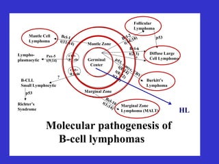
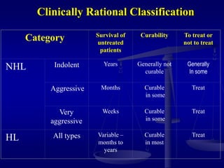
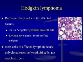
Lymphomas
Dr masoud alzouraiki
Malignant Lymphoma
 Lymphoma Concept
 Hematopoietic process
 B- and T- cell lymphoma
 Lymphoma Classification
 Epidemiological aspects of Lymphoma
 Approach to Lymphoma Patient
 Hodgkin’s Lymphoma
 Adult
 Paediatric
 Non-Hodgkin’s Lymphoma
 Adult
 Paediatric
05/05/2023
Malignant Lymphoma
 Lymphoma Concept
 Hematopoietic process
 B- and T- cell lymphoma
 Lymphoma Classification
 Epidemiological aspects of Lymphoma
 Approach to Lymphoma Patient
 Hodgkin’s Lymphoma
 Adult
 Paediatric
 Non-Hodgkin’s Lymphoma
 Adult
 Paediatric
05/05/2023
Conceptualizing lymphoma
 Lymphomas are malignant transformations of normal
lymphoid cells which reside predominantly in lymphoid
tissues
 As clonal cell expansions at certain developmental
stages
 Lymphatic system is our bodies main fight against
“foreigners”
 Lymphocytes
 Carried through our lymphatic system to help to fight
infection
 Lymphocytes are carried through the lymph vessels as well as
the blood stream, so cancer can start in nodes and spread
anywhere throughout the body.
Hematopoietic
stem cell
Neutrophils
Eosinophils
Basophils
Monocytes
Platelets
Red cells
Myeloid
progenitor
T-lymphocytes
Plasma
cells
B-lymphocytes
naïve
Pro B
Lymphoid
progenitor
prethymic
B-Cell Lymphoma (80%)
 B-Cells help make antibodies, which are proteins that
attach to and help destroy antigens
 Lymphomas are caused when a mutation arises
during the B-cell life cycle
 Various different lymphomas can occur during
several different stages of the cycle
 Follicular lymphoma, which is a type of B-cell lymphoma
is caused by a gene translocation which results in an over
expressed gene called BCL-2, which blocks apoptosis.
B-Cell Cancers
B-cell development
stem
cell
lymphoid
progenitor
progenitor-B
pre-B
immature
B-cell
memory
B-cell
plasma cell
DLBCL,
FL, HL
ALL
CLL
MM
germinal
center
B-cell
mature
naive
B-cell
Germinal
Center
Mantle Zone
Pax-5
t(9;14)
Marginal Zone
Lympho-
plasmacytic
Mantle Cell
Lymphoma
Bcl-1
t(11;14)
Follicular
Lymphoma
p53
Bcl-2
t(14;18)
Bcl-6
t(2;3)
?
Diffuse Large
Cell Lymphoma
Burkitt’s
Lymphoma
p53, c-myc, EBV
t(8;14)
t(8;22)
t(2;8)
B-CLL
Small Lymphocytic
p53
Richter’s
Syndrome
Marginal Zone
Lymphoma (MALT)
Bcl-10
t(1;14)
CD5+
B Cells
CD5-
B Cells
?
Molecular pathogenesis of
B-cell lymphomas
HL
T-Cell Lymphoma (15%)
 The T-cells are born from stem cells, similar to
that of B-cells, but mature in the thymus.
 They help the immune system work in a
coordinated fashion.
 These types of lymphomas are categorized by how
the cell is affected
 Anaplastic Large cell Lymphoma, T-cell lymphoma
caused by a gene translocation in chromosome 5
Clinically useful
classification
Diseases that have distinct
• clinical features
• natural history
• prognosis
• treatment
Biologically rational
classification
Diseases that have distinct
• morphology
• immunophenotype
• genetic features
• clinical features
Lymphoma Classification
Lymphoma classification
(2001 WHO)
Biologically rational classification
 B-cell neoplasms
 precursor
 mature
 T-cell & NK-cell neoplasms
 precursor
 mature
Non-
Hodgkin
Lymphomas
Hodgkin lymphoma
Clinically Rational Classification
Category Survival of
untreated
patients
Curability To treat or
not to treat
NHL Indolent Years Generally not
curable
Generally
In some
Aggressive Months Curable
in some
Treat
Very
aggressive
Weeks Curable
in some
Treat
HL All types Variable –
months to
years
Curable
in most
Treat
Epidemiology of Lymphoma
 Hodgkin Lymphoma
14% of malignant lymphomas
0.5% of all malignancies
~ 8000 new cases/yr in US
~ 1500 deaths/yr
over past 30 years
 age adjusted incidence rates declined appreciably
 mortality rates declined substantially
 Non-Hodgkin Lymphoma
 Most common hematologic malignancy
 60,000 new cases annually
 6th leading cause of cancer death
 incidence rising
 overall incidence up by 73% since 1973
 “epidemic”
 2nd most rapidly rising malignancy
 Why the increase?
 Increase noted in farming states (?? herbicides, insecticides. etc.)
 immunodeficiency dis.(AIDS, post-transplant, genetic)
 autoimmune diseases (Sjogrens, Sprue)
 Infections (H. pylori, EBV)
Approach to Lymphoma Patient
HL
approach dictated mainly
by where the disease is
stage
 the exact histologic
subtype
NHL
 approach is dictated
mainly by the
 histologic subtype
 then staging
Diagnosis-Basics
 As always a good H&P is priceless
 CBC, diff, plts
 ESR, LDH, albumin, LFT’s, Cr
 CT scans Neck/chest/abd/pelvis
 **PET or gallium scan**
 Lymph node bx (again an entire intact LN is
preferable)
 bone marrow bx whom?
 **lymphangiogram or laparotomy**
Diagnosis requires an adequate
biopsy
 Diagnosis should be biopsy-proven before
treatment is initiated
 Need enough tissue to assess cells and
architecture
 open (whole node) bx vs core needle bx vs FNA
Staging-Basics
Ann Arbor Staging System
 Stage I: single lymph node region (I) or
single extralymphatic organ or site (IE)
 Stage II: > 2 lymph node regions on same
side of diaphragm (II) or with limited,
contiguous extra lymphatic tissue
involvement (IIE)
 Stage III: both sides of diaphragm
involved, may include spleen (IIIS) or
local tissue involvement (IIIE)
 Stage IV: multiple/disseminated foci
involved with > 1 extralymphatic organs
(i.e. bone marrow)
Modified Ann Arbor Staging
 “E” designation for extra-lymphatic disease
 B symptoms
 recurrent drenching night sweats during previous month
 unexplained weight loss of more than 10% of the body
weight during the previous 6 months
 unexplained, persistent, or recurrent fever with temps
above 38 C during the previous month
 Criteria for bulk
 10 cm nodal mass
 mediastinal mass > 1/3 thorax diameter
05/05/2023
Radiation fields in Lymphoma
 Extended-field radiation EFRT:
Radiation treatment volume includes the clinically involved nodes+
adjacent, clinically uninvolved sites (eg, mantle or inverted-Y field,
TNI, STNI).Radiation Therapy EFRT for lymphoma treatment
 Involved-field radiation IFRT:
Radiation treatment volume encompasses all of the clinically
involved regions (eg, mediastinal plus low bilateral supraclavicular
field which covers the entire mediastinum)Radiation Therapy IFRT
for lymphoma threatment
 Involved-node radiation INRT:
Radiation treatment volume includes pre- and post-chemotherapy
Radiation Therapy EFRT for
lymphoma treatment
05/05/2023
Radiation Therapy IFRT for
lymphoma threatment
05/05/2023
Hodgkin lymphoma
•first described in 1832
by Dr. Thomas Hodgkin
•classified as an
infectious disease until
1950’s
Classical Hodgkin Lymphoma
Clinical Presentation
 Bimodal distribution: peak in 20’s and a second peak at 50’s
 overall M>F
 Most will present with asymptomatic lymphadenopathy
often in the neck
 Can manifest as mediastinal mass on CXR. (can cause
cough, retrosternal cp or MS
 contiguous spread
 extranodal sites is uncommon except in advanced disease
 “B” symptoms
Diagnosis
 As always a good H&P is priceless
 CBC, diff, plts
 ESR, LDH, albumin, LFT’s, Cr
 CT scans Neck/chest/abd/pelvis
 **PET or gallium scan**
 Lymph node bx (again an entire intact LN is
preferable)
 bone marrow Bx if pt has
 B symptoms,
 clinical stage II-IV,
 anemia, leukopenia or thrombocytopenia
 **lymphangiogram or laparotomy**
Hodgkin lymphoma
 Reed-Sternberg cells in the affected
tissues
 RS is a “crippled” germinal center B cell
 does not have normal B cell surface
antigens
 most cells in affected lymph node are
polyclonal reactive lymphoid cells, not
neoplastic cells
Associated (etiological?) factors
 EBV infection
 smaller family size
 higher socio-economic status
 caucasian > non-caucasian
 possible genetic predisposition
 other: HIV? occupation? herbicides?
HL Histologic subtypes
1. Classical Hodgkin’s lymphoma:
*Reed-Sternberg Cells
*(CD30+/CD15+/CD45-/panB -panT antigen negative)
a. Lymphocyte-rich
b. Nodular sclerosis
c. Mixed cellularity
d. Lymphocyte depleted
2. Nodular Lymphocyte Predominant
*non-classical RS cells
*(CD30-/CD15-/pan-Bcell +)
1.a Nodular Sclerosis Classical HL
 Most common subtype
 Most common in women, adolescents and young
adults
 often will have a mediastinal mass, lower cervical,
supraclavicular L.N. w/ and orderly pattern of spread
 Good Prognosis
1.b Mixed Cellularity Classical HL
 More common in males
 More aggressive, but still curable
 Pts usually older and more likely to have B
symptoms
 More commonly in underdeveloped countries
1.c Lymphocyte depleted Classical HL
 Least common subtype
 Older men and HIV infected pts
 Less peripheral adenopathy, more abdominal
adenopathy.
 BM often involved
1.d Lymphocyte-rich Classical HL
 Older patients usually
 More frequently present w/ mediastinal mass
 Late relapses less common, but more fatal
2. Nodular Lymphocyte Predominant HL
 Only 3-8% of HL
 More common in adults (median age 34)
 More often localized disease
 More common in men
 Slowly progressive w/ very favorable outcomes
 Can progress to large B-cell NHL
*(CD30-/CD15-/pan-Bcell +)
*non-classical RS cells
Overview of Treatment
 HL is highly curable even after relapse
 Over the past century, HL has been converted
from a uniformly fatal disease to one that is
curable in greater than 75 %of patients worldwide
 Stage and prognostic factors will determine high
vs. low risk disease and will drive treatment
choices
EVOLUTION OF "STANDARD"
TREATMENT
 1960’s and early 1970’s:
 Staging laparotomy
 I-II RTH alone cured early stage(TNRT, EFRT, 40-44 Gy)
 III-IV MOPP  late 70’s ABVD
 1980’s:
 Trend to clinical staging (radiology)
 ABVD
 1990’s:
 Only clinical staging
 No more EFRT
 Combined different CTH + Less RTH volume/dose
 2000’s
 ABVD ± IFRT less dose
 radiotherapy is rarely, if ever, used currently as the sole treatment of
Hodgkin lymphoma
Chemotherapy regimens
 MOPP (mechlorethamine, vincristine, procarbazine, and
prednisone) : first CTH regimen to establish the curability of
patients with advanced stage HL obsolete.
 ABVD (doxorubicin, bleomycin, vinblastine, and dacarbazine)
:the gold standard regimen
 Stanford V (doxorubicin, vinblastine, mechlorethamine,
vincristine, bleomycin, etoposide, and prednisone) is a
combined modality treatment that incorporates RTH for the
great majority of patients.
 Escalated or standard BEACOPP (bleomycin, etoposide,
doxorubicin, cyclophosphamide, vincristine, procarbazine, and
prednisone) is increasingly being used worldwide
05/05/2023
Adverse prognostic Factors
EORTC definitions
 Adverse Px factors identified in CS I-II pts. Used to
define tx for CS I-II HD
 Defined as follows:
 Large mediastinal adenopathy
 Age over 50
 B symptoms
 >4 LN regions involved
 B Symptoms + ESR>30 or ESR >50 w/o B symptoms
Favorable CS Stage I-II
 2-4 cycles ABVD  IFRT
 Stanford V for 8wks  IFRT
 Ongoing trials are attempting to identify newer regimens
and determine the optimal number of chemotherapy cycles
to administer to obtain the lowest relapse rate and improve
overall survival
Unfavorable Stage I-II
 3 poor prognostic factors
 4-6 cycles ABVD  IFRT
 Treat 2 cycles past maximum response as assessed
on imaging studies to max. 8 cycles
 Stanford V for 12 wks followed by IFRT
Advanced Stage III-IV HL
 6-8 cycles of ABVD most common regimen used
 Hybrid regimens tested, but not better than ABVD
 BEACOPP is alt. regimen
 The role of IFRT after CTH for advanced stage
HL is controversial for bulky sites
 Stanford V for 12 wks followed by IFRT
“essential part of the protocol” also being tested
Treatment and Prognosis
Stage Treatment 5-year RFS 5-year OS
I,II Fav. 3- 4 ABVD
IFRT 30Gy
>90% >95%
I,II UnFav. 4-6 ABVD
IFRT 36Gy
>85% >90%
III,IV 6-8 ABVD
?IFRT to
bulky sites
60-70% 70-80%
Bulky sites 36Gy
Residual LN 39Gy
Indication for RTH
 IFRT
 Following CTH in CR patients
 Favourable CS I-II 30 Gy/ 15 f
 Unfavourable CS I-II 36 Gy/18 f
 Advanced CS IIIB-IV ?? Bulky sites
 In PR – SD after 2nd line CTH
 38-40 Gy/ 18-20f
 Palliative RTH
 40Gy/20f----5Gy x 4f
Relapsed/Refractory HL
 Refractory (resistant) disease is defined as failure to
obtain an at least partial response with initial treatment (at
least 50 % decrease in lesion size with no new lesions.)
 Relapse reappearance of HL > 3 ms after the
attainment of CR after initial treatment
 Bx area of relapse to prove pt has truly relapsed and not
developed an infection/other malignancy
 If tx w/ XRT only can still salvage w/ chemo
 If late (>12mo) relapse after chemo, can use different
regimen or autologous transplant
HL
Role for Stem Cell Transplantation
 clinical trials show benefit for patients who
receive high dose chemotherapy followed by
SCT for patients
 who have relapsed after initial therapy or
 for patients are primary refractory
Hodgkin Lymphoma
Late Complications
 depends upon treatment modality utilized
 XRT vs. MOPP vs. ABVD vs. CMT
 issues depends upon the age of patient
 relative risks higher in younger patients
 absolute risks higher in older patients
 major focus of current clinical trials to maintain
high cure rate while minimizing late complication
 shorter courses of chemotherapy with lower radiation
doses in smaller fields
 elimination of radiotherapy
Long term
complications of treatment
 depends upon treatment modality used RTH / MOPP/ ABVD/ CMT
 infertility
 MOPP > ABVD; males > females
 sperm banking should be discussed
 premature menopause
 secondary malignancy
 skin, AML, lung, MDS, NHL, thyroid, breast...
 cardiac disease
 radiation-induced hypothyroidism
 major focus of current clinical trials to to maintain high cure rate
while minimizing late complication
 shorter courses of chemotherapy with lower radiation doses in smaller fields
 elimination of radiotherapy
Pediatric HL (HD)
05/05/2023
 Children with HL fare better than adults. OS ~85-
95%
 Mediastinal mass is most common (search for)
 Classic (NS-MC-LR-LD) vs LP(NLPHL)
 NS (~40%)&LP(30%) most common (good prognosis),
 LD (~5%) is the least common (poor prognosis).
 Un-favourable prognostic factors:
 B-symptoms
 Bulky lymphadenopathy >6cm/ bulky mediastinum
>1/3 of intra-thoracic diameter.
 >3 (4) sites of LN involvment
 Staging is the same for adults.
 80~85% I-III
 25-30% B-symptoms
 ~20% bulky lymphadenopathy
 ToCure with min. treatment-related toxicity:
 Limit staging procedures.
 Treatment is risk-adapted.
Hodgkin’s Lymphoma Treatment
 Surgery
 No Staging laparotomy
 LN Bx
 BM Bx for B ± III-IV
 Oophoropexy (♀ pt. with pelvic LN why? How?
 RTH or CTH alone should be avoided
Hodgkin’s Lymphoma Treatment
 CTH:
 MOPP is almost obsolete.
 ABVD, ABVE-PC or OPPA/COP(P) are most commonly
used.
 Localized (involved field) RTH
 risk &CTH response adapted
 IF: protocol specific, but generally = initially involved LN
region(s).
Treatment outline
Favourable CS I-II
2 ABVD
CR
2
ABVD
LD-IFRT
PR
2
ABVD
CR
2
ABVD
HD-IFRT
SD Refractory
Treatment outline
Un-favourable CS I-II (Intermediate Risk)
2
ABVD
CR
2-4
ABVD
LD-IFRT
PR
2
ABVD
CR
2
ABVD
HD-IFRT
SD
2
ABVD
CR
2
ABVD HD-IFRT
Refractory
Treatment outline
Advanced CS IIIB-IV (High Rik) COG –AHOD0831
2
ABVE-
PC
CR
2
ABVE-PC
21 Gy
IFRT
PR
2
IFOS/
VINO
CR
2
ABVE-
PC
21 Gy
IFRT
SD
2
ABVE-
PC
CR
2
ABVE-PC
21 Gy
IFRT
Refractory
RTH Guidelines/advanced
 After end of chemotherapy, patients have risk-adapted RTH (21 Gy)
 The indications
 RER
Initial bulky disease defined as
 a contiguous nodal aggregate that measures > 6 cm
 mediastinal mass with tumor diameter is > 1/3 maximal thoracic diameter
 macroscopic splenic nodules.
 Slow early responding (SER)
 Bulky sites
 FDG-PET scan residual avidity after the first 2 cycles of chemotherapy.
 residual lesions ≥ 2.5 cm on CT scan after completion of all CTH even if FDG-
PET negative
 parenchymal masses 2.5 cm or greater after 2 cycles of chemotherapy
Diagnostic tests
 Lymph node biopsy
 Preferably to have an entire lymph node over core bx
 determine pattern of involvement (germ center/mantle/marginal)
 allows for enough tissue for immunologic and molecular testing
 Bone marrow bx for all pts
 This is to determine stage
 Controversial whether bilateral or unilateral bx’s
 CBC, LDH, Uric acid, LFT, KFT
 CT chest/abd/pelvis
 PET scan
 Aggressive is recommended
 Indolent ????

lymphoma 16-3-2023 dr masoud.ppt

  • 1.
  • 2.
    Malignant Lymphoma  LymphomaConcept  Hematopoietic process  B- and T- cell lymphoma  Lymphoma Classification  Epidemiological aspects of Lymphoma  Approach to Lymphoma Patient  Hodgkin’s Lymphoma  Adult  Paediatric  Non-Hodgkin’s Lymphoma  Adult  Paediatric 05/05/2023
  • 3.
    Malignant Lymphoma  LymphomaConcept  Hematopoietic process  B- and T- cell lymphoma  Lymphoma Classification  Epidemiological aspects of Lymphoma  Approach to Lymphoma Patient  Hodgkin’s Lymphoma  Adult  Paediatric  Non-Hodgkin’s Lymphoma  Adult  Paediatric 05/05/2023
  • 4.
    Conceptualizing lymphoma  Lymphomasare malignant transformations of normal lymphoid cells which reside predominantly in lymphoid tissues  As clonal cell expansions at certain developmental stages  Lymphatic system is our bodies main fight against “foreigners”  Lymphocytes  Carried through our lymphatic system to help to fight infection  Lymphocytes are carried through the lymph vessels as well as the blood stream, so cancer can start in nodes and spread anywhere throughout the body.
  • 5.
  • 6.
    B-Cell Lymphoma (80%) B-Cells help make antibodies, which are proteins that attach to and help destroy antigens  Lymphomas are caused when a mutation arises during the B-cell life cycle  Various different lymphomas can occur during several different stages of the cycle  Follicular lymphoma, which is a type of B-cell lymphoma is caused by a gene translocation which results in an over expressed gene called BCL-2, which blocks apoptosis.
  • 7.
  • 8.
  • 9.
    Germinal Center Mantle Zone Pax-5 t(9;14) Marginal Zone Lympho- plasmacytic MantleCell Lymphoma Bcl-1 t(11;14) Follicular Lymphoma p53 Bcl-2 t(14;18) Bcl-6 t(2;3) ? Diffuse Large Cell Lymphoma Burkitt’s Lymphoma p53, c-myc, EBV t(8;14) t(8;22) t(2;8) B-CLL Small Lymphocytic p53 Richter’s Syndrome Marginal Zone Lymphoma (MALT) Bcl-10 t(1;14) CD5+ B Cells CD5- B Cells ? Molecular pathogenesis of B-cell lymphomas HL
  • 10.
    T-Cell Lymphoma (15%) The T-cells are born from stem cells, similar to that of B-cells, but mature in the thymus.  They help the immune system work in a coordinated fashion.  These types of lymphomas are categorized by how the cell is affected  Anaplastic Large cell Lymphoma, T-cell lymphoma caused by a gene translocation in chromosome 5
  • 11.
    Clinically useful classification Diseases thathave distinct • clinical features • natural history • prognosis • treatment Biologically rational classification Diseases that have distinct • morphology • immunophenotype • genetic features • clinical features Lymphoma Classification
  • 12.
    Lymphoma classification (2001 WHO) Biologicallyrational classification  B-cell neoplasms  precursor  mature  T-cell & NK-cell neoplasms  precursor  mature Non- Hodgkin Lymphomas Hodgkin lymphoma
  • 13.
    Clinically Rational Classification CategorySurvival of untreated patients Curability To treat or not to treat NHL Indolent Years Generally not curable Generally In some Aggressive Months Curable in some Treat Very aggressive Weeks Curable in some Treat HL All types Variable – months to years Curable in most Treat
  • 14.
    Epidemiology of Lymphoma Hodgkin Lymphoma 14% of malignant lymphomas 0.5% of all malignancies ~ 8000 new cases/yr in US ~ 1500 deaths/yr over past 30 years  age adjusted incidence rates declined appreciably  mortality rates declined substantially
  • 15.
     Non-Hodgkin Lymphoma Most common hematologic malignancy  60,000 new cases annually  6th leading cause of cancer death  incidence rising  overall incidence up by 73% since 1973  “epidemic”  2nd most rapidly rising malignancy  Why the increase?  Increase noted in farming states (?? herbicides, insecticides. etc.)  immunodeficiency dis.(AIDS, post-transplant, genetic)  autoimmune diseases (Sjogrens, Sprue)  Infections (H. pylori, EBV)
  • 16.
    Approach to LymphomaPatient HL approach dictated mainly by where the disease is stage  the exact histologic subtype NHL  approach is dictated mainly by the  histologic subtype  then staging
  • 17.
    Diagnosis-Basics  As alwaysa good H&P is priceless  CBC, diff, plts  ESR, LDH, albumin, LFT’s, Cr  CT scans Neck/chest/abd/pelvis  **PET or gallium scan**  Lymph node bx (again an entire intact LN is preferable)  bone marrow bx whom?  **lymphangiogram or laparotomy**
  • 18.
    Diagnosis requires anadequate biopsy  Diagnosis should be biopsy-proven before treatment is initiated  Need enough tissue to assess cells and architecture  open (whole node) bx vs core needle bx vs FNA
  • 19.
    Staging-Basics Ann Arbor StagingSystem  Stage I: single lymph node region (I) or single extralymphatic organ or site (IE)  Stage II: > 2 lymph node regions on same side of diaphragm (II) or with limited, contiguous extra lymphatic tissue involvement (IIE)  Stage III: both sides of diaphragm involved, may include spleen (IIIS) or local tissue involvement (IIIE)  Stage IV: multiple/disseminated foci involved with > 1 extralymphatic organs (i.e. bone marrow)
  • 20.
    Modified Ann ArborStaging  “E” designation for extra-lymphatic disease  B symptoms  recurrent drenching night sweats during previous month  unexplained weight loss of more than 10% of the body weight during the previous 6 months  unexplained, persistent, or recurrent fever with temps above 38 C during the previous month  Criteria for bulk  10 cm nodal mass  mediastinal mass > 1/3 thorax diameter
  • 21.
  • 22.
    Radiation fields inLymphoma  Extended-field radiation EFRT: Radiation treatment volume includes the clinically involved nodes+ adjacent, clinically uninvolved sites (eg, mantle or inverted-Y field, TNI, STNI).Radiation Therapy EFRT for lymphoma treatment  Involved-field radiation IFRT: Radiation treatment volume encompasses all of the clinically involved regions (eg, mediastinal plus low bilateral supraclavicular field which covers the entire mediastinum)Radiation Therapy IFRT for lymphoma threatment  Involved-node radiation INRT: Radiation treatment volume includes pre- and post-chemotherapy
  • 23.
    Radiation Therapy EFRTfor lymphoma treatment 05/05/2023
  • 24.
    Radiation Therapy IFRTfor lymphoma threatment 05/05/2023
  • 25.
    Hodgkin lymphoma •first describedin 1832 by Dr. Thomas Hodgkin •classified as an infectious disease until 1950’s
  • 26.
  • 27.
    Clinical Presentation  Bimodaldistribution: peak in 20’s and a second peak at 50’s  overall M>F  Most will present with asymptomatic lymphadenopathy often in the neck  Can manifest as mediastinal mass on CXR. (can cause cough, retrosternal cp or MS  contiguous spread  extranodal sites is uncommon except in advanced disease  “B” symptoms
  • 28.
    Diagnosis  As alwaysa good H&P is priceless  CBC, diff, plts  ESR, LDH, albumin, LFT’s, Cr  CT scans Neck/chest/abd/pelvis  **PET or gallium scan**  Lymph node bx (again an entire intact LN is preferable)  bone marrow Bx if pt has  B symptoms,  clinical stage II-IV,  anemia, leukopenia or thrombocytopenia  **lymphangiogram or laparotomy**
  • 29.
    Hodgkin lymphoma  Reed-Sternbergcells in the affected tissues  RS is a “crippled” germinal center B cell  does not have normal B cell surface antigens  most cells in affected lymph node are polyclonal reactive lymphoid cells, not neoplastic cells
  • 30.
    Associated (etiological?) factors EBV infection  smaller family size  higher socio-economic status  caucasian > non-caucasian  possible genetic predisposition  other: HIV? occupation? herbicides?
  • 31.
    HL Histologic subtypes 1.Classical Hodgkin’s lymphoma: *Reed-Sternberg Cells *(CD30+/CD15+/CD45-/panB -panT antigen negative) a. Lymphocyte-rich b. Nodular sclerosis c. Mixed cellularity d. Lymphocyte depleted 2. Nodular Lymphocyte Predominant *non-classical RS cells *(CD30-/CD15-/pan-Bcell +)
  • 32.
    1.a Nodular SclerosisClassical HL  Most common subtype  Most common in women, adolescents and young adults  often will have a mediastinal mass, lower cervical, supraclavicular L.N. w/ and orderly pattern of spread  Good Prognosis
  • 33.
    1.b Mixed CellularityClassical HL  More common in males  More aggressive, but still curable  Pts usually older and more likely to have B symptoms  More commonly in underdeveloped countries
  • 34.
    1.c Lymphocyte depletedClassical HL  Least common subtype  Older men and HIV infected pts  Less peripheral adenopathy, more abdominal adenopathy.  BM often involved
  • 35.
    1.d Lymphocyte-rich ClassicalHL  Older patients usually  More frequently present w/ mediastinal mass  Late relapses less common, but more fatal
  • 36.
    2. Nodular LymphocytePredominant HL  Only 3-8% of HL  More common in adults (median age 34)  More often localized disease  More common in men  Slowly progressive w/ very favorable outcomes  Can progress to large B-cell NHL *(CD30-/CD15-/pan-Bcell +) *non-classical RS cells
  • 37.
    Overview of Treatment HL is highly curable even after relapse  Over the past century, HL has been converted from a uniformly fatal disease to one that is curable in greater than 75 %of patients worldwide  Stage and prognostic factors will determine high vs. low risk disease and will drive treatment choices
  • 38.
    EVOLUTION OF "STANDARD" TREATMENT 1960’s and early 1970’s:  Staging laparotomy  I-II RTH alone cured early stage(TNRT, EFRT, 40-44 Gy)  III-IV MOPP  late 70’s ABVD  1980’s:  Trend to clinical staging (radiology)  ABVD  1990’s:  Only clinical staging  No more EFRT  Combined different CTH + Less RTH volume/dose  2000’s  ABVD ± IFRT less dose  radiotherapy is rarely, if ever, used currently as the sole treatment of Hodgkin lymphoma
  • 39.
    Chemotherapy regimens  MOPP(mechlorethamine, vincristine, procarbazine, and prednisone) : first CTH regimen to establish the curability of patients with advanced stage HL obsolete.  ABVD (doxorubicin, bleomycin, vinblastine, and dacarbazine) :the gold standard regimen  Stanford V (doxorubicin, vinblastine, mechlorethamine, vincristine, bleomycin, etoposide, and prednisone) is a combined modality treatment that incorporates RTH for the great majority of patients.  Escalated or standard BEACOPP (bleomycin, etoposide, doxorubicin, cyclophosphamide, vincristine, procarbazine, and prednisone) is increasingly being used worldwide 05/05/2023
  • 40.
    Adverse prognostic Factors EORTCdefinitions  Adverse Px factors identified in CS I-II pts. Used to define tx for CS I-II HD  Defined as follows:  Large mediastinal adenopathy  Age over 50  B symptoms  >4 LN regions involved  B Symptoms + ESR>30 or ESR >50 w/o B symptoms
  • 41.
    Favorable CS StageI-II  2-4 cycles ABVD  IFRT  Stanford V for 8wks  IFRT  Ongoing trials are attempting to identify newer regimens and determine the optimal number of chemotherapy cycles to administer to obtain the lowest relapse rate and improve overall survival
  • 42.
    Unfavorable Stage I-II 3 poor prognostic factors  4-6 cycles ABVD  IFRT  Treat 2 cycles past maximum response as assessed on imaging studies to max. 8 cycles  Stanford V for 12 wks followed by IFRT
  • 43.
    Advanced Stage III-IVHL  6-8 cycles of ABVD most common regimen used  Hybrid regimens tested, but not better than ABVD  BEACOPP is alt. regimen  The role of IFRT after CTH for advanced stage HL is controversial for bulky sites  Stanford V for 12 wks followed by IFRT “essential part of the protocol” also being tested
  • 44.
    Treatment and Prognosis StageTreatment 5-year RFS 5-year OS I,II Fav. 3- 4 ABVD IFRT 30Gy >90% >95% I,II UnFav. 4-6 ABVD IFRT 36Gy >85% >90% III,IV 6-8 ABVD ?IFRT to bulky sites 60-70% 70-80% Bulky sites 36Gy Residual LN 39Gy
  • 45.
    Indication for RTH IFRT  Following CTH in CR patients  Favourable CS I-II 30 Gy/ 15 f  Unfavourable CS I-II 36 Gy/18 f  Advanced CS IIIB-IV ?? Bulky sites  In PR – SD after 2nd line CTH  38-40 Gy/ 18-20f  Palliative RTH  40Gy/20f----5Gy x 4f
  • 46.
    Relapsed/Refractory HL  Refractory(resistant) disease is defined as failure to obtain an at least partial response with initial treatment (at least 50 % decrease in lesion size with no new lesions.)  Relapse reappearance of HL > 3 ms after the attainment of CR after initial treatment  Bx area of relapse to prove pt has truly relapsed and not developed an infection/other malignancy  If tx w/ XRT only can still salvage w/ chemo  If late (>12mo) relapse after chemo, can use different regimen or autologous transplant
  • 47.
    HL Role for StemCell Transplantation  clinical trials show benefit for patients who receive high dose chemotherapy followed by SCT for patients  who have relapsed after initial therapy or  for patients are primary refractory
  • 48.
    Hodgkin Lymphoma Late Complications depends upon treatment modality utilized  XRT vs. MOPP vs. ABVD vs. CMT  issues depends upon the age of patient  relative risks higher in younger patients  absolute risks higher in older patients  major focus of current clinical trials to maintain high cure rate while minimizing late complication  shorter courses of chemotherapy with lower radiation doses in smaller fields  elimination of radiotherapy
  • 49.
    Long term complications oftreatment  depends upon treatment modality used RTH / MOPP/ ABVD/ CMT  infertility  MOPP > ABVD; males > females  sperm banking should be discussed  premature menopause  secondary malignancy  skin, AML, lung, MDS, NHL, thyroid, breast...  cardiac disease  radiation-induced hypothyroidism  major focus of current clinical trials to to maintain high cure rate while minimizing late complication  shorter courses of chemotherapy with lower radiation doses in smaller fields  elimination of radiotherapy
  • 50.
  • 51.
     Children withHL fare better than adults. OS ~85- 95%  Mediastinal mass is most common (search for)  Classic (NS-MC-LR-LD) vs LP(NLPHL)  NS (~40%)&LP(30%) most common (good prognosis),  LD (~5%) is the least common (poor prognosis).  Un-favourable prognostic factors:  B-symptoms  Bulky lymphadenopathy >6cm/ bulky mediastinum >1/3 of intra-thoracic diameter.  >3 (4) sites of LN involvment
  • 52.
     Staging isthe same for adults.  80~85% I-III  25-30% B-symptoms  ~20% bulky lymphadenopathy  ToCure with min. treatment-related toxicity:  Limit staging procedures.  Treatment is risk-adapted.
  • 53.
    Hodgkin’s Lymphoma Treatment Surgery  No Staging laparotomy  LN Bx  BM Bx for B ± III-IV  Oophoropexy (♀ pt. with pelvic LN why? How?  RTH or CTH alone should be avoided
  • 54.
    Hodgkin’s Lymphoma Treatment CTH:  MOPP is almost obsolete.  ABVD, ABVE-PC or OPPA/COP(P) are most commonly used.  Localized (involved field) RTH  risk &CTH response adapted  IF: protocol specific, but generally = initially involved LN region(s).
  • 55.
    Treatment outline Favourable CSI-II 2 ABVD CR 2 ABVD LD-IFRT PR 2 ABVD CR 2 ABVD HD-IFRT SD Refractory
  • 56.
    Treatment outline Un-favourable CSI-II (Intermediate Risk) 2 ABVD CR 2-4 ABVD LD-IFRT PR 2 ABVD CR 2 ABVD HD-IFRT SD 2 ABVD CR 2 ABVD HD-IFRT Refractory
  • 57.
    Treatment outline Advanced CSIIIB-IV (High Rik) COG –AHOD0831 2 ABVE- PC CR 2 ABVE-PC 21 Gy IFRT PR 2 IFOS/ VINO CR 2 ABVE- PC 21 Gy IFRT SD 2 ABVE- PC CR 2 ABVE-PC 21 Gy IFRT Refractory
  • 58.
    RTH Guidelines/advanced  Afterend of chemotherapy, patients have risk-adapted RTH (21 Gy)  The indications  RER Initial bulky disease defined as  a contiguous nodal aggregate that measures > 6 cm  mediastinal mass with tumor diameter is > 1/3 maximal thoracic diameter  macroscopic splenic nodules.  Slow early responding (SER)  Bulky sites  FDG-PET scan residual avidity after the first 2 cycles of chemotherapy.  residual lesions ≥ 2.5 cm on CT scan after completion of all CTH even if FDG- PET negative  parenchymal masses 2.5 cm or greater after 2 cycles of chemotherapy
  • 59.
    Diagnostic tests  Lymphnode biopsy  Preferably to have an entire lymph node over core bx  determine pattern of involvement (germ center/mantle/marginal)  allows for enough tissue for immunologic and molecular testing  Bone marrow bx for all pts  This is to determine stage  Controversial whether bilateral or unilateral bx’s  CBC, LDH, Uric acid, LFT, KFT  CT chest/abd/pelvis  PET scan  Aggressive is recommended  Indolent ????