The document discusses congenital malformations, including:
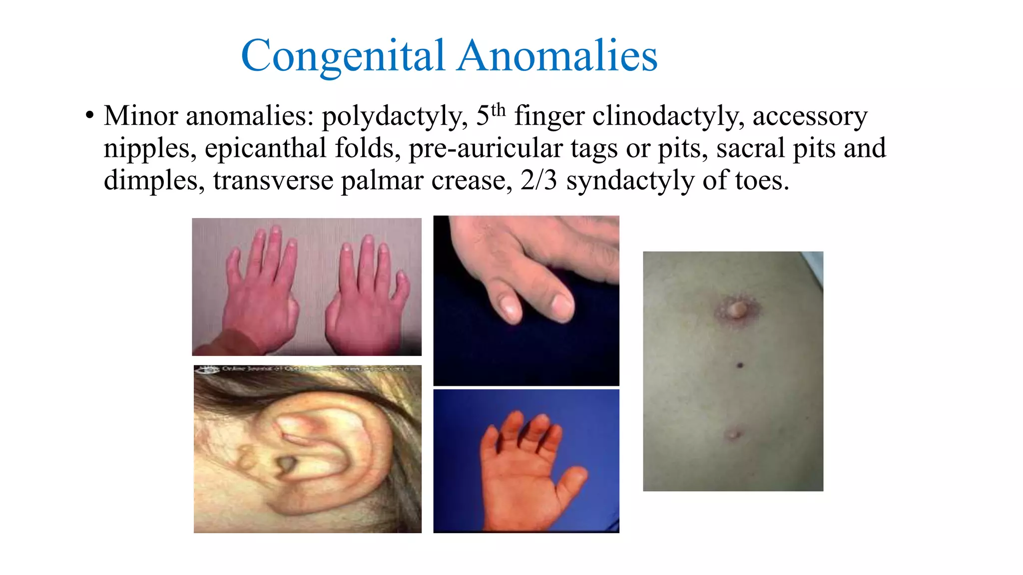
- Types of congenital anomalies like major anomalies that interfere with normal functioning and minor anomalies that have only cosmetic significance.
- Causes of congenital anomalies which can be genetic like chromosomal or single gene defects, or non-genetic like drugs, infections, or maternal illness.
- Stages of normal morphogenesis and how abnormalities can occur if stages are incomplete, take an aberrant form, or functional defects develop. Timing of different malformations is outlined.
- Specific genetic syndromes are mentioned as causes for different malformation patterns. Deletion 22q11 syndrome is discussed in detail as a common microdeletion syndrome.
The Indian Dental Academy is the Leader in continuing dental education , training dentists in all aspects of dentistry and
offering a wide range of dental certified courses in different formats.for more details please visit
www.indiandentalacademy.com
The Indian Dental Academy is the Leader in continuing dental education , training dentists in all aspects of dentistry and
offering a wide range of dental certified courses in different formats.for more details please visit
www.indiandentalacademy.com
Neurofibromatosis cannot be prevented. People with a family history of the disease may choose to undergo genetic testing and counseling to determine if they are at risk for transmitting NF to their offspring.
Dandy–Walker malformation (DWM) encompasses cystic dilatation of the fourth ventricle, complete or partial agenesis of cerebella vermis and enlarged posterior fossa while Dandy–Walker variant (DWV) comprises cystic posterior mass with variable hypoplasia of the cerebella vermis and no enlargement of the posterior fossa.
Craniofacial syndromes /certified fixed orthodontic courses by Indian dental ...Indian dental academy
Indian Dental Academy is the Leader in continuing dental education , training dentists in all aspects of dentistry and offering a wide range of dental certified courses in different formats.
The Indian Dental Academy is the Leader in continuing dental education , training dentists in all aspects of dentistry and
offering a wide range of dental certified courses in different formats.for more details please visit
www.indiandentalacademy.com
263778731218 Abortion Clinic /Pills In Harare ,sisternakatoto
263778731218 Abortion Clinic /Pills In Harare ,ABORTION WOMEN’S CLINIC +27730423979 IN women clinic we believe that every woman should be able to make choices in her pregnancy. Our job is to provide compassionate care, safety,affordable and confidential services. That’s why we have won the trust from all generations of women all over the world. we use non surgical method(Abortion pills) to terminate…Dr.LISA +27730423979women Clinic is committed to providing the highest quality of obstetrical and gynecological care to women of all ages. Our dedicated staff aim to treat each patient and her health concerns with compassion and respect.Our dedicated group ABORTION WOMEN’S CLINIC +27730423979 IN women clinic we believe that every woman should be able to make choices in her pregnancy. Our job is to provide compassionate care, safety,affordable and confidential services. That’s why we have won the trust from all generations of women all over the world. we use non surgical method(Abortion pills) to terminate…Dr.LISA +27730423979women Clinic is committed to providing the highest quality of obstetrical and gynecological care to women of all ages. Our dedicated staff aim to treat each patient and her health concerns with compassion and respect.Our dedicated group of receptionists, nurses, and physicians have worked together as a teamof receptionists, nurses, and physicians have worked together as a team wwww.lisywomensclinic.co.za/
Recomendações da OMS sobre cuidados maternos e neonatais para uma experiência pós-natal positiva.
Em consonância com os ODS – Objetivos do Desenvolvimento Sustentável e a Estratégia Global para a Saúde das Mulheres, Crianças e Adolescentes, e aplicando uma abordagem baseada nos direitos humanos, os esforços de cuidados pós-natais devem expandir-se para além da cobertura e da simples sobrevivência, de modo a incluir cuidados de qualidade.
Estas diretrizes visam melhorar a qualidade dos cuidados pós-natais essenciais e de rotina prestados às mulheres e aos recém-nascidos, com o objetivo final de melhorar a saúde e o bem-estar materno e neonatal.
Uma “experiência pós-natal positiva” é um resultado importante para todas as mulheres que dão à luz e para os seus recém-nascidos, estabelecendo as bases para a melhoria da saúde e do bem-estar a curto e longo prazo. Uma experiência pós-natal positiva é definida como aquela em que as mulheres, pessoas que gestam, os recém-nascidos, os casais, os pais, os cuidadores e as famílias recebem informação consistente, garantia e apoio de profissionais de saúde motivados; e onde um sistema de saúde flexível e com recursos reconheça as necessidades das mulheres e dos bebês e respeite o seu contexto cultural.
Estas diretrizes consolidadas apresentam algumas recomendações novas e já bem fundamentadas sobre cuidados pós-natais de rotina para mulheres e neonatos que recebem cuidados no pós-parto em unidades de saúde ou na comunidade, independentemente dos recursos disponíveis.
É fornecido um conjunto abrangente de recomendações para cuidados durante o período puerperal, com ênfase nos cuidados essenciais que todas as mulheres e recém-nascidos devem receber, e com a devida atenção à qualidade dos cuidados; isto é, a entrega e a experiência do cuidado recebido. Estas diretrizes atualizam e ampliam as recomendações da OMS de 2014 sobre cuidados pós-natais da mãe e do recém-nascido e complementam as atuais diretrizes da OMS sobre a gestão de complicações pós-natais.
O estabelecimento da amamentação e o manejo das principais intercorrências é contemplada.
Recomendamos muito.
Vamos discutir essas recomendações no nosso curso de pós-graduação em Aleitamento no Instituto Ciclos.
Esta publicação só está disponível em inglês até o momento.
Prof. Marcus Renato de Carvalho
www.agostodourado.com
Ethanol (CH3CH2OH), or beverage alcohol, is a two-carbon alcohol
that is rapidly distributed in the body and brain. Ethanol alters many
neurochemical systems and has rewarding and addictive properties. It
is the oldest recreational drug and likely contributes to more morbidity,
mortality, and public health costs than all illicit drugs combined. The
5th edition of the Diagnostic and Statistical Manual of Mental Disorders
(DSM-5) integrates alcohol abuse and alcohol dependence into a single
disorder called alcohol use disorder (AUD), with mild, moderate,
and severe subclassifications (American Psychiatric Association, 2013).
In the DSM-5, all types of substance abuse and dependence have been
combined into a single substance use disorder (SUD) on a continuum
from mild to severe. A diagnosis of AUD requires that at least two of
the 11 DSM-5 behaviors be present within a 12-month period (mild
AUD: 2–3 criteria; moderate AUD: 4–5 criteria; severe AUD: 6–11 criteria).
The four main behavioral effects of AUD are impaired control over
drinking, negative social consequences, risky use, and altered physiological
effects (tolerance, withdrawal). This chapter presents an overview
of the prevalence and harmful consequences of AUD in the U.S.,
the systemic nature of the disease, neurocircuitry and stages of AUD,
comorbidities, fetal alcohol spectrum disorders, genetic risk factors, and
pharmacotherapies for AUD.
HOT NEW PRODUCT! BIG SALES FAST SHIPPING NOW FROM CHINA!! EU KU DB BK substit...GL Anaacs
Contact us if you are interested:
Email / Skype : kefaya1771@gmail.com
Threema: PXHY5PDH
New BATCH Ku !!! MUCH IN DEMAND FAST SALE EVERY BATCH HAPPY GOOD EFFECT BIG BATCH !
Contact me on Threema or skype to start big business!!
Hot-sale products:
NEW HOT EUTYLONE WHITE CRYSTAL!!
5cl-adba precursor (semi finished )
5cl-adba raw materials
ADBB precursor (semi finished )
ADBB raw materials
APVP powder
5fadb/4f-adb
Jwh018 / Jwh210
Eutylone crystal
Protonitazene (hydrochloride) CAS: 119276-01-6
Flubrotizolam CAS: 57801-95-3
Metonitazene CAS: 14680-51-4
Payment terms: Western Union,MoneyGram,Bitcoin or USDT.
Deliver Time: Usually 7-15days
Shipping method: FedEx, TNT, DHL,UPS etc.Our deliveries are 100% safe, fast, reliable and discreet.
Samples will be sent for your evaluation!If you are interested in, please contact me, let's talk details.
We specializes in exporting high quality Research chemical, medical intermediate, Pharmaceutical chemicals and so on. Products are exported to USA, Canada, France, Korea, Japan,Russia, Southeast Asia and other countries.
- Video recording of this lecture in English language: https://youtu.be/lK81BzxMqdo
- Video recording of this lecture in Arabic language: https://youtu.be/Ve4P0COk9OI
- Link to download the book free: https://nephrotube.blogspot.com/p/nephrotube-nephrology-books.html
- Link to NephroTube website: www.NephroTube.com
- Link to NephroTube social media accounts: https://nephrotube.blogspot.com/p/join-nephrotube-on-social-media.html
New Directions in Targeted Therapeutic Approaches for Older Adults With Mantl...i3 Health
i3 Health is pleased to make the speaker slides from this activity available for use as a non-accredited self-study or teaching resource.
This slide deck presented by Dr. Kami Maddocks, Professor-Clinical in the Division of Hematology and
Associate Division Director for Ambulatory Operations
The Ohio State University Comprehensive Cancer Center, will provide insight into new directions in targeted therapeutic approaches for older adults with mantle cell lymphoma.
STATEMENT OF NEED
Mantle cell lymphoma (MCL) is a rare, aggressive B-cell non-Hodgkin lymphoma (NHL) accounting for 5% to 7% of all lymphomas. Its prognosis ranges from indolent disease that does not require treatment for years to very aggressive disease, which is associated with poor survival (Silkenstedt et al, 2021). Typically, MCL is diagnosed at advanced stage and in older patients who cannot tolerate intensive therapy (NCCN, 2022). Although recent advances have slightly increased remission rates, recurrence and relapse remain very common, leading to a median overall survival between 3 and 6 years (LLS, 2021). Though there are several effective options, progress is still needed towards establishing an accepted frontline approach for MCL (Castellino et al, 2022). Treatment selection and management of MCL are complicated by the heterogeneity of prognosis, advanced age and comorbidities of patients, and lack of an established standard approach for treatment, making it vital that clinicians be familiar with the latest research and advances in this area. In this activity chaired by Michael Wang, MD, Professor in the Department of Lymphoma & Myeloma at MD Anderson Cancer Center, expert faculty will discuss prognostic factors informing treatment, the promising results of recent trials in new therapeutic approaches, and the implications of treatment resistance in therapeutic selection for MCL.
Target Audience
Hematology/oncology fellows, attending faculty, and other health care professionals involved in the treatment of patients with mantle cell lymphoma (MCL).
Learning Objectives
1.) Identify clinical and biological prognostic factors that can guide treatment decision making for older adults with MCL
2.) Evaluate emerging data on targeted therapeutic approaches for treatment-naive and relapsed/refractory MCL and their applicability to older adults
3.) Assess mechanisms of resistance to targeted therapies for MCL and their implications for treatment selection
NVBDCP.pptx Nation vector borne disease control programSapna Thakur
NVBDCP was launched in 2003-2004 . Vector-Borne Disease: Disease that results from an infection transmitted to humans and other animals by blood-feeding arthropods, such as mosquitoes, ticks, and fleas. Examples of vector-borne diseases include Dengue fever, West Nile Virus, Lyme disease, and malaria.
Title: Sense of Smell
Presenter: Dr. Faiza, Assistant Professor of Physiology
Qualifications:
MBBS (Best Graduate, AIMC Lahore)
FCPS Physiology
ICMT, CHPE, DHPE (STMU)
MPH (GC University, Faisalabad)
MBA (Virtual University of Pakistan)
Learning Objectives:
Describe the primary categories of smells and the concept of odor blindness.
Explain the structure and location of the olfactory membrane and mucosa, including the types and roles of cells involved in olfaction.
Describe the pathway and mechanisms of olfactory signal transmission from the olfactory receptors to the brain.
Illustrate the biochemical cascade triggered by odorant binding to olfactory receptors, including the role of G-proteins and second messengers in generating an action potential.
Identify different types of olfactory disorders such as anosmia, hyposmia, hyperosmia, and dysosmia, including their potential causes.
Key Topics:
Olfactory Genes:
3% of the human genome accounts for olfactory genes.
400 genes for odorant receptors.
Olfactory Membrane:
Located in the superior part of the nasal cavity.
Medially: Folds downward along the superior septum.
Laterally: Folds over the superior turbinate and upper surface of the middle turbinate.
Total surface area: 5-10 square centimeters.
Olfactory Mucosa:
Olfactory Cells: Bipolar nerve cells derived from the CNS (100 million), with 4-25 olfactory cilia per cell.
Sustentacular Cells: Produce mucus and maintain ionic and molecular environment.
Basal Cells: Replace worn-out olfactory cells with an average lifespan of 1-2 months.
Bowman’s Gland: Secretes mucus.
Stimulation of Olfactory Cells:
Odorant dissolves in mucus and attaches to receptors on olfactory cilia.
Involves a cascade effect through G-proteins and second messengers, leading to depolarization and action potential generation in the olfactory nerve.
Quality of a Good Odorant:
Small (3-20 Carbon atoms), volatile, water-soluble, and lipid-soluble.
Facilitated by odorant-binding proteins in mucus.
Membrane Potential and Action Potential:
Resting membrane potential: -55mV.
Action potential frequency in the olfactory nerve increases with odorant strength.
Adaptation Towards the Sense of Smell:
Rapid adaptation within the first second, with further slow adaptation.
Psychological adaptation greater than receptor adaptation, involving feedback inhibition from the central nervous system.
Primary Sensations of Smell:
Camphoraceous, Musky, Floral, Pepperminty, Ethereal, Pungent, Putrid.
Odor Detection Threshold:
Examples: Hydrogen sulfide (0.0005 ppm), Methyl-mercaptan (0.002 ppm).
Some toxic substances are odorless at lethal concentrations.
Characteristics of Smell:
Odor blindness for single substances due to lack of appropriate receptor protein.
Behavioral and emotional influences of smell.
Transmission of Olfactory Signals:
From olfactory cells to glomeruli in the olfactory bulb, involving lateral inhibition.
Primitive, less old, and new olfactory systems with different path
Ozempic: Preoperative Management of Patients on GLP-1 Receptor Agonists Saeid Safari
Preoperative Management of Patients on GLP-1 Receptor Agonists like Ozempic and Semiglutide
ASA GUIDELINE
NYSORA Guideline
2 Case Reports of Gastric Ultrasound
2. Congenital Malformations
• Incidence
• Types of Congenital anomalies/ Classification
• Terminologies
Basic Concepts
• Normal Morphogenesis
• Normal Stages in Morphogenesis
• Abnormal Morphogenesis
• Developmental Pathology - various systems
Causes
GENETIC
Chromosomal
Single gene
Multifactorial
NON GENETIC
Drugs
Infections
Maternal Illness
3. Introduction
More than 5000 dysmorphic and multiple congenital anomaly
syndromes are described in the London Dysmorphology Database-
single gene disorders
sporadic and non-genetic condition
teratogenic conditions
5. Incidence
Spontaneous First-Trimester Pregnancy Loss
Chromosome abnormalities (trisomy, monosomy, or triploidy) - 50% of all
spontaneous abortions.
60% - gross structural abnormality is present
Perinatal deaths: 25% to 30% - serious structural abnormality
80% of these cases genetic factors can be implicated.
Newborns: 2% to 3% - one major abnormality.
The true incidence - 5%.
Minor abnormalities - 10% of all newborns.
If two or more minor abnormalities - 10% to 20% risk that the baby will also
have a major malformation.
7. Definition and Classification
A malformation is a morphological abnormality that arises due to a
primary, intrinsic developmental defect.
Caused by genetic/ environmental causes or both; many have a
multifactorial etiology; risk of recurrence accordingly varies
Abnormal processes during organogenesis
8. Types of congenital anomalies
• Congenital anomalies:
A. Major: interfere with the normal functioning of an
individual; pose a significant health/ social burden
B. Minor: do not interfere with the normal functioning
of an individual; usually have only cosmetic
significance and no other significant health problem.
9. Congenital Anomalies
• Major anomalies: Eg. Spina bifida, anencephaly,
congenital heart disease, cleft palate, omphalocele etc.
10. • Minor anomalies: polydactyly, 5th finger clinodactyly, accessory
nipples, epicanthal folds, pre-auricular tags or pits, sacral pits and
dimples, transverse palmar crease, 2/3 syndactyly of toes.
Congenital Anomalies
11. Common Single primary defects in development
• Malformation-
• Cleft lip +/- Palate
• Cleft palate
• Cardiac Septal defects
• Defect in Neural tube closure
• Deformation
• CDH
• CTEV
• Dysplasia
• Achondroplasia
• Tuberous sclerosis
Congenital Anomalies
12. Abnormal Morphogenesis
1. Malformation – poor formation of tissue
2. Deformation – extrinsic forces on normal tissue
3. Disruption – breakdown of previously normal tissue
4. Dysplasia – lack of normal organization of cells into tissue
13. Deformation
• Examples:
• Club feet
• Torticollis
• Plagiocephaly
• Risk of recurrence is minimal unless the same mechanical
forces are operative in the next pregnancy (eg. maternal uterine
malformation)
14. Disruption
• Examples:
• Amniotic band sequence
• Tissue ischaemia
• Risk of recurrence is minimal unless genetic causes are
involved
15. Dysplasia
• Dysplasia is an abnormal cellular organization within
a tissue resulting in structural changes, e.g. within
cartilage and bone in
skeletal dysplasias.
• Almost always genetic in etiology.
16. Syndrome
• A dysmorphic syndrome is a recognized composite pattern of 2
or more anomalies with a common specific etiology.
• Most syndromes are made up of 1 or more major anomalies
together with a variable number of minor anomalies.
• Rarely, any one of these features is diagnostic.
• More often the entire constellation of defects must be taken into
consideration to define the diagnosis.
• Etiology: genetic/ a specific environmental agent eg. teratogen
17. Association
• Association is the non-random occurrence of 2 or more
anomalies that occur more frequently than expected by chance
alone, but for which no specific etiology has been
demonstrated.
• Core anomalies usually consist of 5 or more features but any
individual patient rarely has all the core features.
• When 1 component of an association is noted it prompts the
search for others.
• Example: VACTERL association (vertebral, anal, cardiac,
tracheo-esophageal, renal and limb anomalies)
18. Sequence
• A sequence is a pattern of anomalies that results from a single
primary anomaly or a single mechanical factor.
• The factor or event that initiates the sequence may produce
multiple secondary anomalies or may produce a secondary
anomaly that leads to a tertiary anomaly and so on in a
cascading manner.
• The etiology is often heterogeneous.
20. Field Defects
• Field defects are pattern of multiple anomalies that arise from
disturbance of a single developmental field during embryonic
development.
• Etiology could be defect in a single gene, vascular disturbance
etc.
• Two types:
• polytopic field defects (resultant anomalies are in different
body areas) Eg. Goldenhar syndrome
• Monotopic field defects (resultant anomalies are limited to a
single body area) eg. Holoprosencephaly with midline cleft
palate, sacral agenesis and Poland anomaly
21. Complex & Spectrum
• Complex is a general term used to define a composite of
manifestations. It is sometimes used to describe developmental
field defects eg. sacral agenesis complex.
• Spectrum is sometimes used to describe entities with multiple
features particularly those in which prominent features can be
expressed with considerable variation eg. neurofibromatosis
spectrum
23. Phenomena of Morphogenesis
• Zygote – genetic information that guides morphogenesis and
function.
• After first few divisions - differentiation – by activation and
inactivation of certain genes – diverse roles of cells.
• Programmed timely – sequential order – early morphogenesis – no
scope for error.
24. Normal Morphogenesis
1. Cell Migration – germ cells move from yolk sac ectoderm to
urogenital ridge to form Gonads.
2. Control over Mitotic rate – size and shape
3. Interaction between Adjacent Tissues –
• Optic disc – lens from overlying ectoderm
• Ureteric bud – kidney from metanephric tissue
• Notochord – overlying neural tissue
• Prechordal mesoderm – overlying forebrain
25. Normal Morphogenesis
4. Adhesive Associations of Like Cells – like long bones
5. Controlled Cell Death – finger separation, recanalization of
duodenum
6. Hormonal Influence over Morphogenesis – 8 weeks –Y
chromosome – testosterone - phallus enlargement
7. Mechanical forces – size, form – brain, calvarium, upper face
26. Normal Stages in Morphogenesis
1st week – cell division without much enlargement. Metabolic needs –
ova
Day 8 – outlying trophoblast cells invade the endometrium. Early
placenta – nourishment.
Inner cell mass is bilaminar – ectoderm – amniotic cavity
Endoderm – yolk sac
Day 14 – primitive node develops into ectoderm – embryo has axis.
Mesoderm starts developing
27. Normal Stages in Morphogenesis
3rd week – mesoderm -heart begins to develop. Blood cells produced
in yolk sac.
Paraxial mesoderm segmented into somites - Foregut and hindgut
pouches
28. Types of Malformation
Incomplete Morphogenesis – incomplete stages in the development
of a structure examples:
Lack of development – Renal agenesis secondary to failure of ureter
formation.
Hypoplasia – micrognathia
Incomplete separation - syndactyly
Incomplete closure – cleft palate
Incomplete septation - VSD
Incomplete migration of mesoderm – exostrophy of bladder
Incomplete rotation – malrotation of gut
Incomplete resolution of early form – Meckel diverticulum
Persistence of earlier location - cryptorhidism
29. Types of Malformation
Aberrant form – that never exists in normal morphogenesis
Example pelvic spur in nail patella syndrome
Accessory tissue – presumed to have been initiated at about the same
time as the normal tissue.
Example polydactyly, pre auricular tags, accessory spleens.
Functional defects – clubfoot
30. Relative Timing of Malformations
CNS
Face
Gut
GU system
Heart
Limb
Complex
31. CNS
Anencephaly – prior to 26 days
Defect in Closure of anterior neural tube
Degeneration of forebrain
MMC – prior to 28 days
Defect in Closure of posterior neural tube
80% lumbosacral
32. Face
Cleft Lip - prior to 36 days
Defect in closure of lip
42% associated with cleft palate
Cleft maxillary palate - prior to 10 weeks
Defect in closure of maxillary palatal shelves
Branchial sinus or cyst - prior to 8 weeks
Defect in resolution of branchial cleft
Preauricular and along the line anterior to SCM
33. Cleft Lip/ Palate
22q del (CP)
Kabuki syndrome (CP)
van der Woude syndrome (VWS—CL/P and CP)
Stickler syndrome (CP)
Treacher–Collins syndrome (CP)
34. Cleft Lip/ Palate
Smith–Lemli–Opitz (SLO) syndrome (CP)
XL CP and ankyloglossia
Ectrodactyly, ectodermal dysplasia, and clefting syndrome (EEC; CL/P)
Non-syndromic CL/P due to MSX1
35. Gut
Esophageal atresia - < 30 days
Defect in Lateral septation of foregut into trachea and foregut
Rectal atresia - < 6 weeks
Defect in Lateral septation of cloaca into rectum and urogenital sinus
Duodenal atresia - < 7-8 weeks
Defect in Recanalisation of duodenum
Malrotation of gut - < 10 weeks
Defect in Rotation of intestinal loop so that cecum lies to the right
36. Gut
Omphalocele - < 10 weeks
Defect in return of midgut from yolk sac to abdomen
Meckel diverticulum - < 10 weeks
Defect in Obliteration of vitelline duct
Diaphragmatic hernia - < 6 weeks
Defect in Closure of pleuroperitoneal canal
37. Tracheo-oesophageal fistula or oesophageal atresia
VATER/VACTERL
Goldenhar(facio-auriculo-
vertebral)syndrome.
OEIS
Teratogens
Feingold syndrome.
CHARGE syndrome
Opitz syndrome
Mandibulo-facial dysostosis with
microceph- aly (MFDM).
AEG syndrome.
Martinez–Frias syndrome.
Hydrocephalus with features of VATER.
Disorders of laterality due to mutations in
ZIC3.
39. Genitourinary
Exstrophy of bladder - < 30 days
Defect in Migration of infraumblical mesenchyme
Associated with Mullerian and Wolfian duct defects
Bicornuate uterus - <10 weeks
Defect in Fusion of lower portion of mullrian ducts
Hypospadias - < 12 weeks
Defect in Fusion of urethral folds
Cryptorchidism – 7-9 months
Defect in descent
40. Genitourinary
Smith–Lemli–Opitz syndrome (SLO)
WAGR (Wilms tumour–aniridia–genitourinary anomalies– mental retardation)
Drash syndrome or Wilms tumour and pseudohermaphroditism and Frasier syndrome
Opitz syndrome
Mowat–Wilson syndrome
Hand–foot–genital syndrome
41. Heart
TGV - < 34 days
Directional development of bulbus cordis septum
del 22q11
VSD - < 6 weeks
Perimembranous VSD is the most common cardiac defect seen in trisomy 21;
trisomy 18 and trisomy 13, del 22q11, FAS, maternal diabetes
PDA – 9-10 months
43. Limb
Aplasia of radius – prior to 38 days
Defect in genesis of radial bone
Syndactyly, severe – prior to 6 weeks
Defect in separation of digital rays
48. Limb
Thumb anomalies
Thrombocytopenia–absent radius (tar) syndrome.
Holt–oram syndrome
Okihiro syndrome (duane radial ray syndrome).
Acrofacial dysostosis with limb defects (miller syndrome, post-axial acrofacial
dysostosis syndrome (POADS).
Ulnar–mammary syndrome.
49. Limb
Split hand/foot malformation (SHFM)
Ectrodactyly–ectodermal dysplasia–clefting (EEC) syndrome.
10q duplication
Mesoaxial synostoticc syndactyly (MSSD).
Multiple types of reduction defect
Cornelia de lange syndrome (cdls)
Femur–fibula–ulna complex
Roberts syndrome
50. Complex
Cyclopia, holoprosencephaly – prior to 23 days
Defect in prechordal mesoderm development
Secondary defects of mid and forebrain
Chromosomal imbalance
AD HPE
Environmental causes
Pallister–Hall syndrome
Smith–Lemli–Opitz (SLO) syndrome
Pseudotrisomy 13 or HPE polydactyly syndrome
Fetal akinesia sequence
52. Deletion 22q11 syndrome
• DiGeorge/ Sedláčková syndrome/ Velocardiofacial Syndrome
• Most common of the micro- deletion syndrome- approximately 1:4000 births,
• Usually sporadic - autosomal dominant inheritance in some families.
• The molecular defect is a 3-Mb microdeletion on chromosome 22 (22q11.2).
deleted DNA segment is still called the DiGeorge Critical Region - phenotypic
features may be due largely to haploinsufficiency for the TBX1 gene
• Characterized by heart malformations (particularly those involving the cardiac
outflow tract), thymic and parathyroid hypoplasia, cleft palate and typical
facies.
• Approximately 25% have schizophrenia-like episodes in adult life
54. Neural Tube Defects
PREVALENCE - Southeast Asia (15.8 per 10,000 births)
Differences in genetic predisposition, environmental factors and
application of interventions.
Factors causing decrease in the prevalence of open NTDs-
1. Periconceptional folic acid supplementation, food fortification
with folic acid.
2. prenatal screening for open NTDs combined with access to
pregnancy termination (83 percent of anencephaly and 63 percent
of spina bifida affected pregnancies are terminated)
55. Types of Neural Tube Defects
• Open NTDs (80 percent) - defect is only covered by a membrane or,
rarely, nothing at all
Myelomeningocele (spina bifida)
Meningocele
Encephalocele
Anencephaly
• can be associated with cerebral ventriculomegaly.
56. • Closed NTDs (20%) - defect is covered by skin
Lipomyelomeningocele
Lipomeningocele
• associated with a tuft of hair, dimple, birthmark, lump, or other skin
abnormality at the site of the defect, as well as cerebral
ventriculomegaly.
57. Etiology
• Most isolated open NTDs appear to be caused by folate deficiency,
likely in combination with genetic or other environmental risk factors.
• Open NTDs have also been associated with some fetal syndromes,
amniotic band sequence, hyperthermia in early pregnancy,
pregestational diabetes, and obesity.
58. Screening
• We perform ultrasound screening for NTDs (ideally at 18 to 20 weeks)
because it appears to detect more open NTDs than maternal serum
alpha-fetoprotein (MSAFP) screening.
• If optimal images of the fetal spine or intracranial anatomy are not
obtained (eg, fetal position or maternal obesity) or high quality
ultrasound examination is unavailable, MSAFP screening should be
offered.
59. • For maternal serum alpha-fetoprotein (MSAFP) screening, the
American College of Medical Genetics and Genomics reports –
• the detection rate for anencephaly is ≥95 percent at 2.0 or 2.5
multiples of the median (MoMs).
• open spina bifida, the detection rate at 2.0 MoMs is 75 to 90 percent
versus 65 to 80 percent at 2.5 MoMs, with false-positive rates of 2 to 5
percent and 1 to 3 percent, respectively.
60. Recurrence risk
• Recurrence is likely related to a multifactorial inheritance pattern,
including genetic and environmental risk factors.
• The risk of recurrence for isolated NTDs is approximately 2 to 4
percent with one affected sibling
• With two affected siblings, the risk is approximately 10 percent
• The risk of recurrence appears to be higher in countries such as Ireland
where the occurrence (prevalence) of NTDs is high
61.
62. • The recurrence risk for anencephaly is estimated at 2 to 5 percent.
• may be part of specific genetic syndromes if there are associated
anomalies –the risk of recurrence for an encephalocele with Meckel-
Gruber syndrome (posterior encephalocele, cleft palate, and
polydactyly) is 25 percent secondary to known autosomal recessive
inheritance.
• Testing for MTHFR polymorphisms is not recommended as
routine folic acid supplementation at 0.4 mg/day will adequately
increase red cell and serum folate concentrations whether or not the
woman has a polymorphism.
65. Noonan syndrome
• Prevalence 1 in 1000 to 1 in 2500 live births
• Characteristic features :
• Facial features
• Short stature
• Congenital heart defects
• Autosomal dominant
• Complete penetrance but variable expressivity
• Mutations in PTPN11 on chromosome 12
- Missense (50%), gain of function (48%),
66. • Facial features
•Most characteristic in infancy and early
childhood
•Broad high forehead
•Eyes: hypertelorism, epicanthal folds,
down slanting palpebral fissures, light
colored iris
•Ears: low set, posteriorly rotated, thick
helix
•Lips: thick upper lip, deeply grooved
philtrum
67. • Webbing of neck
• Low posterior hairline
• Chest: (70-95%)
• pectus carinatum superiorly
• Pectus excavatum inferiorly
• Wide spaced nipples
• Cubitus valgus in 50 %
• Joint hyperextensibility in 50%
68. Systemic manifestations
• CVS: seen in 60 to 80 % cases
- ECG – wide QRS, left axis deviation, giant Q
waves, negative pattern in left precordial leads
• Oral- high arched palate, dental malocclusion
• Genitourinary:
• Males: many are infertile
• Cryptorchidism (77%)
• Sertoli cell dysfunction
• Females : mostly fertile
69. Systemic manifestations
• Growth
• Short stature: proportionate (60%)
• Normal birth weight and length
• Respond to an extent to growth hormone
therapy
• Hematological / oncological
• Bleeding disorders(55%):
• Coagulation factor defeciencies
• Thrombocytopenia
• Myeloproliferative disorders
72. Noonan and Other RASopathies
Half cases - protein tyrosine phosphatase, non-receptor-type, 11 (PTPN11) gene.
phenotype-genotype correlation - mutation- positive cases - higher frequency of pulmonary stenosis
facial features are similar.
PTPN11-negative cases:
SOS1, SHOC2, KRAS, RIT1, and MAPZK1 genes
Pathway, known as RAS-MAPK
Protein product of PTPN11 is SHP-2 and this, together with SOS1, positively transduces signals to Ras-
GTP, a downstream effector.
The KRAS mutations in NS appear to lead to K-ras proteins with impaired responsiveness to GTPase
activating proteins
Neurofibromatosis, the most common disorder of this group
73.
74. Genes of the RAS-MAPK Pathway and Associated
Syndromes
75. Noonan and Other RASopathies
spectrum of disorders - overlapping features – NS, cardio- facio-
cutaneous, Costello syndromes
RAS-MAPK pathway - for RAS to bind GTP, which results in
activation of the pathway (gain-of-function)- increase in solid tumors
in Costello syndrome,
cellular proliferation in some tissues in cardio-facio-cutaneous
syndrome (e.g., hyperkeratosis).
tumor suppressor - Neurofibromin is a GTPase activating protein
78. Take home messages
• Understanding the type of Congenital Malformation and classifying it,
helps in identifying the etiology.
• Further management of index patient and genetic counselling
regarding risk of recurrence depends on the underlying cause.
79. References:
1. Smith’s recognizable patterns of malformation (7th edition)
2. Emery and Rimoin’s Principles and Practice of Medical Genetics
(6th edition)
3. Oxford Desk Reference Clinical Genetics & Genomics (2nd
edition)