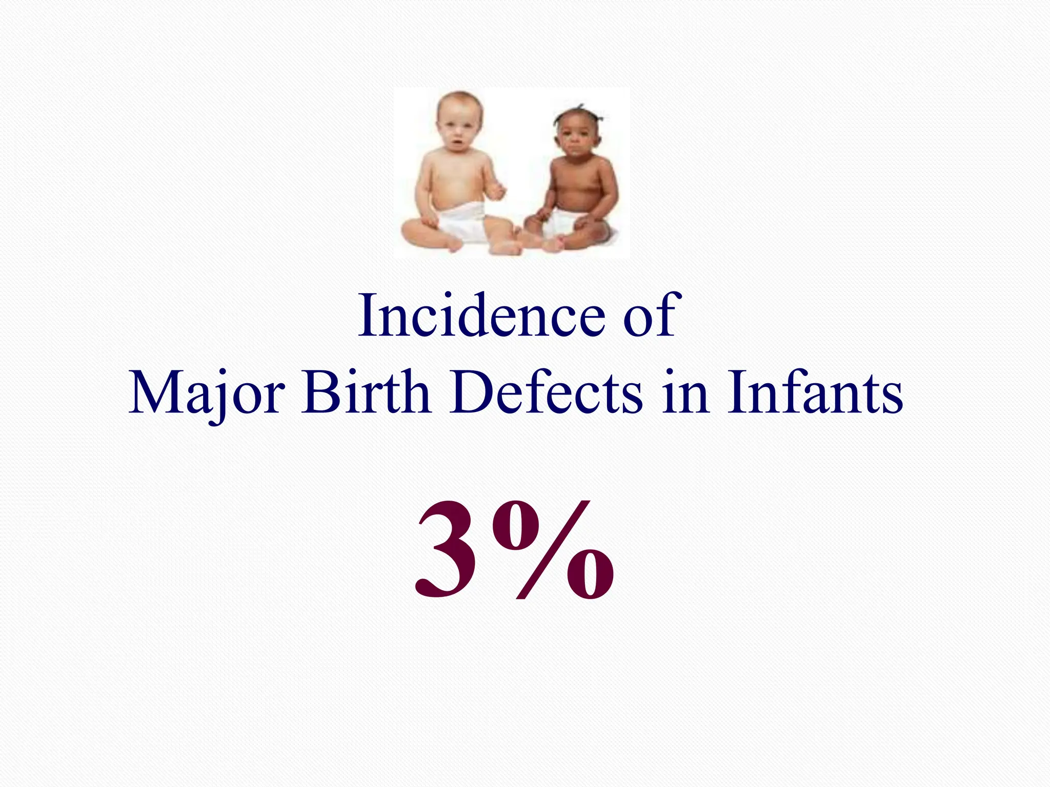
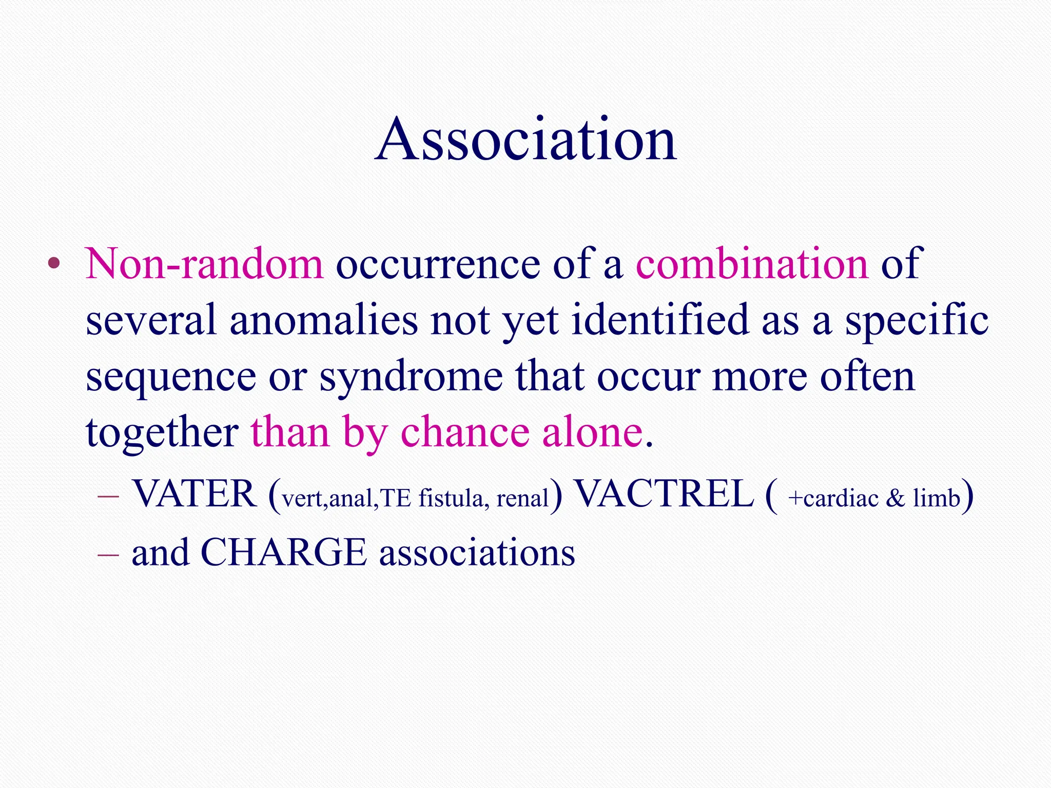
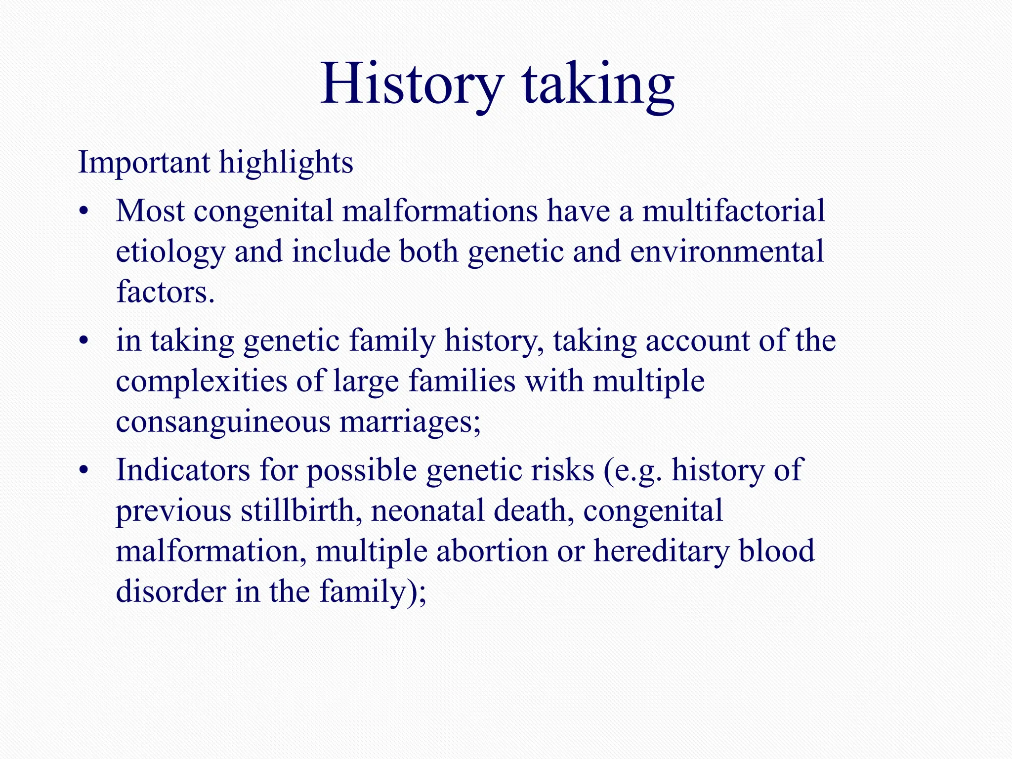
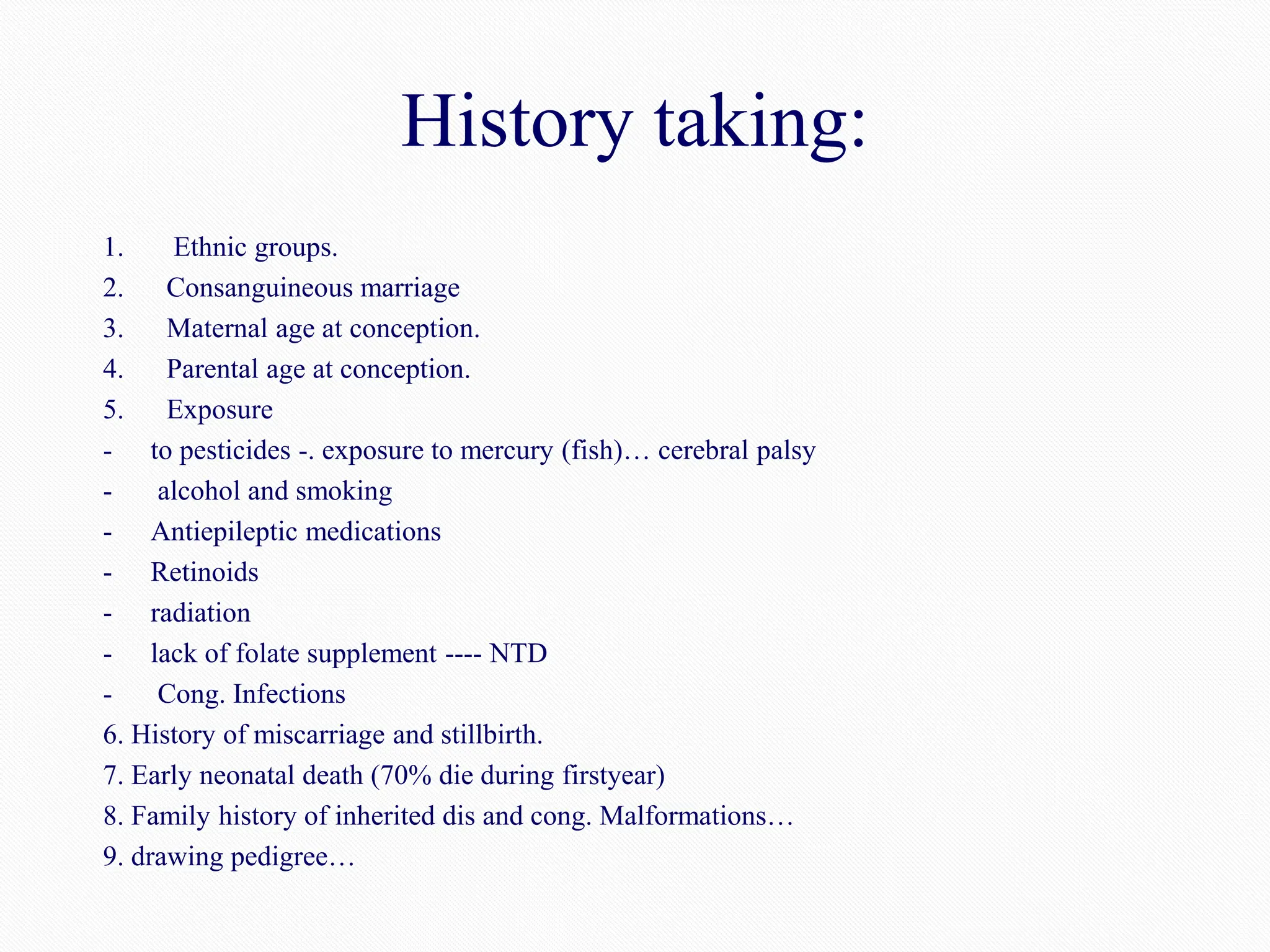
Basic Concepts of Dysmorphology
DR. KHALED IBRAHIM
MBBCH, MSc, PHd, MRCPCH, FRCPCH
Objectives
• Definition of dysmorphology
• Know that defects are common and have a major impact on health
• Recognize normal variants and dysmorphic features and know
they may serve as clues to diagnoses
• Know that four basic different kinds of errors of morphogenesis
can occur. (Malformation, Deformation, Disruption and
Dysplasia)
• Know what is meant by syndrome, sequence, and association
• Approach to a dysmorphic individual:
Suspicion & analysis
Systematic physical examination
Confirmation of diagnosis
Intervention
Most of the photos included in this presentation are from
our patients after permission, few figures are retrieved from
literature.
Great Thanks To Those Families Who
Allowed Us To Take Photos Of Their
Babies
For Sake Of Teaching
Definition of dysmorphology
• The term “dysmorphology” was first coined by Dr.
David Smith,USA in 1960.
• “Dys” (Greek)=disordered or abnormal and
“Morph”=shape
• The term “dysmorphic” is used to describe individuals
whose physical features are not usually found in other
individuals with the same age or ethnic background.
Birth defects are the leading cause of infant mortality with
20 - 25% of perinatal deaths due to lethal birth defects
•* CDC annual estimate of 150,000 babies born with birth defects
•Source: National Center for Health Statistics, 1998 final natality and 1998 period linked birth/infant death data.
Prepared by March of Dimes Perinatal Data Center, July 2000
Incidence of
Major Birth Defects in Infants
3%
Just because it’s congenital
it doesn’t mean it’s genetic.
Causes of Birth Defects
• Multifactorial: 20-30%
• Single gene disorders: 10-20%
• Chromosomal: 15%
• Infection: 2.5%
• Maternal diabetes: 1.5%
• Maternal medications: 1-2%
• Rest unknown
Empiric Recurrence Risks (%)
for Selected Birth Defects
Condition Affected Relatives(s)
None 1 sib/parent 2 sibs / sib & parent
Cleft lip/palate 0.1 4 10-11
Neural Tube Defect 0.1 3 8
Heart Defect 0.3 4-5 10-11
The risk of having any one major birth defect is less than
1% but this risk increases significantly if other relatives
have same birth defect
What are the problems?
When did they happen?
How did they arise?
Why did they occur?
What is the diagnosis?
Who else is at risk?
Where can the patient/family get help?
Dysmorphology exam helpful when there is:
• Abnormal or unusual features and birth defects
• Abnormal growth and/or proportions
• Abnormal genitalia and/or puberty
• Psychomotor delays, speech delays, or mental retardation
• Abnormal neuromuscular function
• Bleeding tendencies
• Blindness or deafness
• Metabolic problems (eg. regression in abilities and/or behavior,
unusual body odors, excessive unexplained illness)
Classification of
birth anomalies
• Normal variations
• Minor anomalies
• Major anomalies
Facial features
Normal intercanthal spacing
Epicanthal folds
Unusual head shape
Dolichcephaly Brachycephaly
Trigonocephaly Plagiocephaly
Oxycephaly Cloverleaf skull
Brachycephaly (flat occiput)
Dolichocephaly: inc ant-post dt positional NICU
Trigonocephaly (turricephaly): genetic dt premature closure of metobic suture triangular frontal palpable suture
Oxycephaly: most severe craniocynostosis due to premature clos. of all sutures…tall head with small length and
width…Tower head(RECDL4 gene)8th cr nerve,, optic,mentalR, syndactly
Brachycephaly: flatt back improve by 6th month
Plagiocephaly:flattened at one area dt pressure PRETERM
Cloverleaf skull: sagittal, coronal,lambdoid
Head circumference
e.g. small head “microcephaly” (head circumference below -3SD)
or large head “macrocephaly”) (HC above +3SD)
Microcephaly
Macrocephaly
(Hydrocephalus)
26
Philtrum
Short philtrum
Long flat philtrum
37
Low set posteriorly rotated ears
Beckwith Wiedemann
Macrosomia
„
Macroglossia „
omphalocele „
Visceromegaly „
Embryonal tumors
„
skin
TUBEROUS SCLEROSIS
Ash-leaf macules
AD mapped to chromosome 9q34.3 (TSC1) and 16p13.3 (TSC2).
Neurofibromatosis
Abnormal growth
Limb anomalies
Brachydactyly (short fingers or toes)
Arachnodactyly (long fingers or toes)
39
Polysyndactyly
43
Hands & feet of a patient with Apert syndrome
Polydactyly
Postaxial polydactyly
(pedunculated post minimus) Complete postaxial polydactyly
42
Minor Anomaly
• Minor variations of normal morphological
features of little of no known medical,
surgical, or cosmetic significance
• Observed in < 4% of the population
Major Anomaly
• Abnormality that has medical, surgical, or
cosmetic significance
Types of Morphologic
Abnormalities
• Malformation
• Deformation
• Disruption
• Dysplasia
Malformation
Deformation
Disruption
Dysplasia
Normal
Malformation
• Defect of morphogenesis in an organ or structure due to an
intrinsically abnormal problem with formation, growth, or
differentiation of an organ or structure usually occurring
before 10 weeks of gestation.
• Examples:
– hypoplasia of an organ or structure (microtia),
incomplete closure (NTDs, cleft palate),
– incomplete separation (syndactaly)
Neural Tube Defects
Malformations are Not Specific
• The same morphological defect, or even a
similar pattern of abnormalities, may occur
as:
– An isolated anomaly in an otherwise normal individual
– A feature in a syndrome, sequence, or association
– A feature of a chromosome disorder, a single gene
defect, multifactorial disorder, or secondary to a
teratogenic effect
Deformation
• Abnormal form or position of a body or region of
the body caused by extrinsic non-disruptive
mechanical forces on a normally developing
structure (fetal constraint)
– clubfoot, congenital hip dislocation, craniofacial
asymmetry, over folded ear…..
Disruption
• Defect resulting from a destructive breakdown of,
or interference with, a normally developing
structure resulting in death of cells or tissue
destruction.
• May be secondary to mechanical forces, infections,
or vascular events.
– Loss of digit due to amniotic bands, lack of normal limb
development due to intrauterine vascular accident
Dysplasia
• Error of morphogenesis due to the abnormal cellular
organization or function in a specific type of tissue most
often due to single gene defects
– Achrondroplasia, ectodermal dysplasia, osteogenesis imperfecta,
Recognizable Patterns of
Anomalies
• Syndromes
• Associations
• Sequences or field defects
Consider a genetic condition or
syndrome when...
• Multiple anomalies
• More than 3 minor anomalies
• More than one major anomaly
• One major anomaly and a few minor
anomalies
Down syndrome
Syndrome
From Greek meaning “running together”
• Multiple anomalies in one or more tissues thought
to be pathologically related due to a specific
etiology (chromosome disorder, single gene
defect, environmental agent, or unknown factor).
– Down syndrome, Williams syndrome, FAS, Turner
syndrome….
Turner syndrome
Sequence/Field Defect
• Constellation of defects derived from a cascade of
effects related to a single known, or presumed,
localized abnormality (malformation, deformation,
disruption)
– Potter sequence
• Renal dysplasia, pulmonary hypoplasia, facial dysmorphisms
– Mandibular hypoplasia (Robin sequence)
• Cleft palate
– Meningomyelocele
• Club foot, hip dislocation, hydrocephalus
Association
• Non-random occurrence of a combination of
several anomalies not yet identified as a specific
sequence or syndrome that occur more often
together than by chance alone.
– VATER (vert,anal,TE fistula, renal) VACTREL ( +cardiac & limb)
– and CHARGE associations
Purposes of
Clinical Genetics Evaluations
• Recognize medical problems
• Make accurate diagnosis
• Provide prognosis and natural history information
• Discuss management
• Deliver appropriate medical care
• Minimize related complications
• Optimize quality of life
• Determine and provide recurrence risks
• Offer genetic and psychosocial counseling
• Provide anticipatory guidance and education
Management of Congenital Anomalies
• Conduct careful clinical evaluation
• Review family, prenatal history, and perinatal history
• Obtain diagnostic studies
– Imaging studies: Photographs, X-rays
– Laboratory studies: Chromosome, DNA, biochemical assays
• Provide medical management and genetic counseling
• If deceased, request autopsy and specific pathological analyses
– Name, photograph,obtain hair, memorialize, bury...
• Provide referrals to social work/psychological services and
support groups as appropriate
Management of Congenital Anomalies
• Children affected by congenital malformation are often fragile
patients with special needs that often require a multidisciplinary
and structured clinical approach.
• Conduct careful clinical evaluation
• Review family, prenatal history, and perinatal history
• Obtain diagnostic studies
– Imaging studies: Photographs, X-rays
– Laboratory studies: Chromosome, DNA, biochemical assays
• Provide medical management and genetic counseling
• If deceased, request autopsy and specific pathological analyses
– Name, photograph,obtain hair, memorialize, bury...
• Provide referrals to social work/psychological services and
support groups as appropriate
History taking
Important highlights
• Most congenital malformations have a multifactorial
etiology and include both genetic and environmental
factors.
• in taking genetic family history, taking account of the
complexities of large families with multiple
consanguineous marriages;
• Indicators for possible genetic risks (e.g. history of
previous stillbirth, neonatal death, congenital
malformation, multiple abortion or hereditary blood
disorder in the family);
History taking:
1. Ethnic groups.
2. Consanguineous marriage
3. Maternal age at conception.
4. Parental age at conception.
5. Exposure
- to pesticides -. exposure to mercury (fish)… cerebral palsy
- alcohol and smoking
- Antiepileptic medications
- Retinoids
- radiation
- lack of folate supplement ---- NTD
- Cong. Infections
6. History of miscarriage and stillbirth.
7. Early neonatal death (70% die during firstyear)
8. Family history of inherited dis and cong. Malformations…
9. drawing pedigree…
Making a diagnosis, even if
it’s a bad diagnosis, is more
useful for patients and their
families than having no
diagnosis, but a wrong
diagnosis is the worst.
Thank You

Dysmorphology Student upload Dr.Khaled.ppt

  • 1.
    Basic Concepts ofDysmorphology DR. KHALED IBRAHIM MBBCH, MSc, PHd, MRCPCH, FRCPCH
  • 2.
    Objectives • Definition ofdysmorphology • Know that defects are common and have a major impact on health • Recognize normal variants and dysmorphic features and know they may serve as clues to diagnoses • Know that four basic different kinds of errors of morphogenesis can occur. (Malformation, Deformation, Disruption and Dysplasia) • Know what is meant by syndrome, sequence, and association • Approach to a dysmorphic individual: Suspicion & analysis Systematic physical examination Confirmation of diagnosis Intervention
  • 3.
    Most of thephotos included in this presentation are from our patients after permission, few figures are retrieved from literature.
  • 4.
    Great Thanks ToThose Families Who Allowed Us To Take Photos Of Their Babies For Sake Of Teaching
  • 5.
    Definition of dysmorphology •The term “dysmorphology” was first coined by Dr. David Smith,USA in 1960. • “Dys” (Greek)=disordered or abnormal and “Morph”=shape • The term “dysmorphic” is used to describe individuals whose physical features are not usually found in other individuals with the same age or ethnic background.
  • 6.
    Birth defects arethe leading cause of infant mortality with 20 - 25% of perinatal deaths due to lethal birth defects •* CDC annual estimate of 150,000 babies born with birth defects •Source: National Center for Health Statistics, 1998 final natality and 1998 period linked birth/infant death data. Prepared by March of Dimes Perinatal Data Center, July 2000
  • 7.
    Incidence of Major BirthDefects in Infants 3%
  • 8.
    Just because it’scongenital it doesn’t mean it’s genetic.
  • 9.
    Causes of BirthDefects • Multifactorial: 20-30% • Single gene disorders: 10-20% • Chromosomal: 15% • Infection: 2.5% • Maternal diabetes: 1.5% • Maternal medications: 1-2% • Rest unknown
  • 10.
    Empiric Recurrence Risks(%) for Selected Birth Defects Condition Affected Relatives(s) None 1 sib/parent 2 sibs / sib & parent Cleft lip/palate 0.1 4 10-11 Neural Tube Defect 0.1 3 8 Heart Defect 0.3 4-5 10-11 The risk of having any one major birth defect is less than 1% but this risk increases significantly if other relatives have same birth defect
  • 11.
    What are theproblems? When did they happen? How did they arise? Why did they occur? What is the diagnosis? Who else is at risk? Where can the patient/family get help?
  • 12.
    Dysmorphology exam helpfulwhen there is: • Abnormal or unusual features and birth defects • Abnormal growth and/or proportions • Abnormal genitalia and/or puberty • Psychomotor delays, speech delays, or mental retardation • Abnormal neuromuscular function • Bleeding tendencies • Blindness or deafness • Metabolic problems (eg. regression in abilities and/or behavior, unusual body odors, excessive unexplained illness)
  • 13.
    Classification of birth anomalies •Normal variations • Minor anomalies • Major anomalies
  • 14.
  • 17.
  • 21.
  • 22.
    Unusual head shape DolichcephalyBrachycephaly Trigonocephaly Plagiocephaly Oxycephaly Cloverleaf skull Brachycephaly (flat occiput) Dolichocephaly: inc ant-post dt positional NICU Trigonocephaly (turricephaly): genetic dt premature closure of metobic suture triangular frontal palpable suture Oxycephaly: most severe craniocynostosis due to premature clos. of all sutures…tall head with small length and width…Tower head(RECDL4 gene)8th cr nerve,, optic,mentalR, syndactly Brachycephaly: flatt back improve by 6th month Plagiocephaly:flattened at one area dt pressure PRETERM Cloverleaf skull: sagittal, coronal,lambdoid
  • 23.
    Head circumference e.g. smallhead “microcephaly” (head circumference below -3SD) or large head “macrocephaly”) (HC above +3SD) Microcephaly Macrocephaly (Hydrocephalus) 26
  • 25.
  • 28.
    Low set posteriorlyrotated ears
  • 29.
    Beckwith Wiedemann Macrosomia „ Macroglossia „ omphalocele„ Visceromegaly „ Embryonal tumors „
  • 30.
  • 31.
    TUBEROUS SCLEROSIS Ash-leaf macules ADmapped to chromosome 9q34.3 (TSC1) and 16p13.3 (TSC2).
  • 32.
  • 33.
  • 34.
  • 35.
    Brachydactyly (short fingersor toes) Arachnodactyly (long fingers or toes) 39
  • 36.
  • 37.
    Hands & feetof a patient with Apert syndrome
  • 38.
    Polydactyly Postaxial polydactyly (pedunculated postminimus) Complete postaxial polydactyly 42
  • 39.
    Minor Anomaly • Minorvariations of normal morphological features of little of no known medical, surgical, or cosmetic significance • Observed in < 4% of the population
  • 41.
    Major Anomaly • Abnormalitythat has medical, surgical, or cosmetic significance
  • 42.
    Types of Morphologic Abnormalities •Malformation • Deformation • Disruption • Dysplasia
  • 43.
  • 44.
    Malformation • Defect ofmorphogenesis in an organ or structure due to an intrinsically abnormal problem with formation, growth, or differentiation of an organ or structure usually occurring before 10 weeks of gestation. • Examples: – hypoplasia of an organ or structure (microtia), incomplete closure (NTDs, cleft palate), – incomplete separation (syndactaly)
  • 45.
  • 47.
    Malformations are NotSpecific • The same morphological defect, or even a similar pattern of abnormalities, may occur as: – An isolated anomaly in an otherwise normal individual – A feature in a syndrome, sequence, or association – A feature of a chromosome disorder, a single gene defect, multifactorial disorder, or secondary to a teratogenic effect
  • 48.
    Deformation • Abnormal formor position of a body or region of the body caused by extrinsic non-disruptive mechanical forces on a normally developing structure (fetal constraint) – clubfoot, congenital hip dislocation, craniofacial asymmetry, over folded ear…..
  • 49.
    Disruption • Defect resultingfrom a destructive breakdown of, or interference with, a normally developing structure resulting in death of cells or tissue destruction. • May be secondary to mechanical forces, infections, or vascular events. – Loss of digit due to amniotic bands, lack of normal limb development due to intrauterine vascular accident
  • 50.
    Dysplasia • Error ofmorphogenesis due to the abnormal cellular organization or function in a specific type of tissue most often due to single gene defects – Achrondroplasia, ectodermal dysplasia, osteogenesis imperfecta,
  • 51.
    Recognizable Patterns of Anomalies •Syndromes • Associations • Sequences or field defects
  • 52.
    Consider a geneticcondition or syndrome when... • Multiple anomalies • More than 3 minor anomalies • More than one major anomaly • One major anomaly and a few minor anomalies
  • 53.
  • 54.
    Syndrome From Greek meaning“running together” • Multiple anomalies in one or more tissues thought to be pathologically related due to a specific etiology (chromosome disorder, single gene defect, environmental agent, or unknown factor). – Down syndrome, Williams syndrome, FAS, Turner syndrome….
  • 55.
  • 56.
    Sequence/Field Defect • Constellationof defects derived from a cascade of effects related to a single known, or presumed, localized abnormality (malformation, deformation, disruption) – Potter sequence • Renal dysplasia, pulmonary hypoplasia, facial dysmorphisms – Mandibular hypoplasia (Robin sequence) • Cleft palate – Meningomyelocele • Club foot, hip dislocation, hydrocephalus
  • 57.
    Association • Non-random occurrenceof a combination of several anomalies not yet identified as a specific sequence or syndrome that occur more often together than by chance alone. – VATER (vert,anal,TE fistula, renal) VACTREL ( +cardiac & limb) – and CHARGE associations
  • 58.
    Purposes of Clinical GeneticsEvaluations • Recognize medical problems • Make accurate diagnosis • Provide prognosis and natural history information • Discuss management • Deliver appropriate medical care • Minimize related complications • Optimize quality of life • Determine and provide recurrence risks • Offer genetic and psychosocial counseling • Provide anticipatory guidance and education
  • 59.
    Management of CongenitalAnomalies • Conduct careful clinical evaluation • Review family, prenatal history, and perinatal history • Obtain diagnostic studies – Imaging studies: Photographs, X-rays – Laboratory studies: Chromosome, DNA, biochemical assays • Provide medical management and genetic counseling • If deceased, request autopsy and specific pathological analyses – Name, photograph,obtain hair, memorialize, bury... • Provide referrals to social work/psychological services and support groups as appropriate
  • 60.
    Management of CongenitalAnomalies • Children affected by congenital malformation are often fragile patients with special needs that often require a multidisciplinary and structured clinical approach. • Conduct careful clinical evaluation • Review family, prenatal history, and perinatal history • Obtain diagnostic studies – Imaging studies: Photographs, X-rays – Laboratory studies: Chromosome, DNA, biochemical assays • Provide medical management and genetic counseling • If deceased, request autopsy and specific pathological analyses – Name, photograph,obtain hair, memorialize, bury... • Provide referrals to social work/psychological services and support groups as appropriate
  • 61.
    History taking Important highlights •Most congenital malformations have a multifactorial etiology and include both genetic and environmental factors. • in taking genetic family history, taking account of the complexities of large families with multiple consanguineous marriages; • Indicators for possible genetic risks (e.g. history of previous stillbirth, neonatal death, congenital malformation, multiple abortion or hereditary blood disorder in the family);
  • 62.
    History taking: 1. Ethnicgroups. 2. Consanguineous marriage 3. Maternal age at conception. 4. Parental age at conception. 5. Exposure - to pesticides -. exposure to mercury (fish)… cerebral palsy - alcohol and smoking - Antiepileptic medications - Retinoids - radiation - lack of folate supplement ---- NTD - Cong. Infections 6. History of miscarriage and stillbirth. 7. Early neonatal death (70% die during firstyear) 8. Family history of inherited dis and cong. Malformations… 9. drawing pedigree…
  • 63.
    Making a diagnosis,even if it’s a bad diagnosis, is more useful for patients and their families than having no diagnosis, but a wrong diagnosis is the worst.
  • 64.