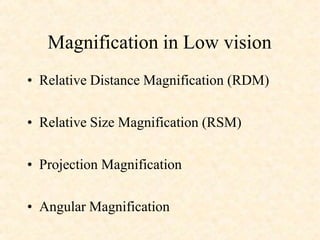
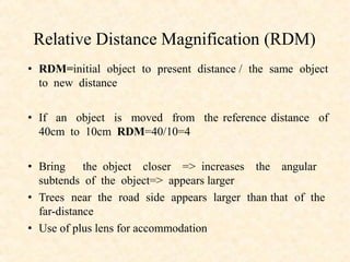
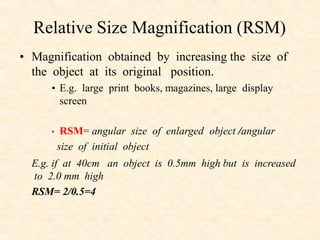
This document discusses different types of magnification used in low vision aids and optical devices. It describes relative distance magnification (RDM) as increasing the apparent size of an object by bringing it closer. Relative size magnification (RSM) increases an object's size while keeping it in the same position. Projection magnification enlarges an image projected on a screen. Angular magnification increases the visual angle subtended by the object. The document also discusses magnification in microscopes, telescopes, and clinical devices like slit lamps, and how magnification is achieved in low vision aids through various combinations of RDM, RSM, and angular magnification.
3. Relative Distance Magnification (RDM)
• RDM=initial object to present distance / the same object
to new distance
• If an object is moved from the reference distance of
40cm to 10cm RDM=40/10=4
• Bring the object closer => increases the angular
subtends of the object=> appears larger
• Trees near the road side appears larger than that of the
far-distance
• Use of plus lens for accommodation
4. Relative Size Magnification (RSM)
• Magnification obtained by increasing the size of
the object at its original position.
• E.g. large print books, magazines, large display
screen
• RSM= angular size of enlarged object /angular
size of initial object
E.g. if at 40cm an object is 0.5mm high but is increased
to 2.0 mm high
RSM= 2/0.5=4
5. Projection magnification
• The magnification produced from the
formation of an enlarged image on a screen ,
of an opaque or transparent object e.g.
overhead projector, CCTV
7. THE MICROSCOPE
1)SIMPLE MICROSCOPE and 2) COMPOUND MICROSCOPE
1)SIMPLE MICROSCOPE- A single convex lens of
short focal length can be used to see magnified image
•
•
of a small object and is called a magnifying glass or simple
microscope
principle -when a small object is placed betn optical centre
& focus of a convex lens, its virtual erect & magnified
image is formed on the same side of the lens
The lens is so held that the image is formed
at the LDDV
8. • Magnifying power = angle subtended at eye by
image produced by lens/ angle subtended at unaided eye
by object at LDDV
• M= 1+D/fe,
D=LDDV, fe=eye piece lens
Uses-
• Jewelers & watch makers
• To see slides
9. • COMPOUND MICROSCOPE
– Objective piece - Short aperture and short focal
length
– Eye piece - short focal length and large aperture
– Principle -
• When a small object is placed just outside the
focus of the object lens its real , inverted and
magnified image is produced in the other side of
the lens between its f and 2f .
10. • The image produced by objective piece acts as
object for eye piece .
• The position of the eye lens is so adjusted that
the final image is formed at LDDV
• Me = 1+D/fe & Mo = v/ u
• M = Mo x Me = v/u (1+D/ fe)
• M= fo/fe= -Doc/D obj
At LDDV-
• M=fo/fe(1+fe/D)
11. TELESCOPE:
1. ASTRONOMICAL TELESCOPE :
– It produces virtual and inverted image
– Used to see heavenly bodies
•Principle -
– The objective forms the real and inverted image of the
distant object at its focal plane
– The position of eye piece is adjusted till the final image is
formed at LDDV.
– Normal adjustment - final image is formed at infinity
• M=fo / fe
– When final image is formed at LDDV -
• M= fo/fe(1+fe/D)
12. 2. TERRESTRIAL TELESCOPE:
– Produces an erect image
– Erecting lens is placed in between objective and eye piece
– Normal adjustment -
• M=fo/fe
– At LDDV-
• M= fo/fe(1+fe/D)
13. 3. GALLILEO’S TELESCOPE :
– It provides an erect image of the distant object by use of two
lenses
– The objective piece ( convex lens )form the real and inverted
image of the distant object on the other side of lens at the focal
plane of objective.
– This image acts as a virtual object for the eye piece(concave
lens) . Final erect image formed at infinity
– The difference between two lens equals to fo-fe
• M= fo/fe
15. • Introduction :
• The ratio of size of image to the size of object
• M = image size / object size
= object vergence / image vergence
= image distance / object distance
• Human eye as the optical system the size of the image on
the retina is being compared with the size of the object of
regard
• Retinal image magnification (RIM) =
• Magnified retinal image size / original retinal size
16. – RIM has three components :
• Relative size magnification (RSM)
• Relative distance magnification (RDM)
• Lens vertex magnification (LVM)
– RSM and RDM can be achieved without the use of lens
where as LVM depends on the kind of lens placed before
the eye and its location
– It allows the use of magnifiers in such a way that image on
the retina are usable and functional although not in perfect
focus
17. • Clinical Significance :
• Direct ophthalmoscopy-
• Image is erect, virtual and
(about 15 times ) magnified in emmetrope ( more in
myopes less in hypermetropes).
• Indirect ophthalmoscopy -
• Image is real ,inverted
and magnified , which depends upon the dioptric
power of the convex lens , position of the lens in
relation to eye ball and refractive status of eye ball.
18. • Slit lamp Bio-microscope - Image is erect ,virtual and
magnification can be adjusted according to need as 10 X , 16 X
and 20
•Low magnification:
–
–
–
–
–
–
7X - 10X : General eye
Lids.
Bulbar conjunctiva/sclera.
Cornea/limbus.
Tears.
Anterior chamber/iris/crystalline lens.
•Medium magnification:
– 20X - 25X : Structure of individual
layers
– Epithelium/epithelial breakdown.
– Stroma.
– Endothelium.
– Contact lens fit/lens condition.
•
•
•
•
X.
High magnification:
– 30X - 40X : Details
Epithelium
– vacuoles
– microcysts
– dystrophies.
Stroma
– striae
– folds.
Endothelium
– Polymegathism
– guttata
– blebs
– cell density.
19. In low vision aids –
• Spectacle Magnifier: RDM
• Hand magnifier :
• Useful in short term visual task
• Magnification depends upon equivalent power and how the
magnifier is used (RDM and Angular Magnification)
• Stand magnifier :
• The fixed focused eye having a fixed distance from object of
regard
• Magnification depends on the power of the magnifier (RDM
and Angular Magnification)
20. •Paper wet magnifier :
• Reading aid in which thick plano- convex lens is held
in contact with the reading material.
• Magnification is relatively low
•In Aniseometropia -
• Contact lens produces low magnification than spectacle
thus removes the aniseokonia .
•In Closed circuit television (CCTV) -
• Projection and relative distance magnification are
used .
•In Telescope -
• Angular magnification is used.