SlideShare a Scribd company logo
Mad Cow Disease and Variant Creutzfeldt Jakob Disease 1


Mad Cow Disease and Variant Creutzfeldt Jakob Disease
Jason C. Potter
Human Disease 3400 sec. 2
Spring Semester 2012
Lynley Rowan, PhD
5 April 2012
	
  
Mad Cow Disease and Variant Creutzfeldt Jakob Disease	


  2	


Mad Cow Disease or Bovine Spongiform Encephalopathy (BSE) emerged in United
Kingdom (UK) in the early 1980’s as a result of cattle being fed a prion infected protein meal. It
was only a matter of time before BSE was transmitted to humans and Variant Creutzfeldt Jakob
Disease (vCJD) developed. VCJD is a rare, degenerative brain disease caused by an abnormal
transmissible protein. Manifestations of vCJD include both psychological and neurological
symptoms, with the prognosis being fatal. The infective agent is a mutated prion protein that is
isolated to neural tissue. Since the development of BSE in the 1980’s, and first reported case of
vCJD in 1996, scientists have been perplexed by the uniqueness of the disease and have
continued their research in the hopes of uncovering more effective identification, diagnosis,
prevention and treatment of vCJD (CDC, 2010).
History
Mad Cow disease was first discovered in 1981-1982 in the UK. BSE is related to a
serious disease known as “scrapie” that has been affecting sheep and goats since the mid-18th
century in Europe. BSE is a zoonotic disease that was transmitted to cattle that were fed with
forms of protein meals containing prion. The protein meal contained prions from sheep infected
with scrapie. The protein meal was made of discarded sheep remains containing neural tissue
(Chachra, Narang, & Narang, 1999). Researchers suspect that around the 1980’s changes in the
rendering process of how the protein-rich meal was processed allowed for the infective agent in
carcasses to survive. This caused a contamination of the protein-rich meal, leading to cattle to
becoming infected and creating the BSE epidemic. (Brown, Will, Bradley, Asher, & Detwiler,
2001, pp. 6-7).
Soon after the discovery of BSE, distress arose regarding the risk of transmission to
humans. At this time, the UK and the European Union began taking measures to ensure BSE did
Mad Cow Disease and Variant Creutzfeldt Jakob Disease	


  3	


not spread to humans. This consisted of a surveillance unit to monitor any cases in humans,
hoping that transmission to humans could be identified quickly (Brown, Will, Bradley, Asher, &
Detwiler, 2001, p. 8). With time, BSE was eventually spread to humans.
From May to October 1995, the surveillance unit was informed of three cases of classic
Creutzfeldt Jakob Disease (CJD) in humans ages 16, 19, and 29. By December 1995, the
surveillance unit had been notified of 10 alleged cases of classic CJD in individuals younger than
50 years of age. Of these ten individuals, some were found to have familial or sporadic CJD or a
different disease. (Brown, Will, Bradley, Asher, & Detwiler, 2001, p. 9) It was no doubt; cases of
CJD were on the rise, whether or not they were considered truly familial, sporadic or infectious.
Researchers reported, “As of January 1, 1996, the relationship between these cases and BSE
began to excite suspicion but remained tentative because critical information judged necessary to
establish a probable connection was still missing” (Brown, Will, Bradley, Asher, & Detwiler,
2001, p. 9).
In February 1996, another case was reported to the surveillance unit with a similar
clinical presentation to the prior cases reported. This confirmed that the recently reported cases
of CJD were undeniably distinctive. On March 4, analysis of the genetics of six reported cases
revealed there was no genetic cause for the syndrome. The following week, two more cases were
neuropathologically confirmed. A report on the entire ten confirmed cases resulted in the
identification of an unrecognized variant of classic CJD, known as vCJD, arising in individuals
under the age of 45 and most likely due to BSE exposure. The link between BSE and vCJD was
firmly established through laboratory and biological testing comparing the biological
characteristics of the pathogenic agent from cattle infected with BSE and vCJD (Brown, Will,
Bradley, Asher, & Detwiler, 2001, pp. 10-11).
Mad Cow Disease and Variant Creutzfeldt Jakob Disease	


  4	


Emergence, Incidence and Prevalence.
Both BSE and vCJD are considered epidemic and initially emerged from the UK. The
Center for Disease Control (CDC) (2010) reported:
From 1986 through 2001, more than 98 % of BSE cases worldwide were reported from
the UK, where the disease was first described. During this same period, the number of
European countries reporting at least one indigenous BSE case increased from 4 through
1993 to 8 through 1998 to18 through 2001. During 2001-2003, three countries outside
Europe (Canada, Japan, and Israel) reported their first indigenous BSE cases.
Although BSE first appeared in the UK, the total number of annual cases being reported
outside the UK has been raising; the CDC (2010) described an increase of 25 % in 2000 and
more than 55 % in 2003. The CDC (2010) confirmed this increase in BSE outside the UK
reflects the “declining large (more than 183,000 total cases) epidemic of BSE in the UK and the
increasing number of other countries with improved surveillance and higher rates of BSE.”
Despite prevention measures to avoid the spread of BSE to Northern America and Canada, the
first case was identified in Canada in 2003. This created a great amount of concern that BSE may
be occurring in North America. In reaction to the reported case of BSE in Canada, the CDC
(2010) reported the US Department of Agriculture (USDA) implemented extra safeguards “to
minimize the risk for human exposure to BSE and on July 1, 2004, initiated a 12- to 18-month-
long intensive testing program for BSE among cattle at relatively high risk for the disease (e.g.,
non-ambulatory cattle).”
According to CDC (2010), only three cases of vCJD have been reported from the United
States. It is important to note, in spite of where an exposure may have occurred, vCJD is
attributed to the country of the individual’s initial onset of symptoms. To date, there has never
been a reported case of vJCD where the individual did not have a history of exposure in a
country where BSE was occurring. Of the three reported cases in the United States, facts imply
Mad Cow Disease and Variant Creutzfeldt Jakob Disease	


  5	


that one of individuals was initially exposed to BSE while residing in the UK and the other
individual while residing Saudi Arabia (CDC, 2010). VCJD was first reported in 1996, and since
then the CDC (2010) reported:
A total of 217 patients with this disease from 11 countries have been identified. As of
October 2009, variant CJD cases have been reported from the following countries: 170
from the United Kingdom, 25 from France, 5 from Spain, 4 from Ireland, 3 from the
United States, 3 in the Netherlands, 2 in Portugal, 2 in Italy, and one each from Canada,
Japan, and Saudi Arabia. Two of the three U.S. cases, two of the four cases from Ireland
and the single cases from Canada and Japan were likely exposed to the BSE agent while
residing in the United Kingdom.
In regards to the year of onset, the CDC (2010) reported the incidence of vCJD in the UK
peaked in 1999, with 1000 cases per week, and since that time has been declining. In contrast,
the number of reported cases in France has been increasing since the beginning of 2005.
Nevertheless, concern is arising regarding the future incidence of vCJD and pattern of this
epidemic. The CDC (2010) reaffirmed a second wave of cases could possibly occur due to in a
subgroup of the population that may be less susceptible in regard to genetics. As of 2003, the UK
and Portugal reported a BSE incidence rate of “more than 100 indigenous cases per million cattle
more than 24 months of age” (CDC, 2010). The CDC (2010) also reported BSE rates per million
cattle over the age of 24 months in regards to the following countries:
58 for the Republic of Ireland, 46 for Spain, 25 for Switzerland, 12 for France, 11 for
Belgium and the Netherlands, 10 for Italy, 9 for Germany, and 7 for Slovakia. The
reported rates for Canada, Czech Republic, Denmark, Japan, Poland, and Slovenia were
between 0.3 and 6 cases per million.
Mad Cow Disease and Variant Creutzfeldt Jakob Disease	


  6	


Below is figure 1 presented by Behgi, et al. (2004), showing the cases of BSE and vCJD in the
UK since 1987:
Figure 1 courtesy of by Behgi, et al. (2004)
The Disease
Etiologic Agent
According to the Journal of Emerging Infectious Diseases, BSE is a zoonotic disease
which becomes fatal to humans eating infected beef. Within weeks of the first reported case of
BSE, concern was raised regarding the risk to humans and the possibility of BSE reaching the
human food chain. BSE is a neurological disease affecting cattle that progressively leads to
death. BSE results from an infective agent called a prion (proteinaceous infectious particles).
Prion diseases are also known as transmissible spongiform encephalopathies (TSEs), affecting
both animals and humans (Brown, Will, Bradley, Asher, & Detwiler, 2001). The CDC (2010)
further explained, “A prion is an abnormal, transmissible agent that is able to induce abnormal
folding of normal cellular prion proteins in the brain, leading to brain damage and the
Mad Cow Disease and Variant Creutzfeldt Jakob Disease	


  7	


characteristics signs and symptoms of the disease.” Unlike bacteria, prions do not contain any
DNA or RNA. Of all the pathogens, prions are considered to be the most resistant to
disinfectants. (Engelkirk & Duben-Engelkirk, 2010, p. 52). The exact mechanism by which
prions cause disease is unclear; however, according to Engelkirk & Duben-Engelkirk (2010), “It
is though that normal protein molecules are converted into nonfunctional ones by causing the
normal molecules to change their shape” (p. 52).
Stages and Symptoms
According to Spencer, Knight and Will (2002), individuals affected by vCJD exhibit
psychiatric symptoms in the early stages of the disease. Common psychiatric symptoms include
dysphoria, withdrawal, uneasiness, insomnia and loss of interest. In the early stages of the vCJD,
neurological symptoms are often times not apparent. However, Spencer, et al .,( 2002) found
that of the first 100 reported cases in the UK, neurological symptoms precede psychological
manifestations in 15% of all the cases and both neurological and psychological symptoms are
present in combination in 22% of all cases from the initial onset of vCJD (p. 1479). During the
disease process, no common early neurological symptoms are present; nevertheless, Spencer, et
al., 2002 stated, “A significant portion of patients do exhibit neurological symptoms within 4
months of clinical onset, including poor memory, pain, sensory symptoms, unsteadiness of gait
and dysarthria” (p. 1479). In the last stages of vCJD, patients experience dementia and
eventually slip into a coma. On average, the duration of the illness is approximately 14 months in
most patients (Behgi, et al., 2004).
Unique Characteristics
VCJD is unique in that most physicians in medical school typically learn about classic
CJD, which is unrelated to the ingestion of contaminated beef. According to the CDC (2010),
Mad Cow Disease and Variant Creutzfeldt Jakob Disease	


  8	


“Classic CJD is not related to mad cow disease. Classic CJD is also distinct from variant CJD,
another prion disease that is related to BSE.” Thus, the epidemiology of vCJD and classic CJD
are quite different and must be carefully diagnosed to distinguish between the two diseases.
Because vJCD is such a unique and new disease, Henley and Herrman (2004) reaffirmed “its
incubation period is unknown, but it is likely to be years or decade” (p 645). In fact, in the UK
individuals were most likely exposed to BSE-contaminated food in 1984-1986. It is likely the
onset of vCJD was not years later until reported cases in 1994-1996 (Henley & Herrmann, 2004).
Diagnosis
Henley and Herrmann (2004) note that diagnosing vCJD includes several diagnostic
tools; however, one must carefully distinguish between vCJD and classic CJD before a final
diagnosis is made. One of the tests performed in diagnosing vCJD is an electroencephalogram
(EEG), which is used to measure electrical activity in the brain. If vCJD is present, there will be
a lack of sharp wave complexes on EEG; in contrast, if classic CJD is present there will be wave
complexes present (p. 645-647). The CDC also provides definitive criteria for the diagnosis of
vCJD in the United States. For a definitive diagnosis examination of the brain tissue must take
place and the following confirming features must be present:
a.   Numerous widespread kuru-type amyloid plaques surrounded by vacuoles in both
the cerebellum and cerebrum - florid plaques.
b.   Spongiform change and extensive prion protein deposition shown by
immunohistochemistry throughout the cerebellum and cerebrum. (Center for
Disease Control and Prevention, 2010, 2011)
These definitive features of diagnosing vCJD differ from the diagnosis of classic CJD
where kuru-type amyliod plaques are rare or absent and prion protein is variable in accumulation
rather than extensive as in vCJD (Henley & Herrmann, 2004, p. 647).
The CDC (2010) also provides criteria for a suspected vCJD diagnosis:
Mad Cow Disease and Variant Creutzfeldt Jakob Disease	


  9	


a.   Current age or age at death <55 years (a brain autopsy is recommended, however,
for all physician-diagnosed CJD cases).
b.   Psychiatric symptoms at illness onset and/or persistent painful sensory symptoms
(frank pain and/or dysesthesia).
c.   Dementia, and development ≥4 months after illness onset of at least two of the
following five neurologic signs: poor coordination, myoclonus, chorea,
hyperreflexia, or visual signs. (If persistent painful sensory symptoms exist, ≥4
month’s delay in the development of the neurologic signs is not required).
d.   A normal or an abnormal EEG, but not the diagnostic EEG changes often seen in
classic CJD.
e.   Duration of illness of over 6 months.
f.   Routine investigations of the patient do not suggest an alternative, non-CJD
diagnosis.
g.   No history of receipt of cadaveric human pituitary growth hormone or a dura
mater graft.
h.   No history of CJD in a first degree relative or prion protein gene mutation in the
patient. (Center for Disease Control and Prevention, 2010, 2011)
Hennley and Herrman (2004) suggest that physicians should advocate for a brain biopsy
or autopsy in patients that may have vCJD or patients that may have died as a result of the
disease. For physicians and health departments whom may need to perform diagnostic testing—
the CDC funds The National Prion Disease Surveillance Center—providing diagnostic services
free of charge. (p. 648).
The Science
Morphology, Genome and Life Cycle
Understanding the science related to vCJD is linked to the causative agent known as a
prion; an infectious agent that consists of a tiny protein that lacks genes, but can mutate, creating
error in gene expression and failure to fold correctly. A prion protein is typically spiral or helical
in shape. The abnormal form of this protein looks much different, containing many flat, sheet-
like structures. These mutations associated with the disease, affecting the shape of the protein,
are thought to cause the clumping of the protein and in turn the accumulation and damage to the
Mad Cow Disease and Variant Creutzfeldt Jakob Disease	


  10	


brain. (Prusiner, 2001). As seen below in Figure 2, brain tissue infected with prions has a spongy
appearance (CDC, 2010).
Figure 2 courtesy of the CDC (2010)
As mentioned, vCJD is caused by ingesting beef containing prion, which eventually
accumulates in the brain leading to death. Diseases affected by prions have long been debated
with uncertainty among scientists; however, recent research conducted by Stanley Prusiner and
Charles Weissmann has confirmed that prion diseases are caused by a toxic form of the prion
protein; this idea is known as the prion hypothesis (Goldberg, 2007, p. 1150).
Golberg (2007) explains:
This protein is able to enter the cells and induce a conformational rearrangement of
normal soluble, monomeric cell constituent, the prion protein (PrPc), into a toxic
transmissible species (PrPsc). In this pathogenic conformation, the prion is primarily
insoluble, is resistant to proteases or detergents. This conformational change and the
resulting accumulation of PrPsc ultimately causes severe loss of neurons, glosis, and
spongiform appearance (p. 1150).
Mad Cow Disease and Variant Creutzfeldt Jakob Disease	


  11	


Below is a simplified diagram (Figure 3) of the prion hypothesis presented by Burthem &
Roberts (2003). Burthen & Roberts explained, “(A and B) Molecules of PrPSc interact directly
with the PrPc causing the structural conversion of the PrPC to form further PrPSc . This
conversion culminates in the formation of polymeric aggregates of PrPSc (C) or in the formation
of further infective material” (p. 223).
Figure 3 courtesy of Burtham & Roberts (2003)
Burthem and Roberts (2003) provide a detailed explanation of the prion hypothesis:
The prion hypothesis requires that, for PrP(Sc)infection to occur, PrP(C)must first be
expressed by the host, i.e. there must be a 'pool' of PrP(C)that can be converted to
PrP(Sc). Immunological surveillance requires that cells of the immune system encounter
and process new proteins entering the body at an early stage after ingestion or
inoculation. Moreover, immune cells migrate widely through the body and establish
frequent and close contact with other cells. This behavior is essential for the effective
immune response to conventional pathogens, but in prion disease this otherwise
beneficial process appears to facilitate the propagation and spread of the abnormal protein
(p. 122).
A	


B	


C	
  
Mad Cow Disease and Variant Creutzfeldt Jakob Disease	


  12	


As discussed, the causative agent in vCLD is a prion; however, Belay (1999) discussed a
specific variation in the sequence of genes has been confirmed. The most significant
polymorphism leads to the formation of two different protein forms. The first form has an amino
acid called methionone at the 129th
amino acid in the protein sequence, and the second form has
an amino acid called valine in the same position (p. 298). Belay (1999) also addressed several
studies that have shown the polymorphism at codon 129 has a significant impact in determining
the susceptibility of the host and the disease expression of familial, iatrogenic (infectious), or
sporadic CJD (p. 298). Individuals that developed the vCJD had a double-methionine form of the
prion protein, but no other mutations. This implies that this particular variation in the gene
somehow influences an individual’s susceptibility to vCJD (Prusiner, 2001). In fact, Brown et
al., (2001) concluded, “The encoding alternatives methionine (met) and valine (Val), are
distributed in the approximate proportions of 50% Met/Val, 40% Met/Met, and 10 % Val/Val.”
In 2001, it was reported that all 76 vCJD patients tested have been homozygous for methionine,
and apparently single infecting strains of BSE may not be able to replicate in any other human
genotype (p. 10). In 2005, Ludman & Turner reported that “ to date in all clinical cases of
variant CJD have occurred in methionine 128 homozygous individuals; it seems likely that
valine homozygous and methionine/valine heterozygous individuals are more resistant to
infection, or if infected, to the development of clinical variant CJD” (p. 16).
Transmission
Bovine to Human.
Research has positively confirmed the transmission of vCJD is directly related to the
biological features of the pathological agent isolated from BSE infected cattle. The source of the
infection appears to have been from beef products contaminated by nervous system tissue
Mad Cow Disease and Variant Creutzfeldt Jakob Disease	


  13	


containing prions (Brown et al., 2001, p. 10). Brown et al., (2001) explained how contamination
originally could have occurred:
Cerebral vascular emboli from cranial stunning instruments used to immobilize cattle
before killing by exsanguination; contact of muscle with brain or spinal cord tissue by
saws or other tools used during slaughter; inclusion of paraspinal ganglia in cuts of meat
containing vertebral tissues; and perhaps most importantly the presence of residual spinal
cord and parasprinal ganglia tissue in the paste of mechanically recovered meat that could
legally be added to cooked meat products such as meat pies, beef sausages, and various
canned meat preparations (p. 10).
According to the CDC (2011), BSE may have originally resulted due to feeding cattle
meat and bone meal that contained products from a randomly occurring case of BSE or scrapie
from possible contaminated sheep products. The CDC (2011) stated, “There is strong evidence
and general agreement that the outbreak was then amplified and spread throughout the United
Kingdom cattle industry by feeding rendered, prion-infected, bovine meat and bone meal to
young calves.” The CDC (2011) confirmed there is solid epidemiologic and laboratory data for a
connecting link between vCJD that was first reported from the United Kingdom in 1996, and the
epidemic of BSE. A unique characteristic of vCJD is the long incubation period. The CDC
(2011) reported similar incubation periods for human forms of prion diseases and the time frame
of initial exposure to BSE and the clinical manifestations of the disease.
Blood Products.
VCJD may also be transmitted through blood products such as transfusions and using
blood products from an infected person with vCJD. Turner & Ludman (2008) addressed the
topic of vCJD by blood transfusion and discussed the direct link of vCJD and blood donation.
Turner and Ludman (2008) reported that, “Eighteen patients with vCJD have, or have has
previously, been blood donors, from whom a total of 66 recipients have been identified, 26 of
whom are still alive. Of those who have died, four cases of transmission of vCJD prions have
Mad Cow Disease and Variant Creutzfeldt Jakob Disease	


  14	


been identified” (p .15) In December 2003, the first case of vCJD linked to a blood transfusion
was indentified. The individual that became infected with vJCD developed the disease 6 years
after receiving a blood transfusion. The blood donor developed signs and symptoms of vJCD 3
years after donating (Turner & Ludman, 2008, p. 15). This data clearly shows that individuals in
the incubation period of vCJD can transmit the disease to another individual through blood
transfusion. No specific immunological reaction related to prion infection has been indentified
nor has specific DNA been found related to the transmission of vCJD. Thus, typical serological
and biological tests in donors have not been possible (Ludlam & Turner, 2005, p. 18).
Iatrogenic.
Transmission from human to human through direct contact has not been confirmed;
however, individuals can be infected with vCJD through iatrogenic transmission. Ludlan &
Turner (2008) reported iatrogenic transmission has been well documented. The transmission
occurs through direct inoculation of the Central Nervous System (CNS) through contaminated
neurosurgical instruments, intracerebral electrodes, dura matter and corneal grafts. Iatrogenic
transmission has also occurred by pituitary growth hormone and gonadotrophins taken from
cadavers and administered to patients by intramuscular injection (Turner & Ludman, 2008, p.
15). Despite technological advanced sterilization techniques, it is possible for vCJD to be
transmitted to another patient. Campbell (2006) reported that stainless steel instruments used in
neurosurgery can transmit vCJD to another patient even if the instrument is thoroughly sterilized
with formaldehyde. It is recommended that only disposable instruments be used in neurological
surgery and tonsil removal to prevent the transmission of vCJD from one patient to another.
According to Ludman & Turner (2005), patients with vCJD have shown evidence of irregular
prion accumulation in follicular dendritic cells and peripheral lymphoid tissue, including the
Mad Cow Disease and Variant Creutzfeldt Jakob Disease	


  15	


tonsils (p. 17). Another concern has been brought up regarding dental surgery involving
removal of an infected root. Since formaldehyde is ineffective in disinfecting instruments, it has
been recommended that dental surgeons use 4% hypochlorine (p.93). Campbell (2006) reiterated
the possibility of direct transmission from human to human through the use of infected surgical
instruments, “The fear is there may be a bank of human carriers passing on the infection to
others” (p. 93).
Port of Entry.
Clinical presentation and specific signs and symptoms of vCJD can vary depending on
the route of infection. CNS transmission is likely to have a shorter incubation period. According
to Ludlam & Turner (2005), the incubation period is roughly two years and the infected
individual develops rapid dementia. Peripherally transmitted cases usually have a longer
incubation period, ranging from 13-15 years and symptoms include ataxia and sensory
disturbances (Ludlam & Turner, 2005, p. 15). Tan, Williams, Khan, Champion & Nielson (1999)
discussed four categories related to certain organs containing more infectious prions than others:
Direct administration into the central nervous system is the most infectious route,
followed by administration in to the blood vessels, and intraperitoneal, intramuscular, and
subcutaneous exposure. Oral ingestion is less efficient than the parenteral routes. Finally,
the dose of infectious material is an important determinant of transmissibility (p. 2333).
Fate of Victim
According to Ludman & Turner (2005), after transmission of the disease, the fate of the
victim infected with vCJD is a poor prognosis that eventually leads to death. Ludman and Turner
(2005) stated in the end stages of the disease patients develop progressive dementia and
myoclonus “with an average clinical course to death of 6 months—2 years” (p. 15).
Mad Cow Disease and Variant Creutzfeldt Jakob Disease	


  16	


Vulnerability and Risk
As reported by the CDC (2010), almost all cases of vCJD have been in persons under the
age of 55, with the median age at death being 28. Ludlam & Turner (2005) also reported the
median age of death has not changed over the first 10 years of the outbreak; this suggests there
may be an age related susceptibility or exposure (p. 17). Reasoning for this specific age
distribution is not fully understood at this time; however, the CDC (2010) suggested that through
the oral route of exposure, older adults are less susceptible to vCJD than young adults and
children. Concern has also been raised that some individuals in the population may be
genetically susceptible to vCJD. The CDC (2010) discussed this concern, presented in the
following quote:
In 2004, a prevalence study of asymptomatic vCJD infections in the UK identified three
positive appendices out of a sample of 12,674 surgically removed tonsils and appendices
that were satisfactory for analysis. Genetic studies completed on two of the appendices
regarded as positive for vCJD revealed that both had a different polymorphism at codon
129 of the prion protein gene than any of the patients with clinical vCJD tested to date,
indicating that more people are genetically susceptible to vCJD infection, although not
necessarily to the disease, than had been previously determined.
The CDC (2010)confirmed the current risk for acquiring vCJD from eating BSE-
contaminated beef products in countries with a possible increase risk of BSE cannot be exactly
determined. The CDC (2010) stated if current public health measures are being implemented,
the risk of being infected with vCJD due to eating contaminated beef is extremely small;
nonetheless, the risk is not zero. The CDC (2010) reported, “A rough estimate of this risk for the
UK in the recent past, for example, was about 1 case per 10 billion servings.”
Individuals receiving blood transfusion are at risk of being infected with vCJD from the
blood of a donor that may still be in the incubation period. Individuals that received blood from
donors in the UK that were subsequently diagnosed with vCJD are considered high risk for
Mad Cow Disease and Variant Creutzfeldt Jakob Disease	


  17	


developing vCJD. These individuals are contacted on a regular basis and offered special follow
up care and counseling. In 2004, in the UK, all patients with hemophilia were sent letters stating
whether or not they had received plasma-derived clotting concentrates between the years of1980-
2001. (Ludlam &Turner, 2008, p. 15, 21). As of 2003, one of case of vCJD in a 62-year old UK
man is thought to have been as a result from a blood transfusion contaminated with the vCJD
infectious agent. Just a year later, another case in the UK was reported that was very suspicious,
likely being transmitted by a blood transfusion. Due to the reported cases of possible
transmission through a blood transfusion, the UK quickly implemented specific safeguards. A
ban was placed on all persons receiving blood transfusions after 1980 in the UK from donating
blood in the future. In January 2002, in order to reduce the risk of bloodborne transmission of
vCJD, the USDA released guidance, which included a geography-based donor deferral policy
(CDC, 2010).
Ludlam &Turner (2008) discussed the potential risk posed to patients undergoing
invasive medical procedures such as neural, tonsil, or oral surgery for an infected root. The risk
increases in these types of surgeries when considering patient to patient transmission due to
surgical instruments that may be harvesting the infective agent associated with vCJD. (15-17).
Prevention
In regards to prevention, the CDC (2010) recommended:
To reduce any risk of acquiring vCJD from food, travelers to Europe or other areas with
indigenous cases of BSE may consider either avoiding beef and beef products altogether
or selecting beef or beef products, such as solid pieces of muscle meat (rather than brains
or beef products like burgers and sausages), that might have a reduced opportunity for
contamination with tissues that may harbor the BSE agent. Milk and milk products from
cows are not believed to pose any risk for transmitting the BSE agent.
In order to prevent BSE from entering the U.S., harsh restrictions were implemented by
the USDA in June of 1997, becoming effective in October 1997. This included restrictions on the
Mad Cow Disease and Variant Creutzfeldt Jakob Disease	


  18	


importing of live cattle, sheep and goats, and certain ruminant products where BSE had been
identified. Later, this restriction was extended to all European countries. This feed ban is
enforced by the FDA through inspections and feed testing programs. In October 2009, the FDA
issued a regulation creating an enhanced BSE-related feed ban in the U.S. This feed ban helped
to synchronize feed control measures in the US and Canada. In July of 2007, an enhanced BSE-
related feed ban was put into effect in Canada in order to quickly eliminate the spread of BSE in
Canada. (CDC, 2010).
To help with prevention measures in relation to vCJD, the CDC continues to monitor
trends and the current incidence in the U.S. through several surveillance methods. The CDC has
placed more attention on the national surveillance of classic CJD in order to more effectively
detect vCJD. This is accomplished through autopsies on patients with possible prion diseases.
This has helped to increase the ability to detect more vCJD cases (CDC, 2010). The CDC also
continues to investigate possible deaths from iatrogenic CJD and vCJD reported by health care
professionals. Most importantly, “in 1996-97, CDC established, in collaboration with the
American Association of Neuropathologists, the National Prion Disease Pathology Surveillance
Center at Case Western Reserve University, which performs special diagnostic tests for prion
diseases, including post-mortem tests that can detect vCJD.” (CDC, 2010).
Treatment
The CDC stated (2010), “As of September 2004, treatment of prion diseases remains
supportive; no specific therapy has been shown to stop the progression of these diseases.”
Becoming infected with vCJD is fatal to the patient. In a study conducted by Vries, Sque, Bryan
& Abu-Saad (2003), the need for palliative care and development of educational initiatives is
addressed. They concluded that caring for patients with vCJD is very a stressful experience in all
Mad Cow Disease and Variant Creutzfeldt Jakob Disease	


  19	


stages of the illness. Patient’s affected by vCJD are often times admitted to psychiatric units,
where they eventually die. Vries, Sque, Bryan & Abu-Saad (2003) argued it is more appropriate
for patients affected by vCJD to be cared for by health care professionals in hospice
environments. Consequently, there should be planning initiated by the healthcare team for
patients affected by illnesses such as vCJD that are terminal and involve dementia (p. 518).
Global Economy
With the outbreak of BSE in the 1990’s and eventual transmission to humans, both the
UK and the U.S. have created measures in response to the crisis. According to the CDC (2010),
the department of Health and Human Services (HHS) in 2001 began to create a department-wide
action plan. This plan helped to outline steps to improve the scientific understanding and
research in regards the BSE and other TESs. The CDC (2010) presented the department-wide
action plan:
Surveillance for human disease is primarily the responsibility of CDC.
Protection is primarily the responsibility of the Food and Drug Administration (FDA).
Research is primarily the responsibility of the National Institutes of Health (NIH).
Oversight is primarily the responsibility of the Office of the Secretary of DHHS.
In is no question, both BSE and vCJD have had significant impact on the global
economy. In fact, Campbell (2006) stated, “The BSE crisis has cost the UK government more
than 4 billion pounds alone in compensation to farmers and caused much disquiet in agriculture”
(p.94). Due to the slaughtering of thousands of cattle; ban on importing ruminant products from
all European countries; and severe restrictions that were placed on the importation of cattle,
sheep, and goats; the global market has suffered greatly.
Campbell (2006) also discussed the controversy of scientist being criticized for initially
telling the public the transmission of BSE to humans was only a distant possibility. Campbell
(2006) stated, “There is no doubt that the reputation of scientists has been badly affected by the
Mad Cow Disease and Variant Creutzfeldt Jakob Disease	


  20	


BSA saga and we know that the story is not at an end. The experience with BSE has increased
the resistance of the public to biotechnology in general” (p. 94). Campbell (2006) also addressed
the concern journalists have with scientist when they pose the question “is it safe to eat beef?”
and they do not receive a direct yes or no, but would rather speak about risk factors.
Beghi, et, al (2004) reaffirmed the reality that the future effect on the global economy in
relation to BSE and vCJD is unknown at this time due to the long and ill-defined incubation
period of prion diseases. Thus, we do not know the beginning and the end of the human outbreak
of BSE and the effects it may have on our future economy.
The epidemic of Mad Cow Disease that spread throughout the UK in the 1980’s raised
concern worldwide. With humans coming in contact with contaminated beef, vCJD developed as
a result. Scientists continue to conduct further research to fully understand the impact of a prion
disease such a vCJD. The research is focused on finding a more effective way of identifying
potential carriers early on, and preventing transmission that can occur through blood transfusions
and invasive medical procedures. In addition to research taking place, government agencies in
the UK and U.S. are continually implementing preventative measures to stop infected beef
products from reaching humans. Due to the uniqueness of this disease and the long incubation
period, scientists, researchers, and government agencies fear there could be a large portion of the
population carrying the disease and unknowingly spreading it to others, creating a second wave
of the BSE outbreak.
Mad Cow Disease and Variant Creutzfeldt Jakob Disease	


  21	


References
Behgi, E., Ferrarese, C., Fratolla, L., Gandolfo, C., Mancardi, G., Rizzuto, N., et al. (2004).
Bovine spongiform encephalopathy and Creutzfeldt-Jakob disease: facts and uncertainties
underlying the casual link between animal and human diseases. Neurological Science , 122-129.
Belay, E. D. (1999). Transmissible Spongiform Encephalopathies in Humans. Annual Review of
Microbiology , 283-314.
Brown, P., Will, R. G., Bradley, R., Asher, D. M., & Detwiler, L. (2001). Bovine Spongiform
Encepjalopathy and Variant Creutzfelt-Jacob Disease; Background, Evolution, and Current
Conerns. Emerging Infectious Diseases , 6-13.
Burthan, J., & Roberts, D. (2003). The Pathology of Variant CreutzFeldt-Jakob Diseasae: the
hypothesis behind conerns for blood components and products. British Journal of Haematology ,
122-127.
Campbell, P. N. (2006). Mad cow disease. Acta Biologica Szegediensis , 89-95.
Center for Disease Control and Prevention. (2010, 2011). Retrieved Feburary 9, 2012, from
http://www.cdc.gov/ncidod/dvrd/vcjd/
Chachra, A., Narang, D., & Narang, R. (1999). Mad Cow Disease. Resonance , 42-44.
Engelkirk, P. G., & Duben-Engelkirk, J. (2010). Burton's Microbiology for Health Sciences.
Lippincott Williams & Wilkins.
Goldberg, A. L. (2007). On Prion, Proteasomes and Mad Cows. The New England Journal of
Medicine , 1150-1152.
Henley, E., & Herrmann, J. (2004). Mad Cow Disease: Dealing sensibly with a new conern. The
Journal of Family Practice , 645-648.
Ludlam, C. A. (2008). An update on the assessment and management of the risk of transmission
of variant Creutzfedldt Jakob Disease and plasma products. British Journal of Haematology , 14-
23.
Ludlam, C. A., & Turner, M. L. (2005). Managing the risk of transmission of variant Creutzfeldt
Jakob disease by blood prosucts. Brithish Journal of Haematology , 13-24.
Prusiner, S. (2001). Shattuck Lecture: Neurodegernaerative diseases and prions. New England
Journal of Medicine , 1516-1526.
Mad Cow Disease and Variant Creutzfeldt Jakob Disease	


  22	


Spencer, M. D., Knight, R. S., & Will, R. G. (2002). First hundred cases of variant Creutzfelt-
Jakob disease: retrospective case note review of early psychiatric and neurological features.
British Medical Journal , 1479-1482.
Tan, L., Williams, M. L., K. M., Champion, H. C., & Nielsen, N. H. (1999). Risk of
Transmission of Bovine Spongiform Encepalopathy to Humans in the United States. Jourmal of
the American Medical Asssocation , 2230-2339.
Vries, K. d., Sque, M., Bryan, K., & Abu-Saad, H. (2003). Variant Creutzfeldt Jakob Disease:
need for mental health and palliative care team collaboration. International Journal of Palliative
Nursing , 512-520.

More Related Content

What's hot

Abdul wajid seminar
Abdul wajid seminarAbdul wajid seminar
Abdul wajid seminar
ismailzai
 
Placental and Decidual Expression of Zika Virus Receptors: The Route to Infec...
Placental and Decidual Expression of Zika Virus Receptors: The Route to Infec...Placental and Decidual Expression of Zika Virus Receptors: The Route to Infec...
Placental and Decidual Expression of Zika Virus Receptors: The Route to Infec...
UCSFGlobalHealthSciences
 
Brucellosis: Epidemiology and Control in india
Brucellosis: Epidemiology and Control in indiaBrucellosis: Epidemiology and Control in india
Brucellosis: Epidemiology and Control in india
Bhoj Raj Singh
 
MAD ./ \. DOG
MAD ./ \. DOGMAD ./ \. DOG
MAD ./ \. DOG
Dikshan
 
rabies 2
rabies 2rabies 2
rabies 2
Dikshan
 

What's hot (19)

Abdul wajid seminar
Abdul wajid seminarAbdul wajid seminar
Abdul wajid seminar
 
Bell tolling for the Swine Flu
Bell tolling for the Swine FluBell tolling for the Swine Flu
Bell tolling for the Swine Flu
 
Immunocompromised hosts and transplant recipients
Immunocompromised hosts and transplant recipientsImmunocompromised hosts and transplant recipients
Immunocompromised hosts and transplant recipients
 
Message map: Listeria monocytogenes
Message map: Listeria monocytogenesMessage map: Listeria monocytogenes
Message map: Listeria monocytogenes
 
Jenner and disease
Jenner and diseaseJenner and disease
Jenner and disease
 
Placental and Decidual Expression of Zika Virus Receptors: The Route to Infec...
Placental and Decidual Expression of Zika Virus Receptors: The Route to Infec...Placental and Decidual Expression of Zika Virus Receptors: The Route to Infec...
Placental and Decidual Expression of Zika Virus Receptors: The Route to Infec...
 
Fazd equine piroplasmosis
Fazd equine piroplasmosis  Fazd equine piroplasmosis
Fazd equine piroplasmosis
 
Biosciences Nairobi – Spread of tetracycline resistance gene tetM via TN916-l...
Biosciences Nairobi – Spread of tetracycline resistance gene tetM via TN916-l...Biosciences Nairobi – Spread of tetracycline resistance gene tetM via TN916-l...
Biosciences Nairobi – Spread of tetracycline resistance gene tetM via TN916-l...
 
Brucellosis: Epidemiology and Control in india
Brucellosis: Epidemiology and Control in indiaBrucellosis: Epidemiology and Control in india
Brucellosis: Epidemiology and Control in india
 
Edward Jenner
Edward JennerEdward Jenner
Edward Jenner
 
Final jbk seminar 27.9.18 prions
Final jbk seminar 27.9.18 prionsFinal jbk seminar 27.9.18 prions
Final jbk seminar 27.9.18 prions
 
pulse polio
pulse poliopulse polio
pulse polio
 
Ebola
EbolaEbola
Ebola
 
The discovery of vaccination
The discovery of vaccinationThe discovery of vaccination
The discovery of vaccination
 
Dr. David R. Smith - What We know (and don’t know) About Pneumonia in Beef Ca...
Dr. David R. Smith - What We know (and don’t know) About Pneumonia in Beef Ca...Dr. David R. Smith - What We know (and don’t know) About Pneumonia in Beef Ca...
Dr. David R. Smith - What We know (and don’t know) About Pneumonia in Beef Ca...
 
Woodstock Vet Clinic-Hookworms
Woodstock Vet Clinic-HookwormsWoodstock Vet Clinic-Hookworms
Woodstock Vet Clinic-Hookworms
 
Zika Virus and Microcephaly
Zika Virus and MicrocephalyZika Virus and Microcephaly
Zika Virus and Microcephaly
 
MAD ./ \. DOG
MAD ./ \. DOGMAD ./ \. DOG
MAD ./ \. DOG
 
rabies 2
rabies 2rabies 2
rabies 2
 

Viewers also liked

Antibiotic Resistanace
Antibiotic ResistanaceAntibiotic Resistanace
Antibiotic Resistanace
shabeel pn
 
Antibiotic resistant bacteria
Antibiotic resistant bacteriaAntibiotic resistant bacteria
Antibiotic resistant bacteria
Nicole Glenn
 
Madcowdisease hk
Madcowdisease hkMadcowdisease hk
Madcowdisease hk
yalesig2012
 
Nosocomial infection
Nosocomial infectionNosocomial infection
Nosocomial infection
Nursing Path
 
Infection control powerpoint 1
Infection control powerpoint 1Infection control powerpoint 1
Infection control powerpoint 1
gregoryjnewman
 
Nosocomial Infection
Nosocomial InfectionNosocomial Infection
Nosocomial Infection
Zahoor Ahmed
 

Viewers also liked (20)

Staphylococcus
StaphylococcusStaphylococcus
Staphylococcus
 
Nosocomial infections
Nosocomial infectionsNosocomial infections
Nosocomial infections
 
Antibiotic Resistanace
Antibiotic ResistanaceAntibiotic Resistanace
Antibiotic Resistanace
 
Antibiotic resistant bacteria
Antibiotic resistant bacteriaAntibiotic resistant bacteria
Antibiotic resistant bacteria
 
Nosocomial infection
Nosocomial infectionNosocomial infection
Nosocomial infection
 
Madcowdisease hk
Madcowdisease hkMadcowdisease hk
Madcowdisease hk
 
Antibiotics resistant infections
Antibiotics resistant infectionsAntibiotics resistant infections
Antibiotics resistant infections
 
Nosocomial infection
Nosocomial infectionNosocomial infection
Nosocomial infection
 
Specific Multi-drug Resistance MRSA-VRSA-ESBL-KPC
 Specific Multi-drug Resistance MRSA-VRSA-ESBL-KPC Specific Multi-drug Resistance MRSA-VRSA-ESBL-KPC
Specific Multi-drug Resistance MRSA-VRSA-ESBL-KPC
 
Vancomycin Resistant Enterococci
Vancomycin Resistant EnterococciVancomycin Resistant Enterococci
Vancomycin Resistant Enterococci
 
Nosocomial infection
Nosocomial infectionNosocomial infection
Nosocomial infection
 
Superbugs
Superbugs Superbugs
Superbugs
 
Antibiotic resistance
Antibiotic resistanceAntibiotic resistance
Antibiotic resistance
 
MRSA
MRSAMRSA
MRSA
 
MRSA
MRSAMRSA
MRSA
 
Nosocomial infection & control
Nosocomial infection & controlNosocomial infection & control
Nosocomial infection & control
 
Mrsa ppt
Mrsa pptMrsa ppt
Mrsa ppt
 
Infection control powerpoint 1
Infection control powerpoint 1Infection control powerpoint 1
Infection control powerpoint 1
 
Nosocomial Infection
Nosocomial InfectionNosocomial Infection
Nosocomial Infection
 
Vancomycin resistance in staphylococci
Vancomycin resistance in staphylococciVancomycin resistance in staphylococci
Vancomycin resistance in staphylococci
 

Similar to Mad Cow_Disease Research_JP

history of HIV
history of HIVhistory of HIV
history of HIV
MIN ZAW
 
AIDS†lyr in the 1980's
AIDS†lyr in the 1980'sAIDS†lyr in the 1980's
AIDS†lyr in the 1980's
Racoat
 

Similar to Mad Cow_Disease Research_JP (20)

Vetrugno, 2016
Vetrugno, 2016Vetrugno, 2016
Vetrugno, 2016
 
2009 swine flu outbreak
2009 swine flu outbreak2009 swine flu outbreak
2009 swine flu outbreak
 
history of HIV
history of HIVhistory of HIV
history of HIV
 
Vaccination history
Vaccination historyVaccination history
Vaccination history
 
Zombies Medical Mystery... or Reality?
Zombies Medical Mystery... or Reality?Zombies Medical Mystery... or Reality?
Zombies Medical Mystery... or Reality?
 
Tuberculosis: The Invisible Threat
Tuberculosis: The Invisible ThreatTuberculosis: The Invisible Threat
Tuberculosis: The Invisible Threat
 
Co relation of csf and neurological findings in hiv positive patients
Co relation of csf and neurological findings in hiv positive patientsCo relation of csf and neurological findings in hiv positive patients
Co relation of csf and neurological findings in hiv positive patients
 
Prion diseases
Prion diseasesPrion diseases
Prion diseases
 
Flu through the ages
Flu through the ages Flu through the ages
Flu through the ages
 
Nids: tse and cattle mutilatrions full article
Nids: tse and cattle mutilatrions  full articleNids: tse and cattle mutilatrions  full article
Nids: tse and cattle mutilatrions full article
 
NIDS - TSE and cattle mutilations full article
NIDS - TSE and cattle mutilations  full articleNIDS - TSE and cattle mutilations  full article
NIDS - TSE and cattle mutilations full article
 
Medium com
Medium comMedium com
Medium com
 
zoonotic disease .ppt
zoonotic disease .pptzoonotic disease .ppt
zoonotic disease .ppt
 
Presentation 6 pryon diseases
Presentation 6 pryon diseasesPresentation 6 pryon diseases
Presentation 6 pryon diseases
 
Table Top Exercise.Linkedin
Table Top Exercise.LinkedinTable Top Exercise.Linkedin
Table Top Exercise.Linkedin
 
Monkey pox. (Anosh Nathaniel)
Monkey pox. (Anosh Nathaniel)Monkey pox. (Anosh Nathaniel)
Monkey pox. (Anosh Nathaniel)
 
AIDS†lyr in the 1980's
AIDS†lyr in the 1980'sAIDS†lyr in the 1980's
AIDS†lyr in the 1980's
 
Influenza Virus
Influenza VirusInfluenza Virus
Influenza Virus
 
Monkeypox Outbreak 2022.pptx
Monkeypox Outbreak 2022.pptxMonkeypox Outbreak 2022.pptx
Monkeypox Outbreak 2022.pptx
 
The unknown facts of polio
The unknown facts of polioThe unknown facts of polio
The unknown facts of polio
 

Mad Cow_Disease Research_JP

  • 1. Mad Cow Disease and Variant Creutzfeldt Jakob Disease 1             Mad Cow Disease and Variant Creutzfeldt Jakob Disease Jason C. Potter Human Disease 3400 sec. 2 Spring Semester 2012 Lynley Rowan, PhD 5 April 2012  
  • 2. Mad Cow Disease and Variant Creutzfeldt Jakob Disease                                                                                                                                              2     Mad Cow Disease or Bovine Spongiform Encephalopathy (BSE) emerged in United Kingdom (UK) in the early 1980’s as a result of cattle being fed a prion infected protein meal. It was only a matter of time before BSE was transmitted to humans and Variant Creutzfeldt Jakob Disease (vCJD) developed. VCJD is a rare, degenerative brain disease caused by an abnormal transmissible protein. Manifestations of vCJD include both psychological and neurological symptoms, with the prognosis being fatal. The infective agent is a mutated prion protein that is isolated to neural tissue. Since the development of BSE in the 1980’s, and first reported case of vCJD in 1996, scientists have been perplexed by the uniqueness of the disease and have continued their research in the hopes of uncovering more effective identification, diagnosis, prevention and treatment of vCJD (CDC, 2010). History Mad Cow disease was first discovered in 1981-1982 in the UK. BSE is related to a serious disease known as “scrapie” that has been affecting sheep and goats since the mid-18th century in Europe. BSE is a zoonotic disease that was transmitted to cattle that were fed with forms of protein meals containing prion. The protein meal contained prions from sheep infected with scrapie. The protein meal was made of discarded sheep remains containing neural tissue (Chachra, Narang, & Narang, 1999). Researchers suspect that around the 1980’s changes in the rendering process of how the protein-rich meal was processed allowed for the infective agent in carcasses to survive. This caused a contamination of the protein-rich meal, leading to cattle to becoming infected and creating the BSE epidemic. (Brown, Will, Bradley, Asher, & Detwiler, 2001, pp. 6-7). Soon after the discovery of BSE, distress arose regarding the risk of transmission to humans. At this time, the UK and the European Union began taking measures to ensure BSE did
  • 3. Mad Cow Disease and Variant Creutzfeldt Jakob Disease                                                                                                                                              3     not spread to humans. This consisted of a surveillance unit to monitor any cases in humans, hoping that transmission to humans could be identified quickly (Brown, Will, Bradley, Asher, & Detwiler, 2001, p. 8). With time, BSE was eventually spread to humans. From May to October 1995, the surveillance unit was informed of three cases of classic Creutzfeldt Jakob Disease (CJD) in humans ages 16, 19, and 29. By December 1995, the surveillance unit had been notified of 10 alleged cases of classic CJD in individuals younger than 50 years of age. Of these ten individuals, some were found to have familial or sporadic CJD or a different disease. (Brown, Will, Bradley, Asher, & Detwiler, 2001, p. 9) It was no doubt; cases of CJD were on the rise, whether or not they were considered truly familial, sporadic or infectious. Researchers reported, “As of January 1, 1996, the relationship between these cases and BSE began to excite suspicion but remained tentative because critical information judged necessary to establish a probable connection was still missing” (Brown, Will, Bradley, Asher, & Detwiler, 2001, p. 9). In February 1996, another case was reported to the surveillance unit with a similar clinical presentation to the prior cases reported. This confirmed that the recently reported cases of CJD were undeniably distinctive. On March 4, analysis of the genetics of six reported cases revealed there was no genetic cause for the syndrome. The following week, two more cases were neuropathologically confirmed. A report on the entire ten confirmed cases resulted in the identification of an unrecognized variant of classic CJD, known as vCJD, arising in individuals under the age of 45 and most likely due to BSE exposure. The link between BSE and vCJD was firmly established through laboratory and biological testing comparing the biological characteristics of the pathogenic agent from cattle infected with BSE and vCJD (Brown, Will, Bradley, Asher, & Detwiler, 2001, pp. 10-11).
  • 4. Mad Cow Disease and Variant Creutzfeldt Jakob Disease                                                                                                                                              4     Emergence, Incidence and Prevalence. Both BSE and vCJD are considered epidemic and initially emerged from the UK. The Center for Disease Control (CDC) (2010) reported: From 1986 through 2001, more than 98 % of BSE cases worldwide were reported from the UK, where the disease was first described. During this same period, the number of European countries reporting at least one indigenous BSE case increased from 4 through 1993 to 8 through 1998 to18 through 2001. During 2001-2003, three countries outside Europe (Canada, Japan, and Israel) reported their first indigenous BSE cases. Although BSE first appeared in the UK, the total number of annual cases being reported outside the UK has been raising; the CDC (2010) described an increase of 25 % in 2000 and more than 55 % in 2003. The CDC (2010) confirmed this increase in BSE outside the UK reflects the “declining large (more than 183,000 total cases) epidemic of BSE in the UK and the increasing number of other countries with improved surveillance and higher rates of BSE.” Despite prevention measures to avoid the spread of BSE to Northern America and Canada, the first case was identified in Canada in 2003. This created a great amount of concern that BSE may be occurring in North America. In reaction to the reported case of BSE in Canada, the CDC (2010) reported the US Department of Agriculture (USDA) implemented extra safeguards “to minimize the risk for human exposure to BSE and on July 1, 2004, initiated a 12- to 18-month- long intensive testing program for BSE among cattle at relatively high risk for the disease (e.g., non-ambulatory cattle).” According to CDC (2010), only three cases of vCJD have been reported from the United States. It is important to note, in spite of where an exposure may have occurred, vCJD is attributed to the country of the individual’s initial onset of symptoms. To date, there has never been a reported case of vJCD where the individual did not have a history of exposure in a country where BSE was occurring. Of the three reported cases in the United States, facts imply
  • 5. Mad Cow Disease and Variant Creutzfeldt Jakob Disease                                                                                                                                              5     that one of individuals was initially exposed to BSE while residing in the UK and the other individual while residing Saudi Arabia (CDC, 2010). VCJD was first reported in 1996, and since then the CDC (2010) reported: A total of 217 patients with this disease from 11 countries have been identified. As of October 2009, variant CJD cases have been reported from the following countries: 170 from the United Kingdom, 25 from France, 5 from Spain, 4 from Ireland, 3 from the United States, 3 in the Netherlands, 2 in Portugal, 2 in Italy, and one each from Canada, Japan, and Saudi Arabia. Two of the three U.S. cases, two of the four cases from Ireland and the single cases from Canada and Japan were likely exposed to the BSE agent while residing in the United Kingdom. In regards to the year of onset, the CDC (2010) reported the incidence of vCJD in the UK peaked in 1999, with 1000 cases per week, and since that time has been declining. In contrast, the number of reported cases in France has been increasing since the beginning of 2005. Nevertheless, concern is arising regarding the future incidence of vCJD and pattern of this epidemic. The CDC (2010) reaffirmed a second wave of cases could possibly occur due to in a subgroup of the population that may be less susceptible in regard to genetics. As of 2003, the UK and Portugal reported a BSE incidence rate of “more than 100 indigenous cases per million cattle more than 24 months of age” (CDC, 2010). The CDC (2010) also reported BSE rates per million cattle over the age of 24 months in regards to the following countries: 58 for the Republic of Ireland, 46 for Spain, 25 for Switzerland, 12 for France, 11 for Belgium and the Netherlands, 10 for Italy, 9 for Germany, and 7 for Slovakia. The reported rates for Canada, Czech Republic, Denmark, Japan, Poland, and Slovenia were between 0.3 and 6 cases per million.
  • 6. Mad Cow Disease and Variant Creutzfeldt Jakob Disease                                                                                                                                              6     Below is figure 1 presented by Behgi, et al. (2004), showing the cases of BSE and vCJD in the UK since 1987: Figure 1 courtesy of by Behgi, et al. (2004) The Disease Etiologic Agent According to the Journal of Emerging Infectious Diseases, BSE is a zoonotic disease which becomes fatal to humans eating infected beef. Within weeks of the first reported case of BSE, concern was raised regarding the risk to humans and the possibility of BSE reaching the human food chain. BSE is a neurological disease affecting cattle that progressively leads to death. BSE results from an infective agent called a prion (proteinaceous infectious particles). Prion diseases are also known as transmissible spongiform encephalopathies (TSEs), affecting both animals and humans (Brown, Will, Bradley, Asher, & Detwiler, 2001). The CDC (2010) further explained, “A prion is an abnormal, transmissible agent that is able to induce abnormal folding of normal cellular prion proteins in the brain, leading to brain damage and the
  • 7. Mad Cow Disease and Variant Creutzfeldt Jakob Disease                                                                                                                                              7     characteristics signs and symptoms of the disease.” Unlike bacteria, prions do not contain any DNA or RNA. Of all the pathogens, prions are considered to be the most resistant to disinfectants. (Engelkirk & Duben-Engelkirk, 2010, p. 52). The exact mechanism by which prions cause disease is unclear; however, according to Engelkirk & Duben-Engelkirk (2010), “It is though that normal protein molecules are converted into nonfunctional ones by causing the normal molecules to change their shape” (p. 52). Stages and Symptoms According to Spencer, Knight and Will (2002), individuals affected by vCJD exhibit psychiatric symptoms in the early stages of the disease. Common psychiatric symptoms include dysphoria, withdrawal, uneasiness, insomnia and loss of interest. In the early stages of the vCJD, neurological symptoms are often times not apparent. However, Spencer, et al .,( 2002) found that of the first 100 reported cases in the UK, neurological symptoms precede psychological manifestations in 15% of all the cases and both neurological and psychological symptoms are present in combination in 22% of all cases from the initial onset of vCJD (p. 1479). During the disease process, no common early neurological symptoms are present; nevertheless, Spencer, et al., 2002 stated, “A significant portion of patients do exhibit neurological symptoms within 4 months of clinical onset, including poor memory, pain, sensory symptoms, unsteadiness of gait and dysarthria” (p. 1479). In the last stages of vCJD, patients experience dementia and eventually slip into a coma. On average, the duration of the illness is approximately 14 months in most patients (Behgi, et al., 2004). Unique Characteristics VCJD is unique in that most physicians in medical school typically learn about classic CJD, which is unrelated to the ingestion of contaminated beef. According to the CDC (2010),
  • 8. Mad Cow Disease and Variant Creutzfeldt Jakob Disease                                                                                                                                              8     “Classic CJD is not related to mad cow disease. Classic CJD is also distinct from variant CJD, another prion disease that is related to BSE.” Thus, the epidemiology of vCJD and classic CJD are quite different and must be carefully diagnosed to distinguish between the two diseases. Because vJCD is such a unique and new disease, Henley and Herrman (2004) reaffirmed “its incubation period is unknown, but it is likely to be years or decade” (p 645). In fact, in the UK individuals were most likely exposed to BSE-contaminated food in 1984-1986. It is likely the onset of vCJD was not years later until reported cases in 1994-1996 (Henley & Herrmann, 2004). Diagnosis Henley and Herrmann (2004) note that diagnosing vCJD includes several diagnostic tools; however, one must carefully distinguish between vCJD and classic CJD before a final diagnosis is made. One of the tests performed in diagnosing vCJD is an electroencephalogram (EEG), which is used to measure electrical activity in the brain. If vCJD is present, there will be a lack of sharp wave complexes on EEG; in contrast, if classic CJD is present there will be wave complexes present (p. 645-647). The CDC also provides definitive criteria for the diagnosis of vCJD in the United States. For a definitive diagnosis examination of the brain tissue must take place and the following confirming features must be present: a.   Numerous widespread kuru-type amyloid plaques surrounded by vacuoles in both the cerebellum and cerebrum - florid plaques. b.   Spongiform change and extensive prion protein deposition shown by immunohistochemistry throughout the cerebellum and cerebrum. (Center for Disease Control and Prevention, 2010, 2011) These definitive features of diagnosing vCJD differ from the diagnosis of classic CJD where kuru-type amyliod plaques are rare or absent and prion protein is variable in accumulation rather than extensive as in vCJD (Henley & Herrmann, 2004, p. 647). The CDC (2010) also provides criteria for a suspected vCJD diagnosis:
  • 9. Mad Cow Disease and Variant Creutzfeldt Jakob Disease                                                                                                                                              9     a.   Current age or age at death <55 years (a brain autopsy is recommended, however, for all physician-diagnosed CJD cases). b.   Psychiatric symptoms at illness onset and/or persistent painful sensory symptoms (frank pain and/or dysesthesia). c.   Dementia, and development ≥4 months after illness onset of at least two of the following five neurologic signs: poor coordination, myoclonus, chorea, hyperreflexia, or visual signs. (If persistent painful sensory symptoms exist, ≥4 month’s delay in the development of the neurologic signs is not required). d.   A normal or an abnormal EEG, but not the diagnostic EEG changes often seen in classic CJD. e.   Duration of illness of over 6 months. f.   Routine investigations of the patient do not suggest an alternative, non-CJD diagnosis. g.   No history of receipt of cadaveric human pituitary growth hormone or a dura mater graft. h.   No history of CJD in a first degree relative or prion protein gene mutation in the patient. (Center for Disease Control and Prevention, 2010, 2011) Hennley and Herrman (2004) suggest that physicians should advocate for a brain biopsy or autopsy in patients that may have vCJD or patients that may have died as a result of the disease. For physicians and health departments whom may need to perform diagnostic testing— the CDC funds The National Prion Disease Surveillance Center—providing diagnostic services free of charge. (p. 648). The Science Morphology, Genome and Life Cycle Understanding the science related to vCJD is linked to the causative agent known as a prion; an infectious agent that consists of a tiny protein that lacks genes, but can mutate, creating error in gene expression and failure to fold correctly. A prion protein is typically spiral or helical in shape. The abnormal form of this protein looks much different, containing many flat, sheet- like structures. These mutations associated with the disease, affecting the shape of the protein, are thought to cause the clumping of the protein and in turn the accumulation and damage to the
  • 10. Mad Cow Disease and Variant Creutzfeldt Jakob Disease                                                                                                                                              10     brain. (Prusiner, 2001). As seen below in Figure 2, brain tissue infected with prions has a spongy appearance (CDC, 2010). Figure 2 courtesy of the CDC (2010) As mentioned, vCJD is caused by ingesting beef containing prion, which eventually accumulates in the brain leading to death. Diseases affected by prions have long been debated with uncertainty among scientists; however, recent research conducted by Stanley Prusiner and Charles Weissmann has confirmed that prion diseases are caused by a toxic form of the prion protein; this idea is known as the prion hypothesis (Goldberg, 2007, p. 1150). Golberg (2007) explains: This protein is able to enter the cells and induce a conformational rearrangement of normal soluble, monomeric cell constituent, the prion protein (PrPc), into a toxic transmissible species (PrPsc). In this pathogenic conformation, the prion is primarily insoluble, is resistant to proteases or detergents. This conformational change and the resulting accumulation of PrPsc ultimately causes severe loss of neurons, glosis, and spongiform appearance (p. 1150).
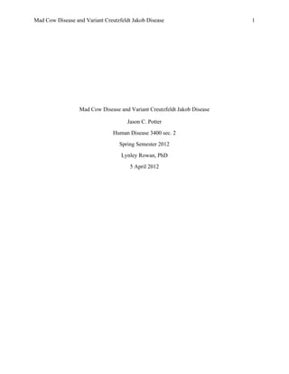
  • 11. Mad Cow Disease and Variant Creutzfeldt Jakob Disease                                                                                                                                              11     Below is a simplified diagram (Figure 3) of the prion hypothesis presented by Burthem & Roberts (2003). Burthen & Roberts explained, “(A and B) Molecules of PrPSc interact directly with the PrPc causing the structural conversion of the PrPC to form further PrPSc . This conversion culminates in the formation of polymeric aggregates of PrPSc (C) or in the formation of further infective material” (p. 223). Figure 3 courtesy of Burtham & Roberts (2003) Burthem and Roberts (2003) provide a detailed explanation of the prion hypothesis: The prion hypothesis requires that, for PrP(Sc)infection to occur, PrP(C)must first be expressed by the host, i.e. there must be a 'pool' of PrP(C)that can be converted to PrP(Sc). Immunological surveillance requires that cells of the immune system encounter and process new proteins entering the body at an early stage after ingestion or inoculation. Moreover, immune cells migrate widely through the body and establish frequent and close contact with other cells. This behavior is essential for the effective immune response to conventional pathogens, but in prion disease this otherwise beneficial process appears to facilitate the propagation and spread of the abnormal protein (p. 122). A         B         C  
  • 12. Mad Cow Disease and Variant Creutzfeldt Jakob Disease                                                                                                                                              12     As discussed, the causative agent in vCLD is a prion; however, Belay (1999) discussed a specific variation in the sequence of genes has been confirmed. The most significant polymorphism leads to the formation of two different protein forms. The first form has an amino acid called methionone at the 129th amino acid in the protein sequence, and the second form has an amino acid called valine in the same position (p. 298). Belay (1999) also addressed several studies that have shown the polymorphism at codon 129 has a significant impact in determining the susceptibility of the host and the disease expression of familial, iatrogenic (infectious), or sporadic CJD (p. 298). Individuals that developed the vCJD had a double-methionine form of the prion protein, but no other mutations. This implies that this particular variation in the gene somehow influences an individual’s susceptibility to vCJD (Prusiner, 2001). In fact, Brown et al., (2001) concluded, “The encoding alternatives methionine (met) and valine (Val), are distributed in the approximate proportions of 50% Met/Val, 40% Met/Met, and 10 % Val/Val.” In 2001, it was reported that all 76 vCJD patients tested have been homozygous for methionine, and apparently single infecting strains of BSE may not be able to replicate in any other human genotype (p. 10). In 2005, Ludman & Turner reported that “ to date in all clinical cases of variant CJD have occurred in methionine 128 homozygous individuals; it seems likely that valine homozygous and methionine/valine heterozygous individuals are more resistant to infection, or if infected, to the development of clinical variant CJD” (p. 16). Transmission Bovine to Human. Research has positively confirmed the transmission of vCJD is directly related to the biological features of the pathological agent isolated from BSE infected cattle. The source of the infection appears to have been from beef products contaminated by nervous system tissue
  • 13. Mad Cow Disease and Variant Creutzfeldt Jakob Disease                                                                                                                                              13     containing prions (Brown et al., 2001, p. 10). Brown et al., (2001) explained how contamination originally could have occurred: Cerebral vascular emboli from cranial stunning instruments used to immobilize cattle before killing by exsanguination; contact of muscle with brain or spinal cord tissue by saws or other tools used during slaughter; inclusion of paraspinal ganglia in cuts of meat containing vertebral tissues; and perhaps most importantly the presence of residual spinal cord and parasprinal ganglia tissue in the paste of mechanically recovered meat that could legally be added to cooked meat products such as meat pies, beef sausages, and various canned meat preparations (p. 10). According to the CDC (2011), BSE may have originally resulted due to feeding cattle meat and bone meal that contained products from a randomly occurring case of BSE or scrapie from possible contaminated sheep products. The CDC (2011) stated, “There is strong evidence and general agreement that the outbreak was then amplified and spread throughout the United Kingdom cattle industry by feeding rendered, prion-infected, bovine meat and bone meal to young calves.” The CDC (2011) confirmed there is solid epidemiologic and laboratory data for a connecting link between vCJD that was first reported from the United Kingdom in 1996, and the epidemic of BSE. A unique characteristic of vCJD is the long incubation period. The CDC (2011) reported similar incubation periods for human forms of prion diseases and the time frame of initial exposure to BSE and the clinical manifestations of the disease. Blood Products. VCJD may also be transmitted through blood products such as transfusions and using blood products from an infected person with vCJD. Turner & Ludman (2008) addressed the topic of vCJD by blood transfusion and discussed the direct link of vCJD and blood donation. Turner and Ludman (2008) reported that, “Eighteen patients with vCJD have, or have has previously, been blood donors, from whom a total of 66 recipients have been identified, 26 of whom are still alive. Of those who have died, four cases of transmission of vCJD prions have
  • 14. Mad Cow Disease and Variant Creutzfeldt Jakob Disease                                                                                                                                              14     been identified” (p .15) In December 2003, the first case of vCJD linked to a blood transfusion was indentified. The individual that became infected with vJCD developed the disease 6 years after receiving a blood transfusion. The blood donor developed signs and symptoms of vJCD 3 years after donating (Turner & Ludman, 2008, p. 15). This data clearly shows that individuals in the incubation period of vCJD can transmit the disease to another individual through blood transfusion. No specific immunological reaction related to prion infection has been indentified nor has specific DNA been found related to the transmission of vCJD. Thus, typical serological and biological tests in donors have not been possible (Ludlam & Turner, 2005, p. 18). Iatrogenic. Transmission from human to human through direct contact has not been confirmed; however, individuals can be infected with vCJD through iatrogenic transmission. Ludlan & Turner (2008) reported iatrogenic transmission has been well documented. The transmission occurs through direct inoculation of the Central Nervous System (CNS) through contaminated neurosurgical instruments, intracerebral electrodes, dura matter and corneal grafts. Iatrogenic transmission has also occurred by pituitary growth hormone and gonadotrophins taken from cadavers and administered to patients by intramuscular injection (Turner & Ludman, 2008, p. 15). Despite technological advanced sterilization techniques, it is possible for vCJD to be transmitted to another patient. Campbell (2006) reported that stainless steel instruments used in neurosurgery can transmit vCJD to another patient even if the instrument is thoroughly sterilized with formaldehyde. It is recommended that only disposable instruments be used in neurological surgery and tonsil removal to prevent the transmission of vCJD from one patient to another. According to Ludman & Turner (2005), patients with vCJD have shown evidence of irregular prion accumulation in follicular dendritic cells and peripheral lymphoid tissue, including the
  • 15. Mad Cow Disease and Variant Creutzfeldt Jakob Disease                                                                                                                                              15     tonsils (p. 17). Another concern has been brought up regarding dental surgery involving removal of an infected root. Since formaldehyde is ineffective in disinfecting instruments, it has been recommended that dental surgeons use 4% hypochlorine (p.93). Campbell (2006) reiterated the possibility of direct transmission from human to human through the use of infected surgical instruments, “The fear is there may be a bank of human carriers passing on the infection to others” (p. 93). Port of Entry. Clinical presentation and specific signs and symptoms of vCJD can vary depending on the route of infection. CNS transmission is likely to have a shorter incubation period. According to Ludlam & Turner (2005), the incubation period is roughly two years and the infected individual develops rapid dementia. Peripherally transmitted cases usually have a longer incubation period, ranging from 13-15 years and symptoms include ataxia and sensory disturbances (Ludlam & Turner, 2005, p. 15). Tan, Williams, Khan, Champion & Nielson (1999) discussed four categories related to certain organs containing more infectious prions than others: Direct administration into the central nervous system is the most infectious route, followed by administration in to the blood vessels, and intraperitoneal, intramuscular, and subcutaneous exposure. Oral ingestion is less efficient than the parenteral routes. Finally, the dose of infectious material is an important determinant of transmissibility (p. 2333). Fate of Victim According to Ludman & Turner (2005), after transmission of the disease, the fate of the victim infected with vCJD is a poor prognosis that eventually leads to death. Ludman and Turner (2005) stated in the end stages of the disease patients develop progressive dementia and myoclonus “with an average clinical course to death of 6 months—2 years” (p. 15).
  • 16. Mad Cow Disease and Variant Creutzfeldt Jakob Disease                                                                                                                                              16     Vulnerability and Risk As reported by the CDC (2010), almost all cases of vCJD have been in persons under the age of 55, with the median age at death being 28. Ludlam & Turner (2005) also reported the median age of death has not changed over the first 10 years of the outbreak; this suggests there may be an age related susceptibility or exposure (p. 17). Reasoning for this specific age distribution is not fully understood at this time; however, the CDC (2010) suggested that through the oral route of exposure, older adults are less susceptible to vCJD than young adults and children. Concern has also been raised that some individuals in the population may be genetically susceptible to vCJD. The CDC (2010) discussed this concern, presented in the following quote: In 2004, a prevalence study of asymptomatic vCJD infections in the UK identified three positive appendices out of a sample of 12,674 surgically removed tonsils and appendices that were satisfactory for analysis. Genetic studies completed on two of the appendices regarded as positive for vCJD revealed that both had a different polymorphism at codon 129 of the prion protein gene than any of the patients with clinical vCJD tested to date, indicating that more people are genetically susceptible to vCJD infection, although not necessarily to the disease, than had been previously determined. The CDC (2010)confirmed the current risk for acquiring vCJD from eating BSE- contaminated beef products in countries with a possible increase risk of BSE cannot be exactly determined. The CDC (2010) stated if current public health measures are being implemented, the risk of being infected with vCJD due to eating contaminated beef is extremely small; nonetheless, the risk is not zero. The CDC (2010) reported, “A rough estimate of this risk for the UK in the recent past, for example, was about 1 case per 10 billion servings.” Individuals receiving blood transfusion are at risk of being infected with vCJD from the blood of a donor that may still be in the incubation period. Individuals that received blood from donors in the UK that were subsequently diagnosed with vCJD are considered high risk for
  • 17. Mad Cow Disease and Variant Creutzfeldt Jakob Disease                                                                                                                                              17     developing vCJD. These individuals are contacted on a regular basis and offered special follow up care and counseling. In 2004, in the UK, all patients with hemophilia were sent letters stating whether or not they had received plasma-derived clotting concentrates between the years of1980- 2001. (Ludlam &Turner, 2008, p. 15, 21). As of 2003, one of case of vCJD in a 62-year old UK man is thought to have been as a result from a blood transfusion contaminated with the vCJD infectious agent. Just a year later, another case in the UK was reported that was very suspicious, likely being transmitted by a blood transfusion. Due to the reported cases of possible transmission through a blood transfusion, the UK quickly implemented specific safeguards. A ban was placed on all persons receiving blood transfusions after 1980 in the UK from donating blood in the future. In January 2002, in order to reduce the risk of bloodborne transmission of vCJD, the USDA released guidance, which included a geography-based donor deferral policy (CDC, 2010). Ludlam &Turner (2008) discussed the potential risk posed to patients undergoing invasive medical procedures such as neural, tonsil, or oral surgery for an infected root. The risk increases in these types of surgeries when considering patient to patient transmission due to surgical instruments that may be harvesting the infective agent associated with vCJD. (15-17). Prevention In regards to prevention, the CDC (2010) recommended: To reduce any risk of acquiring vCJD from food, travelers to Europe or other areas with indigenous cases of BSE may consider either avoiding beef and beef products altogether or selecting beef or beef products, such as solid pieces of muscle meat (rather than brains or beef products like burgers and sausages), that might have a reduced opportunity for contamination with tissues that may harbor the BSE agent. Milk and milk products from cows are not believed to pose any risk for transmitting the BSE agent. In order to prevent BSE from entering the U.S., harsh restrictions were implemented by the USDA in June of 1997, becoming effective in October 1997. This included restrictions on the
  • 18. Mad Cow Disease and Variant Creutzfeldt Jakob Disease                                                                                                                                              18     importing of live cattle, sheep and goats, and certain ruminant products where BSE had been identified. Later, this restriction was extended to all European countries. This feed ban is enforced by the FDA through inspections and feed testing programs. In October 2009, the FDA issued a regulation creating an enhanced BSE-related feed ban in the U.S. This feed ban helped to synchronize feed control measures in the US and Canada. In July of 2007, an enhanced BSE- related feed ban was put into effect in Canada in order to quickly eliminate the spread of BSE in Canada. (CDC, 2010). To help with prevention measures in relation to vCJD, the CDC continues to monitor trends and the current incidence in the U.S. through several surveillance methods. The CDC has placed more attention on the national surveillance of classic CJD in order to more effectively detect vCJD. This is accomplished through autopsies on patients with possible prion diseases. This has helped to increase the ability to detect more vCJD cases (CDC, 2010). The CDC also continues to investigate possible deaths from iatrogenic CJD and vCJD reported by health care professionals. Most importantly, “in 1996-97, CDC established, in collaboration with the American Association of Neuropathologists, the National Prion Disease Pathology Surveillance Center at Case Western Reserve University, which performs special diagnostic tests for prion diseases, including post-mortem tests that can detect vCJD.” (CDC, 2010). Treatment The CDC stated (2010), “As of September 2004, treatment of prion diseases remains supportive; no specific therapy has been shown to stop the progression of these diseases.” Becoming infected with vCJD is fatal to the patient. In a study conducted by Vries, Sque, Bryan & Abu-Saad (2003), the need for palliative care and development of educational initiatives is addressed. They concluded that caring for patients with vCJD is very a stressful experience in all
  • 19. Mad Cow Disease and Variant Creutzfeldt Jakob Disease                                                                                                                                              19     stages of the illness. Patient’s affected by vCJD are often times admitted to psychiatric units, where they eventually die. Vries, Sque, Bryan & Abu-Saad (2003) argued it is more appropriate for patients affected by vCJD to be cared for by health care professionals in hospice environments. Consequently, there should be planning initiated by the healthcare team for patients affected by illnesses such as vCJD that are terminal and involve dementia (p. 518). Global Economy With the outbreak of BSE in the 1990’s and eventual transmission to humans, both the UK and the U.S. have created measures in response to the crisis. According to the CDC (2010), the department of Health and Human Services (HHS) in 2001 began to create a department-wide action plan. This plan helped to outline steps to improve the scientific understanding and research in regards the BSE and other TESs. The CDC (2010) presented the department-wide action plan: Surveillance for human disease is primarily the responsibility of CDC. Protection is primarily the responsibility of the Food and Drug Administration (FDA). Research is primarily the responsibility of the National Institutes of Health (NIH). Oversight is primarily the responsibility of the Office of the Secretary of DHHS. In is no question, both BSE and vCJD have had significant impact on the global economy. In fact, Campbell (2006) stated, “The BSE crisis has cost the UK government more than 4 billion pounds alone in compensation to farmers and caused much disquiet in agriculture” (p.94). Due to the slaughtering of thousands of cattle; ban on importing ruminant products from all European countries; and severe restrictions that were placed on the importation of cattle, sheep, and goats; the global market has suffered greatly. Campbell (2006) also discussed the controversy of scientist being criticized for initially telling the public the transmission of BSE to humans was only a distant possibility. Campbell (2006) stated, “There is no doubt that the reputation of scientists has been badly affected by the
  • 20. Mad Cow Disease and Variant Creutzfeldt Jakob Disease                                                                                                                                              20     BSA saga and we know that the story is not at an end. The experience with BSE has increased the resistance of the public to biotechnology in general” (p. 94). Campbell (2006) also addressed the concern journalists have with scientist when they pose the question “is it safe to eat beef?” and they do not receive a direct yes or no, but would rather speak about risk factors. Beghi, et, al (2004) reaffirmed the reality that the future effect on the global economy in relation to BSE and vCJD is unknown at this time due to the long and ill-defined incubation period of prion diseases. Thus, we do not know the beginning and the end of the human outbreak of BSE and the effects it may have on our future economy. The epidemic of Mad Cow Disease that spread throughout the UK in the 1980’s raised concern worldwide. With humans coming in contact with contaminated beef, vCJD developed as a result. Scientists continue to conduct further research to fully understand the impact of a prion disease such a vCJD. The research is focused on finding a more effective way of identifying potential carriers early on, and preventing transmission that can occur through blood transfusions and invasive medical procedures. In addition to research taking place, government agencies in the UK and U.S. are continually implementing preventative measures to stop infected beef products from reaching humans. Due to the uniqueness of this disease and the long incubation period, scientists, researchers, and government agencies fear there could be a large portion of the population carrying the disease and unknowingly spreading it to others, creating a second wave of the BSE outbreak.
  • 21. Mad Cow Disease and Variant Creutzfeldt Jakob Disease                                                                                                                                              21     References Behgi, E., Ferrarese, C., Fratolla, L., Gandolfo, C., Mancardi, G., Rizzuto, N., et al. (2004). Bovine spongiform encephalopathy and Creutzfeldt-Jakob disease: facts and uncertainties underlying the casual link between animal and human diseases. Neurological Science , 122-129. Belay, E. D. (1999). Transmissible Spongiform Encephalopathies in Humans. Annual Review of Microbiology , 283-314. Brown, P., Will, R. G., Bradley, R., Asher, D. M., & Detwiler, L. (2001). Bovine Spongiform Encepjalopathy and Variant Creutzfelt-Jacob Disease; Background, Evolution, and Current Conerns. Emerging Infectious Diseases , 6-13. Burthan, J., & Roberts, D. (2003). The Pathology of Variant CreutzFeldt-Jakob Diseasae: the hypothesis behind conerns for blood components and products. British Journal of Haematology , 122-127. Campbell, P. N. (2006). Mad cow disease. Acta Biologica Szegediensis , 89-95. Center for Disease Control and Prevention. (2010, 2011). Retrieved Feburary 9, 2012, from http://www.cdc.gov/ncidod/dvrd/vcjd/ Chachra, A., Narang, D., & Narang, R. (1999). Mad Cow Disease. Resonance , 42-44. Engelkirk, P. G., & Duben-Engelkirk, J. (2010). Burton's Microbiology for Health Sciences. Lippincott Williams & Wilkins. Goldberg, A. L. (2007). On Prion, Proteasomes and Mad Cows. The New England Journal of Medicine , 1150-1152. Henley, E., & Herrmann, J. (2004). Mad Cow Disease: Dealing sensibly with a new conern. The Journal of Family Practice , 645-648. Ludlam, C. A. (2008). An update on the assessment and management of the risk of transmission of variant Creutzfedldt Jakob Disease and plasma products. British Journal of Haematology , 14- 23. Ludlam, C. A., & Turner, M. L. (2005). Managing the risk of transmission of variant Creutzfeldt Jakob disease by blood prosucts. Brithish Journal of Haematology , 13-24. Prusiner, S. (2001). Shattuck Lecture: Neurodegernaerative diseases and prions. New England Journal of Medicine , 1516-1526.
  • 22. Mad Cow Disease and Variant Creutzfeldt Jakob Disease                                                                                                                                              22     Spencer, M. D., Knight, R. S., & Will, R. G. (2002). First hundred cases of variant Creutzfelt- Jakob disease: retrospective case note review of early psychiatric and neurological features. British Medical Journal , 1479-1482. Tan, L., Williams, M. L., K. M., Champion, H. C., & Nielsen, N. H. (1999). Risk of Transmission of Bovine Spongiform Encepalopathy to Humans in the United States. Jourmal of the American Medical Asssocation , 2230-2339. Vries, K. d., Sque, M., Bryan, K., & Abu-Saad, H. (2003). Variant Creutzfeldt Jakob Disease: need for mental health and palliative care team collaboration. International Journal of Palliative Nursing , 512-520.