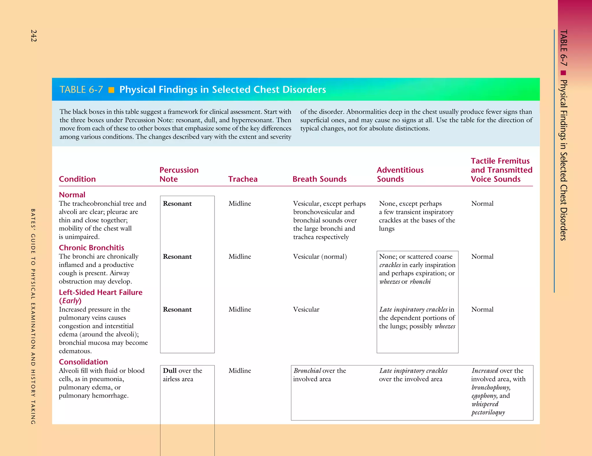
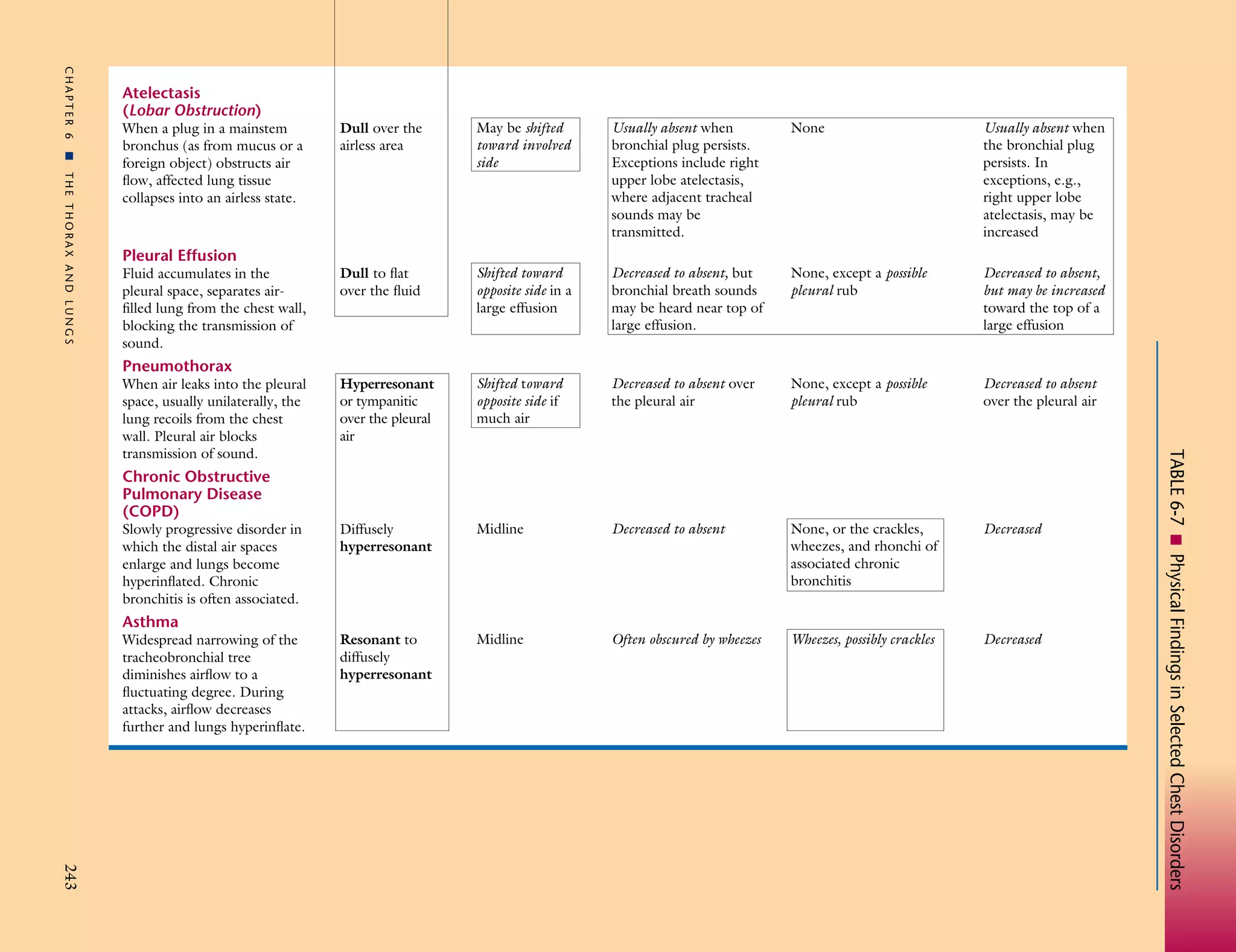
SlideShare a Scribd company logo
The black boxes in this table suggest a framework for clinical assessment. Start with
the three boxes under Percussion Note: resonant, dull, and hyperresonant. Then
move from each of these to other boxes that emphasize some of the key differences
among various conditions. The changes described vary with the extent and severity

Condition
Normal
BATES’ GUIDE TO PHYSICAL EXAMINATION AND HISTORY TAKING

The tracheobronchial tree and
alveoli are clear; pleurae are
thin and close together;
mobility of the chest wall
is unimpaired.

Chronic Bronchitis

The bronchi are chronically
inflamed and a productive
cough is present. Airway
obstruction may develop.

Left-Sided Heart Failure
(Early)

Increased pressure in the
pulmonary veins causes
congestion and interstitial
edema (around the alveoli);
bronchial mucosa may become
edematous.

Consolidation

Alveoli fill with fluid or blood
cells, as in pneumonia,
pulmonary edema, or
pulmonary hemorrhage.

Percussion
Note

of the disorder. Abnormalities deep in the chest usually produce fewer signs than
superficial ones, and may cause no signs at all. Use the table for the direction of
typical changes, not for absolute distinctions.

Adventitious
Sounds

Tactile Fremitus
and Transmitted
Voice Sounds

Trachea

Breath Sounds

Resonant

Midline

Vesicular, except perhaps
bronchovesicular and
bronchial sounds over
the large bronchi and
trachea respectively

None, except perhaps
a few transient inspiratory
crackles at the bases of the
lungs

Normal

Resonant

Midline

Vesicular (normal)

None; or scattered coarse
crackles in early inspiration
and perhaps expiration; or
wheezes or rhonchi

Normal

Resonant

Midline

Vesicular

Late inspiratory crackles in
the dependent portions of
the lungs; possibly wheezes

Normal

Dull over the
airless area

Midline

Bronchial over the
involved area

Late inspiratory crackles
over the involved area

Increased over the
involved area, with
bronchophony,
egophony, and
whispered
pectoriloquy

TABLE 6-7 I Physical Findings in Selected Chest Disorders

242

TABLE 6-7 I Physical Findings in Selected Chest Disorders
CHAPTER 6

Atelectasis
(Lobar Obstruction)

I
THE THORAX AND LUNGS

When a plug in a mainstem
bronchus (as from mucus or a
foreign object) obstructs air
flow, affected lung tissue
collapses into an airless state.

Pleural Effusion

Fluid accumulates in the
pleural space, separates airfilled lung from the chest wall,
blocking the transmission of
sound.

Pneumothorax

Chronic Obstructive
Pulmonary Disease
(COPD)

Slowly progressive disorder in
which the distal air spaces
enlarge and lungs become
hyperinflated. Chronic
bronchitis is often associated.

Asthma

Widespread narrowing of the
tracheobronchial tree
diminishes airflow to a
fluctuating degree. During
attacks, airflow decreases
further and lungs hyperinflate.

May be shifted
toward involved
side

Usually absent when
bronchial plug persists.
Exceptions include right
upper lobe atelectasis,
where adjacent tracheal
sounds may be
transmitted.

None

Usually absent when
the bronchial plug
persists. In
exceptions, e.g.,
right upper lobe
atelectasis, may be
increased

Dull to flat
over the fluid

Shifted toward
opposite side in a
large effusion

Decreased to absent, but
bronchial breath sounds
may be heard near top of
large effusion.

None, except a possible
pleural rub

Decreased to absent,
but may be increased
toward the top of a
large effusion

Hyperresonant
or tympanitic
over the pleural
air

Shifted toward
opposite side if
much air

Decreased to absent over
the pleural air

None, except a possible
pleural rub

Decreased to absent
over the pleural air

Diffusely
hyperresonant

Midline

Decreased to absent

None, or the crackles,
wheezes, and rhonchi of
associated chronic
bronchitis

Decreased

Resonant to
diffusely
hyperresonant

Midline

Often obscured by wheezes

Wheezes, possibly crackles

Decreased

243

TABLE 6-7 I Physical Findings in Selected Chest Disorders

When air leaks into the pleural
space, usually unilaterally, the
lung recoils from the chest
wall. Pleural air blocks
transmission of sound.

Dull over the
airless area

More Related Content

What's hot

Pneumothorax & chest tube final year mbbs lecture
Pneumothorax & chest tube   final year mbbs lecturePneumothorax & chest tube   final year mbbs lecture
Pneumothorax & chest tube final year mbbs lecture
Mr Adeel Abbas
 
Ppt atelectasis
Ppt atelectasisPpt atelectasis
Ppt atelectasis
Debanjali Basu
 
Atlectasis
AtlectasisAtlectasis
Atlectasis
Hasnah Noi
 
Dyspnea
DyspneaDyspnea
Dyspnea
Fatima Awadh
 
Atelectasis (collapse)
Atelectasis (collapse) Atelectasis (collapse)
Atelectasis (collapse)
santusan
 
Atelectasis
Atelectasis   Atelectasis
Atelectasis
OM VERMA
 
DISEASES OF PLEURA WITH DIAGRAMATIC ILLUSTRATION
DISEASES OF PLEURA WITH DIAGRAMATIC ILLUSTRATIONDISEASES OF PLEURA WITH DIAGRAMATIC ILLUSTRATION
DISEASES OF PLEURA WITH DIAGRAMATIC ILLUSTRATION
immad obaid
 
CXR: Pneumothorax / Pleural Thickening
CXR: Pneumothorax / Pleural ThickeningCXR: Pneumothorax / Pleural Thickening
CXR: Pneumothorax / Pleural Thickening
Stanley Medical College, Department of Medicine
 
Pneumothorax
PneumothoraxPneumothorax
Hemoptysis
HemoptysisHemoptysis
Hemoptysis
imangalal
 
Pneumomediastinum
PneumomediastinumPneumomediastinum
Pneumomediastinum
Swati Patanwal
 
Atelectasis 160424141702-converted
Atelectasis 160424141702-convertedAtelectasis 160424141702-converted
Atelectasis 160424141702-converted
Apatel99094
 
Pulmonary Emphysema. Pathomorphology, outcomes, complications
Pulmonary Emphysema. Pathomorphology, outcomes, complications Pulmonary Emphysema. Pathomorphology, outcomes, complications
Pulmonary Emphysema. Pathomorphology, outcomes, complications
Semey State Medical University
 
Pneumothorax & chest tube final year mbbs lecture
Pneumothorax & chest tube   final year mbbs lecturePneumothorax & chest tube   final year mbbs lecture
Pneumothorax & chest tube final year mbbs lecture
Mr Adeel Abbas
 
Pneumothorax
PneumothoraxPneumothorax
Atelectasis IN CHILD
Atelectasis IN CHILD Atelectasis IN CHILD
Atelectasis IN CHILD
lingampelli
 
Management of traumatic pneumothorax and haemothorax
Management of traumatic pneumothorax and haemothoraxManagement of traumatic pneumothorax and haemothorax
Management of traumatic pneumothorax and haemothorax
SachinMalayaiah1
 
Pneumothorax
PneumothoraxPneumothorax
Pneumothorax
Medical Student
 
Pneumothorax
PneumothoraxPneumothorax
Pneumothorax
salman habeeb
 
Approach To Dyspnea
Approach To DyspneaApproach To Dyspnea
Approach To Dyspnea
NoshirwanGazder
 

What's hot (20)

Pneumothorax & chest tube final year mbbs lecture
Pneumothorax & chest tube   final year mbbs lecturePneumothorax & chest tube   final year mbbs lecture
Pneumothorax & chest tube final year mbbs lecture
 
Ppt atelectasis
Ppt atelectasisPpt atelectasis
Ppt atelectasis
 
Atlectasis
AtlectasisAtlectasis
Atlectasis
 
Dyspnea
DyspneaDyspnea
Dyspnea
 
Atelectasis (collapse)
Atelectasis (collapse) Atelectasis (collapse)
Atelectasis (collapse)
 
Atelectasis
Atelectasis   Atelectasis
Atelectasis
 
DISEASES OF PLEURA WITH DIAGRAMATIC ILLUSTRATION
DISEASES OF PLEURA WITH DIAGRAMATIC ILLUSTRATIONDISEASES OF PLEURA WITH DIAGRAMATIC ILLUSTRATION
DISEASES OF PLEURA WITH DIAGRAMATIC ILLUSTRATION
 
CXR: Pneumothorax / Pleural Thickening
CXR: Pneumothorax / Pleural ThickeningCXR: Pneumothorax / Pleural Thickening
CXR: Pneumothorax / Pleural Thickening
 
Pneumothorax
PneumothoraxPneumothorax
Pneumothorax
 
Hemoptysis
HemoptysisHemoptysis
Hemoptysis
 
Pneumomediastinum
PneumomediastinumPneumomediastinum
Pneumomediastinum
 
Atelectasis 160424141702-converted
Atelectasis 160424141702-convertedAtelectasis 160424141702-converted
Atelectasis 160424141702-converted
 
Pulmonary Emphysema. Pathomorphology, outcomes, complications
Pulmonary Emphysema. Pathomorphology, outcomes, complications Pulmonary Emphysema. Pathomorphology, outcomes, complications
Pulmonary Emphysema. Pathomorphology, outcomes, complications
 
Pneumothorax & chest tube final year mbbs lecture
Pneumothorax & chest tube   final year mbbs lecturePneumothorax & chest tube   final year mbbs lecture
Pneumothorax & chest tube final year mbbs lecture
 
Pneumothorax
PneumothoraxPneumothorax
Pneumothorax
 
Atelectasis IN CHILD
Atelectasis IN CHILD Atelectasis IN CHILD
Atelectasis IN CHILD
 
Management of traumatic pneumothorax and haemothorax
Management of traumatic pneumothorax and haemothoraxManagement of traumatic pneumothorax and haemothorax
Management of traumatic pneumothorax and haemothorax
 
Pneumothorax
PneumothoraxPneumothorax
Pneumothorax
 
Pneumothorax
PneumothoraxPneumothorax
Pneumothorax
 
Approach To Dyspnea
Approach To DyspneaApproach To Dyspnea
Approach To Dyspnea
 

Similar to Lung exambates

Approach to respiratory disorderv 3.pptx
Approach to respiratory disorderv 3.pptxApproach to respiratory disorderv 3.pptx
Approach to respiratory disorderv 3.pptx
tesa10
 
Atelecstasis
AtelecstasisAtelecstasis
Atelecstasis
jenishaadhikari
 
PLEURAL EFFUSION.ppt
PLEURAL EFFUSION.pptPLEURAL EFFUSION.ppt
PLEURAL EFFUSION.ppt
AnguaniVictor
 
ATELACTASIS.pptx
ATELACTASIS.pptxATELACTASIS.pptx
ATELACTASIS.pptx
DishaThakur53
 
chest X ray basics and interpretation
chest X ray basics and interpretationchest X ray basics and interpretation
chest X ray basics and interpretation
somaskandan Rajendran
 
ATELECTASIS.pptx
ATELECTASIS.pptxATELECTASIS.pptx
ATELECTASIS.pptx
carolineouma6
 
Chest x ray review.
Chest x ray review.Chest x ray review.
Chest x ray review.
Muhammad Ali
 
congenital lung.pptx
congenital lung.pptxcongenital lung.pptx
congenital lung.pptx
ranjitharadhakrishna3
 
Respiratory Diseases -Obstructive lung diseases.ppt
Respiratory Diseases -Obstructive lung diseases.pptRespiratory Diseases -Obstructive lung diseases.ppt
Respiratory Diseases -Obstructive lung diseases.ppt
ArpitaHalder8
 
Lung Diseases
Lung DiseasesLung Diseases
Lung Diseases
Ashley Nicole
 
2.CXR.ppt
2.CXR.ppt2.CXR.ppt
2.CXR.ppt
syedWahaj16
 
Dyspnea
DyspneaDyspnea
Dyspnea
cairo1957
 
Reading chest X-ray
Reading chest X-rayReading chest X-ray
Reading chest X-ray
Mohammad Ihmeidan
 
Respiratory System Physical Examination
Respiratory System Physical ExaminationRespiratory System Physical Examination
Respiratory System Physical Examination
Saneesh P J
 
Tetralogy of Fallot (TOF)
 Tetralogy of Fallot (TOF) Tetralogy of Fallot (TOF)
Tetralogy of Fallot (TOF)
Syed Kamrul Hasan
 
4. Pneumothorax, COPD, CVDs.pdf cardiovascular system
4. Pneumothorax, COPD, CVDs.pdf cardiovascular system4. Pneumothorax, COPD, CVDs.pdf cardiovascular system
4. Pneumothorax, COPD, CVDs.pdf cardiovascular system
akoeljames8543
 
Patttern of lung disease on chest x ray
Patttern of lung disease on   chest x rayPatttern of lung disease on   chest x ray
Patttern of lung disease on chest x ray
Vrishit Saraswat
 
Chest X-Ray in Pediatrics.pptx
Chest X-Ray in Pediatrics.pptxChest X-Ray in Pediatrics.pptx
Chest X-Ray in Pediatrics.pptx
HurshidaShia
 
Acute resp failure.pptx
Acute resp failure.pptxAcute resp failure.pptx
Acute resp failure.pptx
Dipali Dumbre
 
belajar cle.pptx
belajar cle.pptxbelajar cle.pptx
belajar cle.pptx
KeziaPane1
 

Similar to Lung exambates (20)

Approach to respiratory disorderv 3.pptx
Approach to respiratory disorderv 3.pptxApproach to respiratory disorderv 3.pptx
Approach to respiratory disorderv 3.pptx
 
Atelecstasis
AtelecstasisAtelecstasis
Atelecstasis
 
PLEURAL EFFUSION.ppt
PLEURAL EFFUSION.pptPLEURAL EFFUSION.ppt
PLEURAL EFFUSION.ppt
 
ATELACTASIS.pptx
ATELACTASIS.pptxATELACTASIS.pptx
ATELACTASIS.pptx
 
chest X ray basics and interpretation
chest X ray basics and interpretationchest X ray basics and interpretation
chest X ray basics and interpretation
 
ATELECTASIS.pptx
ATELECTASIS.pptxATELECTASIS.pptx
ATELECTASIS.pptx
 
Chest x ray review.
Chest x ray review.Chest x ray review.
Chest x ray review.
 
congenital lung.pptx
congenital lung.pptxcongenital lung.pptx
congenital lung.pptx
 
Respiratory Diseases -Obstructive lung diseases.ppt
Respiratory Diseases -Obstructive lung diseases.pptRespiratory Diseases -Obstructive lung diseases.ppt
Respiratory Diseases -Obstructive lung diseases.ppt
 
Lung Diseases
Lung DiseasesLung Diseases
Lung Diseases
 
2.CXR.ppt
2.CXR.ppt2.CXR.ppt
2.CXR.ppt
 
Dyspnea
DyspneaDyspnea
Dyspnea
 
Reading chest X-ray
Reading chest X-rayReading chest X-ray
Reading chest X-ray
 
Respiratory System Physical Examination
Respiratory System Physical ExaminationRespiratory System Physical Examination
Respiratory System Physical Examination
 
Tetralogy of Fallot (TOF)
 Tetralogy of Fallot (TOF) Tetralogy of Fallot (TOF)
Tetralogy of Fallot (TOF)
 
4. Pneumothorax, COPD, CVDs.pdf cardiovascular system
4. Pneumothorax, COPD, CVDs.pdf cardiovascular system4. Pneumothorax, COPD, CVDs.pdf cardiovascular system
4. Pneumothorax, COPD, CVDs.pdf cardiovascular system
 
Patttern of lung disease on chest x ray
Patttern of lung disease on   chest x rayPatttern of lung disease on   chest x ray
Patttern of lung disease on chest x ray
 
Chest X-Ray in Pediatrics.pptx
Chest X-Ray in Pediatrics.pptxChest X-Ray in Pediatrics.pptx
Chest X-Ray in Pediatrics.pptx
 
Acute resp failure.pptx
Acute resp failure.pptxAcute resp failure.pptx
Acute resp failure.pptx
 
belajar cle.pptx
belajar cle.pptxbelajar cle.pptx
belajar cle.pptx
 

More from kantemur

Pneumonia subtypes
Pneumonia subtypesPneumonia subtypes
Pneumonia subtypes
kantemur
 
Tumor cell morphology
Tumor cell morphologyTumor cell morphology
Tumor cell morphology
kantemur
 
Inflammation adel-1
Inflammation adel-1Inflammation adel-1
Inflammation adel-1
kantemur
 
Neoplasia
NeoplasiaNeoplasia
Neoplasia
kantemur
 
History taking
History takingHistory taking
History taking
kantemur
 
Introduction to auscultation
Introduction to auscultationIntroduction to auscultation
Introduction to auscultation
kantemur
 
Spinal cord reflexes
Spinal cord reflexesSpinal cord reflexes
Spinal cord reflexes
kantemur
 

More from kantemur (7)

Pneumonia subtypes
Pneumonia subtypesPneumonia subtypes
Pneumonia subtypes
 
Tumor cell morphology
Tumor cell morphologyTumor cell morphology
Tumor cell morphology
 
Inflammation adel-1
Inflammation adel-1Inflammation adel-1
Inflammation adel-1
 
Neoplasia
NeoplasiaNeoplasia
Neoplasia
 
History taking
History takingHistory taking
History taking
 
Introduction to auscultation
Introduction to auscultationIntroduction to auscultation
Introduction to auscultation
 
Spinal cord reflexes
Spinal cord reflexesSpinal cord reflexes
Spinal cord reflexes
 

Lung exambates

  • 1. The black boxes in this table suggest a framework for clinical assessment. Start with the three boxes under Percussion Note: resonant, dull, and hyperresonant. Then move from each of these to other boxes that emphasize some of the key differences among various conditions. The changes described vary with the extent and severity Condition Normal BATES’ GUIDE TO PHYSICAL EXAMINATION AND HISTORY TAKING The tracheobronchial tree and alveoli are clear; pleurae are thin and close together; mobility of the chest wall is unimpaired. Chronic Bronchitis The bronchi are chronically inflamed and a productive cough is present. Airway obstruction may develop. Left-Sided Heart Failure (Early) Increased pressure in the pulmonary veins causes congestion and interstitial edema (around the alveoli); bronchial mucosa may become edematous. Consolidation Alveoli fill with fluid or blood cells, as in pneumonia, pulmonary edema, or pulmonary hemorrhage. Percussion Note of the disorder. Abnormalities deep in the chest usually produce fewer signs than superficial ones, and may cause no signs at all. Use the table for the direction of typical changes, not for absolute distinctions. Adventitious Sounds Tactile Fremitus and Transmitted Voice Sounds Trachea Breath Sounds Resonant Midline Vesicular, except perhaps bronchovesicular and bronchial sounds over the large bronchi and trachea respectively None, except perhaps a few transient inspiratory crackles at the bases of the lungs Normal Resonant Midline Vesicular (normal) None; or scattered coarse crackles in early inspiration and perhaps expiration; or wheezes or rhonchi Normal Resonant Midline Vesicular Late inspiratory crackles in the dependent portions of the lungs; possibly wheezes Normal Dull over the airless area Midline Bronchial over the involved area Late inspiratory crackles over the involved area Increased over the involved area, with bronchophony, egophony, and whispered pectoriloquy TABLE 6-7 I Physical Findings in Selected Chest Disorders 242 TABLE 6-7 I Physical Findings in Selected Chest Disorders
  • 2. CHAPTER 6 Atelectasis (Lobar Obstruction) I THE THORAX AND LUNGS When a plug in a mainstem bronchus (as from mucus or a foreign object) obstructs air flow, affected lung tissue collapses into an airless state. Pleural Effusion Fluid accumulates in the pleural space, separates airfilled lung from the chest wall, blocking the transmission of sound. Pneumothorax Chronic Obstructive Pulmonary Disease (COPD) Slowly progressive disorder in which the distal air spaces enlarge and lungs become hyperinflated. Chronic bronchitis is often associated. Asthma Widespread narrowing of the tracheobronchial tree diminishes airflow to a fluctuating degree. During attacks, airflow decreases further and lungs hyperinflate. May be shifted toward involved side Usually absent when bronchial plug persists. Exceptions include right upper lobe atelectasis, where adjacent tracheal sounds may be transmitted. None Usually absent when the bronchial plug persists. In exceptions, e.g., right upper lobe atelectasis, may be increased Dull to flat over the fluid Shifted toward opposite side in a large effusion Decreased to absent, but bronchial breath sounds may be heard near top of large effusion. None, except a possible pleural rub Decreased to absent, but may be increased toward the top of a large effusion Hyperresonant or tympanitic over the pleural air Shifted toward opposite side if much air Decreased to absent over the pleural air None, except a possible pleural rub Decreased to absent over the pleural air Diffusely hyperresonant Midline Decreased to absent None, or the crackles, wheezes, and rhonchi of associated chronic bronchitis Decreased Resonant to diffusely hyperresonant Midline Often obscured by wheezes Wheezes, possibly crackles Decreased 243 TABLE 6-7 I Physical Findings in Selected Chest Disorders When air leaks into the pleural space, usually unilaterally, the lung recoils from the chest wall. Pleural air blocks transmission of sound. Dull over the airless area