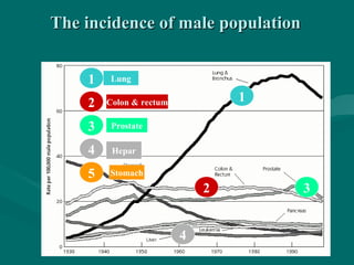
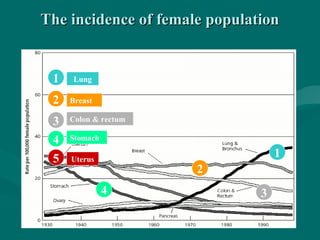
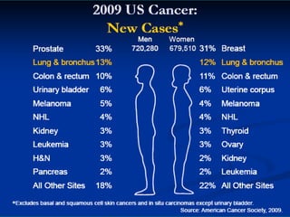
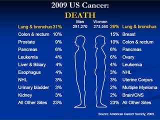
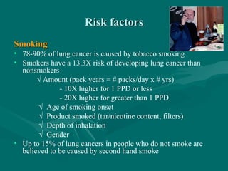
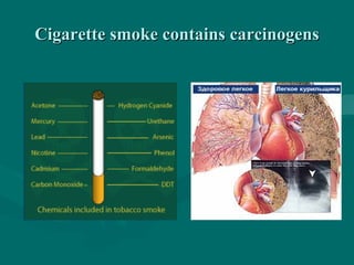
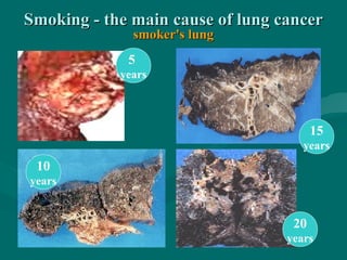
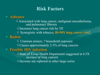
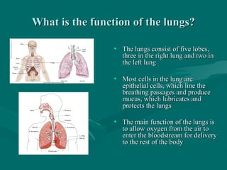
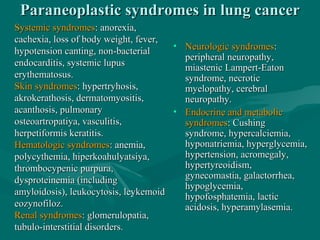
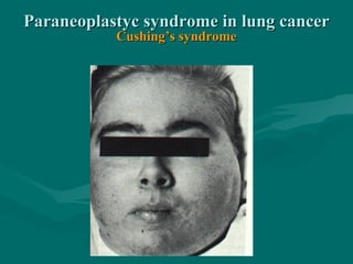
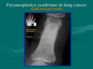
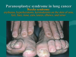
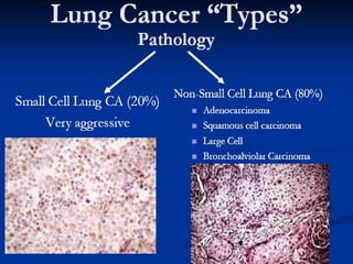
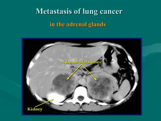
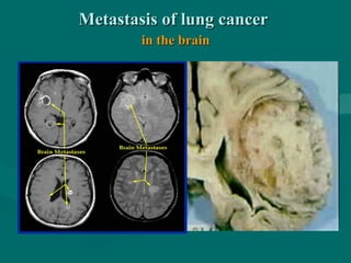
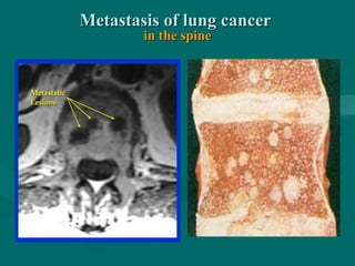
LUNG CANCERLUNG CANCER
Cancer morbidityCancer morbidity
(patients per year)(patients per year)
Lungs 1 350 000 (12,3%)Lungs 1 350 000 (12,3%)
Breast 1 050 000 (10,4%)Breast 1 050 000 (10,4%)
Colon and rectum 945 000 (9,4%)Colon and rectum 945 000 (9,4%)
Stomach 876 000 (8,7%)Stomach 876 000 (8,7%)
Hepar 564 000 (5,6%)Hepar 564 000 (5,6%)
Prostate 543 000 (5,4%)Prostate 543 000 (5,4%)
What is lung cancer?What is lung cancer?
• The leading cause of cancer death among men and
women
• Begins when cells in the lung grow out of control and
form a tumor
• There are two main types of lung cancer: non-small
cell and small cell
Famous people who died of lung cancerFamous people who died of lung cancer
Georg VIGeorg VI, (1895-1952)
King of Great Britain
Rudolf AbelRudolf Abel (Fisher), (1903-1971)
secret service agent
Sergej ParadjanovSergej Paradjanov, (1924- 1990)
film producer
Walter Elias "Walt" Disney"Walt" Disney, (1901–1966)
screenwriter
Dmitri ShostakovichDmitri Shostakovich, (1906-1975)
composer
SponsoredSponsored
Medical Lecture Notes –Medical Lecture Notes – All SubjectsAll Subjects
USMLE Exam (America) –USMLE Exam (America) – PracticePractice
Lung cancer in the world in recent yearsLung cancer in the world in recent years
the incidence for men increased by 44%, women - 75%the incidence for men increased by 44%, women - 75%
Маly 5,3 1,5
Brasil 67,5 14,4
USA, white
USA, afro-american
61,3
99,1
33,8
38,5
China
Japan
India
56,1
39,6
14,5
18,2
10,3
3,7
France
Germany
Italy
Ukraine
67,4
70,9
82,7
77,6
7,2
10,3
14,9
13,8
New Zeland,
Māori
46,5
99,7
18,2
72,9
manman womanwoman
The incidence of male populationThe incidence of male population
1
5
2 3
4
1
2
3
4
Lung
Colon & rectum
Prostate
Hepar
Stomach
The incidence of female populationThe incidence of female population
3
2
2
5
1
1
Lung
Uterus
Stomach
Colon & rectum
Breast
4
34
Lung cancerLung cancer
• Lung cancer is the #1 cancer killer worldwideLung cancer is the #1 cancer killer worldwide
• World Health Organization estimates 1 million newWorld Health Organization estimates 1 million new
cases each yearcases each year
• 200,000 cases annually in U.S.200,000 cases annually in U.S.
• More than breast, prostate and colon combinedMore than breast, prostate and colon combined
Risk factorsRisk factors
SmokingSmoking
• 78-90% of lung cancer is caused by tobacco smoking
• Smokers have a 13.3X risk of developing lung cancer than
nonsmokers
√ Amount (pack years = # packs/day x # yrs)
- 10X higher for 1 PPD or less
- 20X higher for greater than 1 PPD
√ Age of smoking onset
√ Product smoked (tar/nicotine content, filters)
√ Depth of inhalation
√ Gender
• Up to 15% of lung cancers in people who do not smoke are
believed to be caused by second hand smoke
Tobacco’s toll in U.S.Tobacco’s toll in U.S.
• 􀂄􀂄 Leading preventable cause of death, killingLeading preventable cause of death, killing
>400,000/yr>400,000/yr
• 􀂄􀂄 Tobacco kills more people than from AIDS,Tobacco kills more people than from AIDS,
alcohol, car accidents, murders, suicides, andalcohol, car accidents, murders, suicides, and firesfires
COMBINEDCOMBINED
• 􀂄􀂄 Up to 90% of lung cancer cases, 1/3 of total cancerUp to 90% of lung cancer cases, 1/3 of total cancer
deaths, and 1 in 5 deaths from heart disease aredeaths, and 1 in 5 deaths from heart disease are
tobacco relatedtobacco related
• 􀂄􀂄 Tobacco results in $89 billion in annual health careTobacco results in $89 billion in annual health care
costscosts
Cigarette smoke contains carcinogensCigarette smoke contains carcinogens
Smoking - the main cause of lung cancerSmoking - the main cause of lung cancer
smoker's lungsmoker's lung
5
years
10
years
15
years
20
years
Risk Factors
• AsbestosAsbestos
√ Associated with lung cancer, malignant mesothelioma,
and pulmonary fibrosis
√ Increases lung cancer risk by 5X
√ Synergistic with tobacco, 80-90X lung cancer risk!
• RadonRadon
√ Uranium miners, ? household exposure
√ Causes approximately 2-3% of lung cancers
• Possibly HIV infectionPossibly HIV infection
√ State of Texas Health Department suggested at 6.5X
increase of lung cancers
√ Increase not reported in other large series
Risk Factors
• Other implicated environmental agentsenvironmental agents
√ Aromatic polycyclic hydrocarbons
√ Beryllium
√ Nickel
√ Copper
√ Chromium
√ Cadmium
√ Diesel exhaust
• Possible protective effectPossible protective effect
√ Dietary fiber
√ Vegetables
The main periods of development ofThe main periods of development of
malignant lung tumorsmalignant lung tumors
• LatencyLatency of the tumor - the time from the beginning of theof the tumor - the time from the beginning of the
carcinogen to the appearance of tumor, diagnosed, lastscarcinogen to the appearance of tumor, diagnosed, lasts 10-1710-17
years, depending on the intensity of division ofyears, depending on the intensity of division of cancer cells,cancer cells,
which determine its malignancy. Thenwhich determine its malignancy. Then continuedcontinued
asymptomaticasymptomatic preclinical periodpreclinical period of tumor development,of tumor development,
and with the clinical symptoms appearand with the clinical symptoms appear clinical periodclinical period begins.begins.
• On average, for the development of tumor diameter of 1-2 mmOn average, for the development of tumor diameter of 1-2 mm
(1-2 million tumor cells) to about 20 doubling of tumor mass(1-2 million tumor cells) to about 20 doubling of tumor mass..
• In half of cases of lung cancer tumor grows to a diameter of 1 cmIn half of cases of lung cancer tumor grows to a diameter of 1 cm
(1 billion tumor cells) for 7 or more years.(1 billion tumor cells) for 7 or more years.
What is the function of the lungs?What is the function of the lungs?
• The lungs consist of five lobes,The lungs consist of five lobes,
three in the right lung and two inthree in the right lung and two in
the left lungthe left lung
• Most cells in the lung areMost cells in the lung are
epithelial cells, which line theepithelial cells, which line the
breathing passages and producebreathing passages and produce
mucus, which lubricates andmucus, which lubricates and
protects the lungsprotects the lungs
• The main function of the lungs isThe main function of the lungs is
to allow oxygen from the air toto allow oxygen from the air to
enter the bloodstream for deliveryenter the bloodstream for delivery
to the rest of the bodyto the rest of the body
Paraneoplastic syndromes in lung cancerParaneoplastic syndromes in lung cancer
Systemic syndromesSystemic syndromes: anorexia,: anorexia,
cachexia, loss of body weight, fever,cachexia, loss of body weight, fever,
hypotension canting, non-bacterialhypotension canting, non-bacterial
endocarditis, systemic lupusendocarditis, systemic lupus
erythematosus.erythematosus.
Skin syndromesSkin syndromes: hypertryhosis,: hypertryhosis,
akrokerathosis, dermatomyositis,akrokerathosis, dermatomyositis,
acanthosis, pulmonaryacanthosis, pulmonary
osteoartropatiya, vasculitis,osteoartropatiya, vasculitis,
herpetiformis keratitis.herpetiformis keratitis.
Hematologic syndromesHematologic syndromes: anemia,: anemia,
polycythemia, hiperkoahulyatsiya,polycythemia, hiperkoahulyatsiya,
thrombocypenic purpura,thrombocypenic purpura,
dysproteinemia (includingdysproteinemia (including
amyloidosis), leukocytosis, leykemoidamyloidosis), leukocytosis, leykemoid
eozynofiloz.eozynofiloz.
Renal syndromesRenal syndromes: glomerulopatia,: glomerulopatia,
tubulo-interstitial disorders.tubulo-interstitial disorders.
• Neurologic syndromesNeurologic syndromes::
peripheral neuropathy,peripheral neuropathy,
miastenic Lampert-Eatonmiastenic Lampert-Eaton
syndrome, necroticsyndrome, necrotic
myelopathy, cerebralmyelopathy, cerebral
neuropathy.neuropathy.
• Endocrine and metabolicEndocrine and metabolic
syndromessyndromes: Cushing: Cushing
syndrome, hypercalciemia,syndrome, hypercalciemia,
hyponatriemia, hyperglycemia,hyponatriemia, hyperglycemia,
hypertension, acromegaly,hypertension, acromegaly,
hypertyreoidism,hypertyreoidism,
gynecomastia, galactorrhea,gynecomastia, galactorrhea,
hypoglycemia,hypoglycemia,
hypofosphatemia, lactichypofosphatemia, lactic
acidosis, hyperamylasemia.acidosis, hyperamylasemia.
Paraneoplastyc syndrome in lung cancerParaneoplastyc syndrome in lung cancer
nail deformity phalanxes as "drum sticks"nail deformity phalanxes as "drum sticks"
Paraneoplastyc syndrome in lung cancerParaneoplastyc syndrome in lung cancer
Cushing’s syndromeCushing’s syndrome
Paraneoplastyc syndrome in lung cancerParaneoplastyc syndrome in lung cancer
ossificated periostitisossificated periostitis
Paraneoplastyc syndrome in lung cancerParaneoplastyc syndrome in lung cancer
Bazeks syndromeBazeks syndrome::
erythema, hyperkeratosis, keratodermia on the skin of arm,erythema, hyperkeratosis, keratodermia on the skin of arm,
feet, face, nose, ears, knees, elbows, and torsofeet, face, nose, ears, knees, elbows, and torso
Paraneoplastyc syndrome in lung cancerParaneoplastyc syndrome in lung cancer
Bazeks syndromeBazeks syndrome::
erythema, hyperkeratosis, keratodermia on the skin oferythema, hyperkeratosis, keratodermia on the skin of
arm, feet, face, nose, ears, knees, elbows, and torsoarm, feet, face, nose, ears, knees, elbows, and torso
Paraneoplastyc syndrome in lung cancerParaneoplastyc syndrome in lung cancer
black acantosisblack acantosis
History and presentation of lung cancerHistory and presentation of lung cancer
Natural historyNatural history
· Multiple areas of carcinoma in situ have been found· Multiple areas of carcinoma in situ have been found
in the bronchi of cigarette smokersin the bronchi of cigarette smokers
· Lung carcinoma, especially small-cell, metastatizes· Lung carcinoma, especially small-cell, metastatizes
early and widely, most commonly to brain, bone,early and widely, most commonly to brain, bone,
lung, liver, adrenals, and skinlung, liver, adrenals, and skin
· Initial spread is usually first to the hilar nodes,· Initial spread is usually first to the hilar nodes,
secondly to the mediastinal nodes, and thirdly tosecondly to the mediastinal nodes, and thirdly to
the scalene nodesthe scalene nodes
Lung cancer and early detectionLung cancer and early detection
• No tests are recommended for screening the generalNo tests are recommended for screening the general
populationpopulation
• A low-dose helical computerized tomography (CT orA low-dose helical computerized tomography (CT or
CAT) scan is currently being studied for this purposeCAT) scan is currently being studied for this purpose
• Any person who is at increased risk due to smokingAny person who is at increased risk due to smoking
or asbestos exposure should discuss the benefits andor asbestos exposure should discuss the benefits and
limitations of a screening CT scan with his or herlimitations of a screening CT scan with his or her
doctordoctor
What are the symptoms of lung cancer?
• Fatigue (tiredness)Fatigue (tiredness)
• CoughCough
• Shortness of breathShortness of breath
• Chest painChest pain
• Loss of appetiteLoss of appetite
• Coughing up phlegmCoughing up phlegm
• Hemoptysis (coughing up blood)Hemoptysis (coughing up blood)
• If cancer has spread, symptoms include bone pain,If cancer has spread, symptoms include bone pain,
difficulty breathing, abdominal pain, headache,difficulty breathing, abdominal pain, headache,
weakness, and confusionweakness, and confusion
Clinical featuresClinical features
• Asymptomatic: any mass noted on CXR is cancerAsymptomatic: any mass noted on CXR is cancer
until proven otherwiseuntil proven otherwise
· Cough (most common - 75%)· Cough (most common - 75%)
· Anorexia and weight loss (70%)· Anorexia and weight loss (70%)
· Dyspnea (60%)· Dyspnea (60%)
· Chest pain (50%)· Chest pain (50%)
· Hemoptysis (33%)· Hemoptysis (33%)
· Other: wheezing, bone pain, pneumonia, pleural· Other: wheezing, bone pain, pneumonia, pleural
effusion, hoarseness, Cushing's syndrome,effusion, hoarseness, Cushing's syndrome,
inappropriate ADH secretion, eosinophilia (tumorinappropriate ADH secretion, eosinophilia (tumor
necrosis) and neuromyopathiesnecrosis) and neuromyopathies
How is lung cancer evaluated?How is lung cancer evaluated?
• Because almost all patients will have a tumor in the lung, aBecause almost all patients will have a tumor in the lung, a
chest x-ray or CT scan of the chest is performedchest x-ray or CT scan of the chest is performed
• The diagnosis must be confirmed with a biopsyThe diagnosis must be confirmed with a biopsy
• The location(s) of all sites of cancer is determined byThe location(s) of all sites of cancer is determined by
additional CT scans, PET (positron emission tomography)additional CT scans, PET (positron emission tomography)
scans, and MRI (magnetic resonance imaging)scans, and MRI (magnetic resonance imaging)
• It is important to find out if cancer started in the lung orIt is important to find out if cancer started in the lung or
somewhere else in the body. Cancer arising in other parts ofsomewhere else in the body. Cancer arising in other parts of
the body can spread to the lung as wellthe body can spread to the lung as well
Diagnosis and stagingDiagnosis and staging
• CXR with review of previous filmsCXR with review of previous films
· Chest CT· Chest CT
· Sputum cytology· Sputum cytology
· Bronchoscopy - washings, brushings, and· Bronchoscopy - washings, brushings, and
transbronchial biopsy: definitive diagnosis intransbronchial biopsy: definitive diagnosis in
about 85% of patientsabout 85% of patients
· CT-transthoracic needle aspiration: near-100%· CT-transthoracic needle aspiration: near-100%
specificity, false-negative rate 15%specificity, false-negative rate 15%
· Open lung biopsy (VATS or thoracotomy)· Open lung biopsy (VATS or thoracotomy)
TNM staging overviewTNM staging overview
Stage IStage I
All T1 tumors and T2 tumors without lymph nodeAll T1 tumors and T2 tumors without lymph node
metastasismetastasis
Stage IIStage II
T1 and T2 tumors with involvement of ipsilateralT1 and T2 tumors with involvement of ipsilateral
hilar nodeshilar nodes
Stage IIIaStage IIIa Ipsilateral mediastinal diseaseIpsilateral mediastinal disease
Stage IIIbStage IIIb More widespread diseaseMore widespread disease
Stage IVStage IV Unresectable diseaseUnresectable disease
TNM staging for lung carcinomaTNM staging for lung carcinoma
Primary Tumor (T)Primary Tumor (T)
TXTX:: Primary tumor cannot be assessed, or tumorPrimary tumor cannot be assessed, or tumor
proven by the presence of malignant cells in sputumproven by the presence of malignant cells in sputum
or bronchial washings but not visualized by imagingor bronchial washings but not visualized by imaging
or bronchoscopyor bronchoscopy
Т1
Т2
Т2
Т1
T0T0 No evidence of primary tumorNo evidence of primary tumor
TisTis : Carcinoma in situ: Carcinoma in situ
T1T1 : Tumor 3 cm or less in greatest dimension, surrounded by lung or: Tumor 3 cm or less in greatest dimension, surrounded by lung or
visceral pleura, without bronchoscopic evidence of invasion morevisceral pleura, without bronchoscopic evidence of invasion more
proximal than the lobar bronchusproximal than the lobar bronchus
T2T2 : Tumor with any of the: Tumor with any of the
following features of size orfollowing features of size or
extent:extent:
· More than 3 cm in greatest· More than 3 cm in greatest
dimensiondimension
· Involving main bronchus, 2· Involving main bronchus, 2
cm or more distal to the carinacm or more distal to the carina
· Invading the visceral pleura· Invading the visceral pleura
· Associated with atelectasis or· Associated with atelectasis or
obstructive pneumonitis thatobstructive pneumonitis that
extends to the hilar region butextends to the hilar region but
does not involve the entiredoes not involve the entire
lunglung
Primary tumor (T)Primary tumor (T)
Т3
Т3
Т3
Т3
T3T3 : Tumor of any size that:: Tumor of any size that:
· directly invades the chest wall (including superior sulcus· directly invades the chest wall (including superior sulcus
tumors), diaphragm, mediastinal pleura, or parietal pericardiumtumors), diaphragm, mediastinal pleura, or parietal pericardium
· is located in the main bronchus less than 2 cm distal to the· is located in the main bronchus less than 2 cm distal to the
carina but without involvement of the carinacarina but without involvement of the carina
· is associated with atelectasis or obstructive pneumonitis of the· is associated with atelectasis or obstructive pneumonitis of the
entire lungentire lung
Primary tumor (T)Primary tumor (T)
Т4
Т4
T4T4 : Tumor of any size that:: Tumor of any size that:
· invades the mediastinum, heart, great vessels, trachea, esophagus,· invades the mediastinum, heart, great vessels, trachea, esophagus,
vertebral body, or carinavertebral body, or carina
· is associated with a malignant pleural effusion· is associated with a malignant pleural effusion
Primary tumor (T)Primary tumor (T)
N1
Regional Lymph Nodes (N)Regional Lymph Nodes (N)
NXNX: Regional lymph nodes cannot be assessed: Regional lymph nodes cannot be assessed
N0N0: No regional lymph nodes metastasis: No regional lymph nodes metastasis
N1N1: Metastasis in ipsilateral peribronchial and/or ipsilateral: Metastasis in ipsilateral peribronchial and/or ipsilateral
hilar lymph nodes, including direct extensionhilar lymph nodes, including direct extension
TNM staging for lung carcinomaTNM staging for lung carcinoma
N2
N2
Regional Lymph Nodes (N)Regional Lymph Nodes (N)
N2N2:: metastasis in ipsilateral mediastinal, paratracheal,metastasis in ipsilateral mediastinal, paratracheal,
bifurcationalbifurcational and/or subcarinaland/or subcarinal lymph node(s)lymph node(s)
TNM staging for lung carcinomaTNM staging for lung carcinoma
N3
N3
N3
N3N3:: Metastasis in contralateral mediastinal, contralateralMetastasis in contralateral mediastinal, contralateral
hilar, ipsilateral or contralateral scalene or supraclavicularhilar, ipsilateral or contralateral scalene or supraclavicular
lymph node(s)lymph node(s)
Regional Lymph Nodes (N)Regional Lymph Nodes (N)TNM staging for lung carcinomaTNM staging for lung carcinoma
Distant Metastases (M)Distant Metastases (M)
• MXMX: Presence of distant metastasis cannot be assessed: Presence of distant metastasis cannot be assessed
• M0M0: No distant metastasis: No distant metastasis
• M1M1: Distant metastasis: Distant metastasis
TNM staging for lung carcinomaTNM staging for lung carcinoma
Small-cell lung cancerSmall-cell lung cancer
metastatic lesion of skin and face soft tissuemetastatic lesion of skin and face soft tissue
Clinical forms of lung cancerClinical forms of lung cancer
The central lung cancer (40-60%) - a tumor that develops in the
mucosa of large (main, partial, segmental) bronchus and
accompanied by the development of bronchial obtstructive
syndrome: endobronchial cancer; peribronchial nodular cancer;
ramified cancer.
Peripheral lung cancer (40-50%) - a tumor growing in the mucosa
of small and smaller bronchus and grows as a unit, located in
peripheral lung divisions: nodular cancer; pneumonia-look
cancer, apical lung cancer (Pancoast).
Atypical forms of lung cancer (5-10%): mediastinal form -
germination into mediastinum and lesions ofmediastinal lymph
nodes, bone form - extensive metastatic bone lesions, brain
form - metastatic brain with the development of neurologic
symptoms.
Clinical forms of lung cancerClinical forms of lung cancer
atypical “brain” formatypical “brain” form
Clinical forms of lung cancerClinical forms of lung cancer
“vein cava superior compression syndrome”“vein cava superior compression syndrome”
in the mediastinal form of lung cancerin the mediastinal form of lung cancer
Clinical forms of lung cancerClinical forms of lung cancer
atypical mediastinal form withatypical mediastinal form with
“vein cava superior compression syndrome”“vein cava superior compression syndrome”
Clinical forms of lung cancerClinical forms of lung cancer
apical cancer (Pancoast H., 1924)apical cancer (Pancoast H., 1924)
Horner's syndrome - ptosis, miosis, enoftalm on the rightHorner's syndrome - ptosis, miosis, enoftalm on the right
Metastasis of lung cancerMetastasis of lung cancer
• metastasesmetastases in the liverin the liver - 40% of patients- 40% of patients
• metastasesmetastases in the brainin the brain - in the 25-40%- in the 25-40%
• metastasesmetastases in the bonein the bone - in the 15-20%- in the 15-20%
• metastasesmetastases in the adrenal glandsin the adrenal glands - in the 25-30%- in the 25-30%
• metastasesmetastases in the kidneysin the kidneys - in 15-20%- in 15-20%
Metastasis of lung cancerMetastasis of lung cancer
in the adrenal glandsin the adrenal glands
Metastasis of lung cancerMetastasis of lung cancer
in the brainin the brain
Metastasis of lung cancerMetastasis of lung cancer
in the spinein the spine
Forms of growth of the central lung cancerForms of growth of the central lung cancer
1 – endobronchial1 – endobronchial
2 – endoperibronchial2 – endoperibronchial
3 – peribronchial3 – peribronchial
1
23
Central endoperibronchial lung cancerCentral endoperibronchial lung cancer
Central lung cancerCentral lung cancer
Central cancer comes from theCentral cancer comes from the
large bronchus: the main, lobal, orlarge bronchus: the main, lobal, or
segmental.segmental.
Type of growth: endobronchial,Type of growth: endobronchial,
peribronchial, and mixed.peribronchial, and mixed.
Central cancer gradually leads toCentral cancer gradually leads to
the bronchus and the developmentthe bronchus and the development
of obturation atelectasis.of obturation atelectasis.
In the early stages revealed cough,In the early stages revealed cough,
hemoptysis, subfebrility.hemoptysis, subfebrility.
Tumor is not visible on the X-ray,Tumor is not visible on the X-ray,
it can be detected only atit can be detected only at
bronchoscopy.bronchoscopy.
Peripheral lung cancerPeripheral lung cancer
1
3
5
2
4
Peripheral cancer goesPeripheral cancer goes
of small bronchi and grows as aof small bronchi and grows as a
unit in a peripheral pulmonaryunit in a peripheral pulmonary
areas (1)areas (1)
In early stages - asymptomatic,In early stages - asymptomatic,
symptoms (pain, cough,symptoms (pain, cough,
hemoptysis) appear duringhemoptysis) appear during
germination chest wall, largegermination chest wall, large
bronchi and vessels (2, 3)bronchi and vessels (2, 3)
Peripheral tumor detected on filmPeripheral tumor detected on film
in 2 projections but can "hide" forin 2 projections but can "hide" for
shadow of diaphragm, heart, andshadow of diaphragm, heart, and
mediastinum (4, 5)mediastinum (4, 5)
Peripheral lungPeripheral lung
cancercancer
RadiographyRadiography
atelectasis of the upper lobe of right lungatelectasis of the upper lobe of right lung
Radiography and CTRadiography and CT
atelectasis of the lower lobe of the right lungatelectasis of the lower lobe of the right lung
injuried lung volume reduction,
mediastinum shadow dislocates in the injuried side
high standing diaphragm domes
Radiogram and endoscopyRadiogram and endoscopy
atelectasis of the right lung middle lobeatelectasis of the right lung middle lobe
RadiographyRadiography
cancer of the left lung lower lobecancer of the left lung lower lobe
?
Tumor does not seen at the direct X-ray,Tumor does not seen at the direct X-ray,
but clearly visible on the lateral imagebut clearly visible on the lateral image
and CTand CT
RadiographyRadiography
atelectasis of the upper lobe of the left lungatelectasis of the upper lobe of the left lung
RadiographyRadiography
peripheral left lung cancerperipheral left lung cancer
Computed tomography in the diagnosis of lung cancerComputed tomography in the diagnosis of lung cancer
probability of lymph node metastases lesions depending on their size:probability of lymph node metastases lesions depending on their size:
less than 10 mm - 11%, 10 - 19 mm - 32% more than 20mm - 51%less than 10 mm - 11%, 10 - 19 mm - 32% more than 20mm - 51%
Computed tomographyComputed tomography
tumor of left upper lobal bronchustumor of left upper lobal bronchus
Radiography and CTRadiography and CT
peripheral lung cancerperipheral lung cancer
Computed tomographyComputed tomography
Bronchoalveolar carcinomaBronchoalveolar carcinoma
of the lower part of right lungof the lower part of right lung
• Докторna.byДокторna.by
Bronchoscopy
the main method of early
diagnostics of central lung cancer
Bronchoscopy in the diagnosis of lung cancerin the diagnosis of lung cancer
Bronchoscopy in the diagnosis of lung cancerBronchoscopy in the diagnosis of lung cancer
Bronchoscopy in the diagnosis of lung cancerin the diagnosis of lung cancer
Transbronchial bifurcation lymph node biopsyTransbronchial bifurcation lymph node biopsy
Bronchoscopy in the diagnosis of lung cancerin the diagnosis of lung cancer
Endoscopic removal of tracheal fibromaEndoscopic removal of tracheal fibroma
Endoscopic laser removalEndoscopic laser removal
of benign bronchus tumorof benign bronchus tumor
Endoscopic laser recanalizationEndoscopic laser recanalization
of malignant injuried bronchusof malignant injuried bronchus
Transbronhial biopsyTransbronhial biopsy
of peripheral lung tumorsof peripheral lung tumors
Puncture biopsy under CT controlPuncture biopsy under CT control
Transthoracis puncture biopsyTransthoracis puncture biopsy
under CT controlunder CT control
MediastinoscopyMediastinoscopy
evaluation of mediastinal lymph nodesevaluation of mediastinal lymph nodes
Radioisotope scintigraphyRadioisotope scintigraphy
in the diagnosis of skeletal metastases in lung cancerin the diagnosis of skeletal metastases in lung cancer
Most informative diagnostic methods of lung cancerMost informative diagnostic methods of lung cancer
computed tomography, PET scanning, and their combinationcomputed tomography, PET scanning, and their combination
How is lung cancer treated?How is lung cancer treated?
• Treatment depends on the stage and type of lung cancerTreatment depends on the stage and type of lung cancer
• SurgerySurgery
• Radiation therapyRadiation therapy
• Chemotherapy (options include a combination of drugs)Chemotherapy (options include a combination of drugs)
• Targeted therapyTargeted therapy
• Lung cancer is usually treated with a combination of therapiesLung cancer is usually treated with a combination of therapies
Treatment OptionsTreatment Options
Surgery to remove tumors, chemotherapy, and radiation - inSurgery to remove tumors, chemotherapy, and radiation - in
combination or alone - are common treatments for lung cancer.combination or alone - are common treatments for lung cancer.
SurgerySurgery –an operation to remove cancer cells.–an operation to remove cancer cells.
Radiation therapyRadiation therapy – uses high-energy rays to shrink or kill– uses high-energy rays to shrink or kill
cancer cells.cancer cells.
ChemotherapyChemotherapy – uses anticancer drugs that attack cancer cells– uses anticancer drugs that attack cancer cells
and normal cells. These drugs are usually givenand normal cells. These drugs are usually given
by injection or by mouth.by injection or by mouth.
Treatment options depend on cancer type and stage of cancer.Treatment options depend on cancer type and stage of cancer.
Cancer treatment: surgeryCancer treatment: surgery
• The tumor and the nearby lymph nodes in the chestThe tumor and the nearby lymph nodes in the chest
are typically removed to offer the best chance for cureare typically removed to offer the best chance for cure
• For non-small cell lung cancer, a lobectomy (removalFor non-small cell lung cancer, a lobectomy (removal
of the entire lobe where the tumor is located), hasof the entire lobe where the tumor is located), has
shown to be most effectiveshown to be most effective
• Surgery may not be possible in some patientsSurgery may not be possible in some patients
Cancer treatment: chemotherapyCancer treatment: chemotherapy
• Drugs used to kill cancer cellsDrugs used to kill cancer cells
• A combination of medications is often usedA combination of medications is often used
• May be prescribed before or after surgery, or before,May be prescribed before or after surgery, or before,
during, or after radiation therapyduring, or after radiation therapy
• Can improve survival and lessen lung cancerCan improve survival and lessen lung cancer
symptoms in all patients, even those with widespreadsymptoms in all patients, even those with widespread
lung cancerlung cancer
Cancer treatment: radiation therapyCancer treatment: radiation therapy
• The use of high-energy x-rays or other particles toThe use of high-energy x-rays or other particles to
destroy cancer cellsdestroy cancer cells
• Side effects include fatigue, malaise (feeling unwell), lossSide effects include fatigue, malaise (feeling unwell), loss
of appetite, and skin irritation at the treatment siteof appetite, and skin irritation at the treatment site
• Radiation pneumonitis is the irritation and inflammationRadiation pneumonitis is the irritation and inflammation
of the lung; occurs in 15% of patientsof the lung; occurs in 15% of patients
• It is important that the radiation treatments avoid theIt is important that the radiation treatments avoid the
healthy parts of the lunghealthy parts of the lung
Lung cancer stagingLung cancer staging
• Staging is a way of describing a cancer, such as theStaging is a way of describing a cancer, such as the
size of the tumor and where it has spreadsize of the tumor and where it has spread
• Staging is the most important tool doctors have toStaging is the most important tool doctors have to
determine a patient’s prognosisdetermine a patient’s prognosis
• The type of treatment a person receives depends onThe type of treatment a person receives depends on
the stage of the cancerthe stage of the cancer
• Staging is different for non-small cell lung cancer andStaging is different for non-small cell lung cancer and
small cell lung cancersmall cell lung cancer
Stage I non-small cell lung cancerStage I non-small cell lung cancer
• Cancer is found only inCancer is found only in
the lungthe lung
• Surgical removalSurgical removal
recommendedrecommended
• Radiation therapy and/orRadiation therapy and/or
chemotherapy may also bechemotherapy may also be
usedused
Stage II non-small cell lung cancerStage II non-small cell lung cancer
• The cancer has spread toThe cancer has spread to
lymph nodes in the lunglymph nodes in the lung
• Treatment is surgery toTreatment is surgery to
remove the tumor andremove the tumor and
nearby lymph nodesnearby lymph nodes
• ChemotherapyChemotherapy
recommended;recommended; rradiationadiation
therapy sometimes giventherapy sometimes given
after chemotherapyafter chemotherapy
Stage III non-small cell lung cancerStage III non-small cell lung cancer
• The cancer has spread to the lymphThe cancer has spread to the lymph
nodes located in the center of the chest,nodes located in the center of the chest,
outside the lungoutside the lung
• Stage IIIAStage IIIA cancer has spread to lymphcancer has spread to lymph
nodes in the chest, on the same sidenodes in the chest, on the same side
where the cancer originatedwhere the cancer originated
• Stage IIIBStage IIIB cancer has spread to lymphcancer has spread to lymph
nodes on the opposite side of the chest,nodes on the opposite side of the chest,
under the collarbone, or the pleuraunder the collarbone, or the pleura
(lining of the chest cavity)(lining of the chest cavity)
• Surgery or radiation therapy withSurgery or radiation therapy with
chemotherapy recommended forchemotherapy recommended for stagestage
IIIAIIIA
• Chemotherapy and sometimes radiationChemotherapy and sometimes radiation
therapy recommended fortherapy recommended for stage IIIBstage IIIB
Stage IV non-small cell lung cancerStage IV non-small cell lung cancer
• The cancer has spreadThe cancer has spread
to different lobes of theto different lobes of the
lung or to other organs,lung or to other organs,
such as the brain,such as the brain,
bones, and liverbones, and liver
• Stage IV non-small cellStage IV non-small cell
lung cancer is treatedlung cancer is treated
with chemotherapywith chemotherapy
Small cell lung cancer – all stagesSmall cell lung cancer – all stages
• Patients with limited stage (confined to one area of thePatients with limited stage (confined to one area of the
chest) small cell lung cancer are treated with simultaneouschest) small cell lung cancer are treated with simultaneous
radiation therapy and chemotherapyradiation therapy and chemotherapy
• Patients with extensive stage (not confined to one area ofPatients with extensive stage (not confined to one area of
the chest) small cell lung cancer are treated withthe chest) small cell lung cancer are treated with
chemotherapy onlychemotherapy only
• Because small cell lung cancer can spread to the brain,Because small cell lung cancer can spread to the brain,
preventative radiation therapy to the brain is routinelypreventative radiation therapy to the brain is routinely
recommended to all patients whose tumors disappearrecommended to all patients whose tumors disappear
following chemotherapy and radiation therapyfollowing chemotherapy and radiation therapy
Survival RatesSurvival Rates
• 5-year localized survival rate is 49%5-year localized survival rate is 49%
– Localized cancer is cancer that, at the time of diagnosis, hadLocalized cancer is cancer that, at the time of diagnosis, had
not spread to additional sites within the body. Typically, thenot spread to additional sites within the body. Typically, the
earlier a cancer is detected and diagnosed, the more successfulearlier a cancer is detected and diagnosed, the more successful
the treatment, thus enhancing the survival rate. Only 16% ofthe treatment, thus enhancing the survival rate. Only 16% of
lung cancers are diagnosed at this early stagelung cancers are diagnosed at this early stage
• 5-year overall survival rate is 15%5-year overall survival rate is 15%
– The 5-year survival rates represent persons who are living 5The 5-year survival rates represent persons who are living 5
years after diagnosis, whether disease-free, in remission, oryears after diagnosis, whether disease-free, in remission, or
under treatment. They do not imply that 5-year survivors haveunder treatment. They do not imply that 5-year survivors have
been permanently cured of cancer.been permanently cured of cancer.
Living with lung cancerLiving with lung cancer
• Patients with lung cancer face the risk of cancer growing back or thePatients with lung cancer face the risk of cancer growing back or the
development of a new lung cancer. All patients must follow up withdevelopment of a new lung cancer. All patients must follow up with
their doctors for regular x-rays, scans, and check-upstheir doctors for regular x-rays, scans, and check-ups
• Many people with lung cancer feel that they will not receive asMany people with lung cancer feel that they will not receive as
much support or help from people around them because they believemuch support or help from people around them because they believe
others will think that their behavior caused the diseaseothers will think that their behavior caused the disease
• Doctors and other members of the health-care team can help patientsDoctors and other members of the health-care team can help patients
and families cope with a diagnosis of lung cancerand families cope with a diagnosis of lung cancer
• Patients can take comfort knowing that the advances being made inPatients can take comfort knowing that the advances being made in
the diagnosis and treatment of lung cancer will provide more andthe diagnosis and treatment of lung cancer will provide more and
more patients with a chance for curemore patients with a chance for cure

Lung cancer. Morbidity

  • 1.
  • 2.
    Cancer morbidityCancer morbidity (patientsper year)(patients per year) Lungs 1 350 000 (12,3%)Lungs 1 350 000 (12,3%) Breast 1 050 000 (10,4%)Breast 1 050 000 (10,4%) Colon and rectum 945 000 (9,4%)Colon and rectum 945 000 (9,4%) Stomach 876 000 (8,7%)Stomach 876 000 (8,7%) Hepar 564 000 (5,6%)Hepar 564 000 (5,6%) Prostate 543 000 (5,4%)Prostate 543 000 (5,4%)
  • 3.
    What is lungcancer?What is lung cancer? • The leading cause of cancer death among men and women • Begins when cells in the lung grow out of control and form a tumor • There are two main types of lung cancer: non-small cell and small cell
  • 4.
    Famous people whodied of lung cancerFamous people who died of lung cancer Georg VIGeorg VI, (1895-1952) King of Great Britain Rudolf AbelRudolf Abel (Fisher), (1903-1971) secret service agent Sergej ParadjanovSergej Paradjanov, (1924- 1990) film producer Walter Elias "Walt" Disney"Walt" Disney, (1901–1966) screenwriter Dmitri ShostakovichDmitri Shostakovich, (1906-1975) composer
  • 5.
    SponsoredSponsored Medical Lecture Notes–Medical Lecture Notes – All SubjectsAll Subjects USMLE Exam (America) –USMLE Exam (America) – PracticePractice
  • 6.
    Lung cancer inthe world in recent yearsLung cancer in the world in recent years the incidence for men increased by 44%, women - 75%the incidence for men increased by 44%, women - 75% Маly 5,3 1,5 Brasil 67,5 14,4 USA, white USA, afro-american 61,3 99,1 33,8 38,5 China Japan India 56,1 39,6 14,5 18,2 10,3 3,7 France Germany Italy Ukraine 67,4 70,9 82,7 77,6 7,2 10,3 14,9 13,8 New Zeland, Māori 46,5 99,7 18,2 72,9 manman womanwoman
  • 7.
    The incidence ofmale populationThe incidence of male population 1 5 2 3 4 1 2 3 4 Lung Colon & rectum Prostate Hepar Stomach
  • 8.
    The incidence offemale populationThe incidence of female population 3 2 2 5 1 1 Lung Uterus Stomach Colon & rectum Breast 4 34
  • 11.
    Lung cancerLung cancer •Lung cancer is the #1 cancer killer worldwideLung cancer is the #1 cancer killer worldwide • World Health Organization estimates 1 million newWorld Health Organization estimates 1 million new cases each yearcases each year • 200,000 cases annually in U.S.200,000 cases annually in U.S. • More than breast, prostate and colon combinedMore than breast, prostate and colon combined
  • 12.
    Risk factorsRisk factors SmokingSmoking •78-90% of lung cancer is caused by tobacco smoking • Smokers have a 13.3X risk of developing lung cancer than nonsmokers √ Amount (pack years = # packs/day x # yrs) - 10X higher for 1 PPD or less - 20X higher for greater than 1 PPD √ Age of smoking onset √ Product smoked (tar/nicotine content, filters) √ Depth of inhalation √ Gender • Up to 15% of lung cancers in people who do not smoke are believed to be caused by second hand smoke
  • 13.
    Tobacco’s toll inU.S.Tobacco’s toll in U.S. • 􀂄􀂄 Leading preventable cause of death, killingLeading preventable cause of death, killing >400,000/yr>400,000/yr • 􀂄􀂄 Tobacco kills more people than from AIDS,Tobacco kills more people than from AIDS, alcohol, car accidents, murders, suicides, andalcohol, car accidents, murders, suicides, and firesfires COMBINEDCOMBINED • 􀂄􀂄 Up to 90% of lung cancer cases, 1/3 of total cancerUp to 90% of lung cancer cases, 1/3 of total cancer deaths, and 1 in 5 deaths from heart disease aredeaths, and 1 in 5 deaths from heart disease are tobacco relatedtobacco related • 􀂄􀂄 Tobacco results in $89 billion in annual health careTobacco results in $89 billion in annual health care costscosts
  • 14.
    Cigarette smoke containscarcinogensCigarette smoke contains carcinogens
  • 15.
    Smoking - themain cause of lung cancerSmoking - the main cause of lung cancer smoker's lungsmoker's lung 5 years 10 years 15 years 20 years
  • 16.
    Risk Factors • AsbestosAsbestos √Associated with lung cancer, malignant mesothelioma, and pulmonary fibrosis √ Increases lung cancer risk by 5X √ Synergistic with tobacco, 80-90X lung cancer risk! • RadonRadon √ Uranium miners, ? household exposure √ Causes approximately 2-3% of lung cancers • Possibly HIV infectionPossibly HIV infection √ State of Texas Health Department suggested at 6.5X increase of lung cancers √ Increase not reported in other large series
  • 17.
    Risk Factors • Otherimplicated environmental agentsenvironmental agents √ Aromatic polycyclic hydrocarbons √ Beryllium √ Nickel √ Copper √ Chromium √ Cadmium √ Diesel exhaust • Possible protective effectPossible protective effect √ Dietary fiber √ Vegetables
  • 18.
    The main periodsof development ofThe main periods of development of malignant lung tumorsmalignant lung tumors • LatencyLatency of the tumor - the time from the beginning of theof the tumor - the time from the beginning of the carcinogen to the appearance of tumor, diagnosed, lastscarcinogen to the appearance of tumor, diagnosed, lasts 10-1710-17 years, depending on the intensity of division ofyears, depending on the intensity of division of cancer cells,cancer cells, which determine its malignancy. Thenwhich determine its malignancy. Then continuedcontinued asymptomaticasymptomatic preclinical periodpreclinical period of tumor development,of tumor development, and with the clinical symptoms appearand with the clinical symptoms appear clinical periodclinical period begins.begins. • On average, for the development of tumor diameter of 1-2 mmOn average, for the development of tumor diameter of 1-2 mm (1-2 million tumor cells) to about 20 doubling of tumor mass(1-2 million tumor cells) to about 20 doubling of tumor mass.. • In half of cases of lung cancer tumor grows to a diameter of 1 cmIn half of cases of lung cancer tumor grows to a diameter of 1 cm (1 billion tumor cells) for 7 or more years.(1 billion tumor cells) for 7 or more years.
  • 19.
    What is thefunction of the lungs?What is the function of the lungs? • The lungs consist of five lobes,The lungs consist of five lobes, three in the right lung and two inthree in the right lung and two in the left lungthe left lung • Most cells in the lung areMost cells in the lung are epithelial cells, which line theepithelial cells, which line the breathing passages and producebreathing passages and produce mucus, which lubricates andmucus, which lubricates and protects the lungsprotects the lungs • The main function of the lungs isThe main function of the lungs is to allow oxygen from the air toto allow oxygen from the air to enter the bloodstream for deliveryenter the bloodstream for delivery to the rest of the bodyto the rest of the body
  • 20.
    Paraneoplastic syndromes inlung cancerParaneoplastic syndromes in lung cancer Systemic syndromesSystemic syndromes: anorexia,: anorexia, cachexia, loss of body weight, fever,cachexia, loss of body weight, fever, hypotension canting, non-bacterialhypotension canting, non-bacterial endocarditis, systemic lupusendocarditis, systemic lupus erythematosus.erythematosus. Skin syndromesSkin syndromes: hypertryhosis,: hypertryhosis, akrokerathosis, dermatomyositis,akrokerathosis, dermatomyositis, acanthosis, pulmonaryacanthosis, pulmonary osteoartropatiya, vasculitis,osteoartropatiya, vasculitis, herpetiformis keratitis.herpetiformis keratitis. Hematologic syndromesHematologic syndromes: anemia,: anemia, polycythemia, hiperkoahulyatsiya,polycythemia, hiperkoahulyatsiya, thrombocypenic purpura,thrombocypenic purpura, dysproteinemia (includingdysproteinemia (including amyloidosis), leukocytosis, leykemoidamyloidosis), leukocytosis, leykemoid eozynofiloz.eozynofiloz. Renal syndromesRenal syndromes: glomerulopatia,: glomerulopatia, tubulo-interstitial disorders.tubulo-interstitial disorders. • Neurologic syndromesNeurologic syndromes:: peripheral neuropathy,peripheral neuropathy, miastenic Lampert-Eatonmiastenic Lampert-Eaton syndrome, necroticsyndrome, necrotic myelopathy, cerebralmyelopathy, cerebral neuropathy.neuropathy. • Endocrine and metabolicEndocrine and metabolic syndromessyndromes: Cushing: Cushing syndrome, hypercalciemia,syndrome, hypercalciemia, hyponatriemia, hyperglycemia,hyponatriemia, hyperglycemia, hypertension, acromegaly,hypertension, acromegaly, hypertyreoidism,hypertyreoidism, gynecomastia, galactorrhea,gynecomastia, galactorrhea, hypoglycemia,hypoglycemia, hypofosphatemia, lactichypofosphatemia, lactic acidosis, hyperamylasemia.acidosis, hyperamylasemia.
  • 21.
    Paraneoplastyc syndrome inlung cancerParaneoplastyc syndrome in lung cancer nail deformity phalanxes as "drum sticks"nail deformity phalanxes as "drum sticks"
  • 22.
    Paraneoplastyc syndrome inlung cancerParaneoplastyc syndrome in lung cancer Cushing’s syndromeCushing’s syndrome
  • 23.
    Paraneoplastyc syndrome inlung cancerParaneoplastyc syndrome in lung cancer ossificated periostitisossificated periostitis
  • 24.
    Paraneoplastyc syndrome inlung cancerParaneoplastyc syndrome in lung cancer Bazeks syndromeBazeks syndrome:: erythema, hyperkeratosis, keratodermia on the skin of arm,erythema, hyperkeratosis, keratodermia on the skin of arm, feet, face, nose, ears, knees, elbows, and torsofeet, face, nose, ears, knees, elbows, and torso
  • 25.
    Paraneoplastyc syndrome inlung cancerParaneoplastyc syndrome in lung cancer Bazeks syndromeBazeks syndrome:: erythema, hyperkeratosis, keratodermia on the skin oferythema, hyperkeratosis, keratodermia on the skin of arm, feet, face, nose, ears, knees, elbows, and torsoarm, feet, face, nose, ears, knees, elbows, and torso
  • 26.
    Paraneoplastyc syndrome inlung cancerParaneoplastyc syndrome in lung cancer black acantosisblack acantosis
  • 27.
    History and presentationof lung cancerHistory and presentation of lung cancer Natural historyNatural history · Multiple areas of carcinoma in situ have been found· Multiple areas of carcinoma in situ have been found in the bronchi of cigarette smokersin the bronchi of cigarette smokers · Lung carcinoma, especially small-cell, metastatizes· Lung carcinoma, especially small-cell, metastatizes early and widely, most commonly to brain, bone,early and widely, most commonly to brain, bone, lung, liver, adrenals, and skinlung, liver, adrenals, and skin · Initial spread is usually first to the hilar nodes,· Initial spread is usually first to the hilar nodes, secondly to the mediastinal nodes, and thirdly tosecondly to the mediastinal nodes, and thirdly to the scalene nodesthe scalene nodes
  • 28.
    Lung cancer andearly detectionLung cancer and early detection • No tests are recommended for screening the generalNo tests are recommended for screening the general populationpopulation • A low-dose helical computerized tomography (CT orA low-dose helical computerized tomography (CT or CAT) scan is currently being studied for this purposeCAT) scan is currently being studied for this purpose • Any person who is at increased risk due to smokingAny person who is at increased risk due to smoking or asbestos exposure should discuss the benefits andor asbestos exposure should discuss the benefits and limitations of a screening CT scan with his or herlimitations of a screening CT scan with his or her doctordoctor
  • 29.
    What are thesymptoms of lung cancer? • Fatigue (tiredness)Fatigue (tiredness) • CoughCough • Shortness of breathShortness of breath • Chest painChest pain • Loss of appetiteLoss of appetite • Coughing up phlegmCoughing up phlegm • Hemoptysis (coughing up blood)Hemoptysis (coughing up blood) • If cancer has spread, symptoms include bone pain,If cancer has spread, symptoms include bone pain, difficulty breathing, abdominal pain, headache,difficulty breathing, abdominal pain, headache, weakness, and confusionweakness, and confusion
  • 30.
    Clinical featuresClinical features •Asymptomatic: any mass noted on CXR is cancerAsymptomatic: any mass noted on CXR is cancer until proven otherwiseuntil proven otherwise · Cough (most common - 75%)· Cough (most common - 75%) · Anorexia and weight loss (70%)· Anorexia and weight loss (70%) · Dyspnea (60%)· Dyspnea (60%) · Chest pain (50%)· Chest pain (50%) · Hemoptysis (33%)· Hemoptysis (33%) · Other: wheezing, bone pain, pneumonia, pleural· Other: wheezing, bone pain, pneumonia, pleural effusion, hoarseness, Cushing's syndrome,effusion, hoarseness, Cushing's syndrome, inappropriate ADH secretion, eosinophilia (tumorinappropriate ADH secretion, eosinophilia (tumor necrosis) and neuromyopathiesnecrosis) and neuromyopathies
  • 31.
    How is lungcancer evaluated?How is lung cancer evaluated? • Because almost all patients will have a tumor in the lung, aBecause almost all patients will have a tumor in the lung, a chest x-ray or CT scan of the chest is performedchest x-ray or CT scan of the chest is performed • The diagnosis must be confirmed with a biopsyThe diagnosis must be confirmed with a biopsy • The location(s) of all sites of cancer is determined byThe location(s) of all sites of cancer is determined by additional CT scans, PET (positron emission tomography)additional CT scans, PET (positron emission tomography) scans, and MRI (magnetic resonance imaging)scans, and MRI (magnetic resonance imaging) • It is important to find out if cancer started in the lung orIt is important to find out if cancer started in the lung or somewhere else in the body. Cancer arising in other parts ofsomewhere else in the body. Cancer arising in other parts of the body can spread to the lung as wellthe body can spread to the lung as well
  • 32.
    Diagnosis and stagingDiagnosisand staging • CXR with review of previous filmsCXR with review of previous films · Chest CT· Chest CT · Sputum cytology· Sputum cytology · Bronchoscopy - washings, brushings, and· Bronchoscopy - washings, brushings, and transbronchial biopsy: definitive diagnosis intransbronchial biopsy: definitive diagnosis in about 85% of patientsabout 85% of patients · CT-transthoracic needle aspiration: near-100%· CT-transthoracic needle aspiration: near-100% specificity, false-negative rate 15%specificity, false-negative rate 15% · Open lung biopsy (VATS or thoracotomy)· Open lung biopsy (VATS or thoracotomy)
  • 33.
    TNM staging overviewTNMstaging overview Stage IStage I All T1 tumors and T2 tumors without lymph nodeAll T1 tumors and T2 tumors without lymph node metastasismetastasis Stage IIStage II T1 and T2 tumors with involvement of ipsilateralT1 and T2 tumors with involvement of ipsilateral hilar nodeshilar nodes Stage IIIaStage IIIa Ipsilateral mediastinal diseaseIpsilateral mediastinal disease Stage IIIbStage IIIb More widespread diseaseMore widespread disease Stage IVStage IV Unresectable diseaseUnresectable disease
  • 34.
    TNM staging forlung carcinomaTNM staging for lung carcinoma Primary Tumor (T)Primary Tumor (T) TXTX:: Primary tumor cannot be assessed, or tumorPrimary tumor cannot be assessed, or tumor proven by the presence of malignant cells in sputumproven by the presence of malignant cells in sputum or bronchial washings but not visualized by imagingor bronchial washings but not visualized by imaging or bronchoscopyor bronchoscopy
  • 35.
    Т1 Т2 Т2 Т1 T0T0 No evidenceof primary tumorNo evidence of primary tumor TisTis : Carcinoma in situ: Carcinoma in situ T1T1 : Tumor 3 cm or less in greatest dimension, surrounded by lung or: Tumor 3 cm or less in greatest dimension, surrounded by lung or visceral pleura, without bronchoscopic evidence of invasion morevisceral pleura, without bronchoscopic evidence of invasion more proximal than the lobar bronchusproximal than the lobar bronchus T2T2 : Tumor with any of the: Tumor with any of the following features of size orfollowing features of size or extent:extent: · More than 3 cm in greatest· More than 3 cm in greatest dimensiondimension · Involving main bronchus, 2· Involving main bronchus, 2 cm or more distal to the carinacm or more distal to the carina · Invading the visceral pleura· Invading the visceral pleura · Associated with atelectasis or· Associated with atelectasis or obstructive pneumonitis thatobstructive pneumonitis that extends to the hilar region butextends to the hilar region but does not involve the entiredoes not involve the entire lunglung Primary tumor (T)Primary tumor (T)
  • 36.
    Т3 Т3 Т3 Т3 T3T3 : Tumorof any size that:: Tumor of any size that: · directly invades the chest wall (including superior sulcus· directly invades the chest wall (including superior sulcus tumors), diaphragm, mediastinal pleura, or parietal pericardiumtumors), diaphragm, mediastinal pleura, or parietal pericardium · is located in the main bronchus less than 2 cm distal to the· is located in the main bronchus less than 2 cm distal to the carina but without involvement of the carinacarina but without involvement of the carina · is associated with atelectasis or obstructive pneumonitis of the· is associated with atelectasis or obstructive pneumonitis of the entire lungentire lung Primary tumor (T)Primary tumor (T)
  • 37.
    Т4 Т4 T4T4 : Tumorof any size that:: Tumor of any size that: · invades the mediastinum, heart, great vessels, trachea, esophagus,· invades the mediastinum, heart, great vessels, trachea, esophagus, vertebral body, or carinavertebral body, or carina · is associated with a malignant pleural effusion· is associated with a malignant pleural effusion Primary tumor (T)Primary tumor (T)
  • 38.
    N1 Regional Lymph Nodes(N)Regional Lymph Nodes (N) NXNX: Regional lymph nodes cannot be assessed: Regional lymph nodes cannot be assessed N0N0: No regional lymph nodes metastasis: No regional lymph nodes metastasis N1N1: Metastasis in ipsilateral peribronchial and/or ipsilateral: Metastasis in ipsilateral peribronchial and/or ipsilateral hilar lymph nodes, including direct extensionhilar lymph nodes, including direct extension TNM staging for lung carcinomaTNM staging for lung carcinoma
  • 39.
    N2 N2 Regional Lymph Nodes(N)Regional Lymph Nodes (N) N2N2:: metastasis in ipsilateral mediastinal, paratracheal,metastasis in ipsilateral mediastinal, paratracheal, bifurcationalbifurcational and/or subcarinaland/or subcarinal lymph node(s)lymph node(s) TNM staging for lung carcinomaTNM staging for lung carcinoma
  • 40.
    N3 N3 N3 N3N3:: Metastasis incontralateral mediastinal, contralateralMetastasis in contralateral mediastinal, contralateral hilar, ipsilateral or contralateral scalene or supraclavicularhilar, ipsilateral or contralateral scalene or supraclavicular lymph node(s)lymph node(s) Regional Lymph Nodes (N)Regional Lymph Nodes (N)TNM staging for lung carcinomaTNM staging for lung carcinoma
  • 41.
    Distant Metastases (M)DistantMetastases (M) • MXMX: Presence of distant metastasis cannot be assessed: Presence of distant metastasis cannot be assessed • M0M0: No distant metastasis: No distant metastasis • M1M1: Distant metastasis: Distant metastasis TNM staging for lung carcinomaTNM staging for lung carcinoma
  • 45.
    Small-cell lung cancerSmall-celllung cancer metastatic lesion of skin and face soft tissuemetastatic lesion of skin and face soft tissue
  • 46.
    Clinical forms oflung cancerClinical forms of lung cancer The central lung cancer (40-60%) - a tumor that develops in the mucosa of large (main, partial, segmental) bronchus and accompanied by the development of bronchial obtstructive syndrome: endobronchial cancer; peribronchial nodular cancer; ramified cancer. Peripheral lung cancer (40-50%) - a tumor growing in the mucosa of small and smaller bronchus and grows as a unit, located in peripheral lung divisions: nodular cancer; pneumonia-look cancer, apical lung cancer (Pancoast). Atypical forms of lung cancer (5-10%): mediastinal form - germination into mediastinum and lesions ofmediastinal lymph nodes, bone form - extensive metastatic bone lesions, brain form - metastatic brain with the development of neurologic symptoms.
  • 47.
    Clinical forms oflung cancerClinical forms of lung cancer atypical “brain” formatypical “brain” form
  • 48.
    Clinical forms oflung cancerClinical forms of lung cancer “vein cava superior compression syndrome”“vein cava superior compression syndrome” in the mediastinal form of lung cancerin the mediastinal form of lung cancer
  • 49.
    Clinical forms oflung cancerClinical forms of lung cancer atypical mediastinal form withatypical mediastinal form with “vein cava superior compression syndrome”“vein cava superior compression syndrome”
  • 50.
    Clinical forms oflung cancerClinical forms of lung cancer apical cancer (Pancoast H., 1924)apical cancer (Pancoast H., 1924) Horner's syndrome - ptosis, miosis, enoftalm on the rightHorner's syndrome - ptosis, miosis, enoftalm on the right
  • 51.
    Metastasis of lungcancerMetastasis of lung cancer • metastasesmetastases in the liverin the liver - 40% of patients- 40% of patients • metastasesmetastases in the brainin the brain - in the 25-40%- in the 25-40% • metastasesmetastases in the bonein the bone - in the 15-20%- in the 15-20% • metastasesmetastases in the adrenal glandsin the adrenal glands - in the 25-30%- in the 25-30% • metastasesmetastases in the kidneysin the kidneys - in 15-20%- in 15-20%
  • 52.
    Metastasis of lungcancerMetastasis of lung cancer in the adrenal glandsin the adrenal glands
  • 53.
    Metastasis of lungcancerMetastasis of lung cancer in the brainin the brain
  • 54.
    Metastasis of lungcancerMetastasis of lung cancer in the spinein the spine
  • 55.
    Forms of growthof the central lung cancerForms of growth of the central lung cancer 1 – endobronchial1 – endobronchial 2 – endoperibronchial2 – endoperibronchial 3 – peribronchial3 – peribronchial 1 23
  • 56.
    Central endoperibronchial lungcancerCentral endoperibronchial lung cancer
  • 57.
    Central lung cancerCentrallung cancer Central cancer comes from theCentral cancer comes from the large bronchus: the main, lobal, orlarge bronchus: the main, lobal, or segmental.segmental. Type of growth: endobronchial,Type of growth: endobronchial, peribronchial, and mixed.peribronchial, and mixed. Central cancer gradually leads toCentral cancer gradually leads to the bronchus and the developmentthe bronchus and the development of obturation atelectasis.of obturation atelectasis. In the early stages revealed cough,In the early stages revealed cough, hemoptysis, subfebrility.hemoptysis, subfebrility. Tumor is not visible on the X-ray,Tumor is not visible on the X-ray, it can be detected only atit can be detected only at bronchoscopy.bronchoscopy.
  • 58.
    Peripheral lung cancerPeripherallung cancer 1 3 5 2 4 Peripheral cancer goesPeripheral cancer goes of small bronchi and grows as aof small bronchi and grows as a unit in a peripheral pulmonaryunit in a peripheral pulmonary areas (1)areas (1) In early stages - asymptomatic,In early stages - asymptomatic, symptoms (pain, cough,symptoms (pain, cough, hemoptysis) appear duringhemoptysis) appear during germination chest wall, largegermination chest wall, large bronchi and vessels (2, 3)bronchi and vessels (2, 3) Peripheral tumor detected on filmPeripheral tumor detected on film in 2 projections but can "hide" forin 2 projections but can "hide" for shadow of diaphragm, heart, andshadow of diaphragm, heart, and mediastinum (4, 5)mediastinum (4, 5)
  • 59.
  • 60.
    RadiographyRadiography atelectasis of theupper lobe of right lungatelectasis of the upper lobe of right lung
  • 61.
    Radiography and CTRadiographyand CT atelectasis of the lower lobe of the right lungatelectasis of the lower lobe of the right lung injuried lung volume reduction, mediastinum shadow dislocates in the injuried side high standing diaphragm domes
  • 62.
    Radiogram and endoscopyRadiogramand endoscopy atelectasis of the right lung middle lobeatelectasis of the right lung middle lobe
  • 63.
    RadiographyRadiography cancer of theleft lung lower lobecancer of the left lung lower lobe ? Tumor does not seen at the direct X-ray,Tumor does not seen at the direct X-ray, but clearly visible on the lateral imagebut clearly visible on the lateral image and CTand CT
  • 64.
    RadiographyRadiography atelectasis of theupper lobe of the left lungatelectasis of the upper lobe of the left lung
  • 65.
    RadiographyRadiography peripheral left lungcancerperipheral left lung cancer
  • 66.
    Computed tomography inthe diagnosis of lung cancerComputed tomography in the diagnosis of lung cancer probability of lymph node metastases lesions depending on their size:probability of lymph node metastases lesions depending on their size: less than 10 mm - 11%, 10 - 19 mm - 32% more than 20mm - 51%less than 10 mm - 11%, 10 - 19 mm - 32% more than 20mm - 51%
  • 67.
    Computed tomographyComputed tomography tumorof left upper lobal bronchustumor of left upper lobal bronchus
  • 68.
    Radiography and CTRadiographyand CT peripheral lung cancerperipheral lung cancer
  • 69.
    Computed tomographyComputed tomography BronchoalveolarcarcinomaBronchoalveolar carcinoma of the lower part of right lungof the lower part of right lung
  • 70.
    • Докторna.byДокторna.by Bronchoscopy the mainmethod of early diagnostics of central lung cancer
  • 71.
    Bronchoscopy in thediagnosis of lung cancerin the diagnosis of lung cancer
  • 72.
    Bronchoscopy in thediagnosis of lung cancerBronchoscopy in the diagnosis of lung cancer
  • 73.
    Bronchoscopy in thediagnosis of lung cancerin the diagnosis of lung cancer Transbronchial bifurcation lymph node biopsyTransbronchial bifurcation lymph node biopsy
  • 74.
    Bronchoscopy in thediagnosis of lung cancerin the diagnosis of lung cancer Endoscopic removal of tracheal fibromaEndoscopic removal of tracheal fibroma
  • 75.
    Endoscopic laser removalEndoscopiclaser removal of benign bronchus tumorof benign bronchus tumor
  • 76.
    Endoscopic laser recanalizationEndoscopiclaser recanalization of malignant injuried bronchusof malignant injuried bronchus
  • 77.
    Transbronhial biopsyTransbronhial biopsy ofperipheral lung tumorsof peripheral lung tumors
  • 78.
    Puncture biopsy underCT controlPuncture biopsy under CT control
  • 79.
    Transthoracis puncture biopsyTransthoracispuncture biopsy under CT controlunder CT control
  • 80.
    MediastinoscopyMediastinoscopy evaluation of mediastinallymph nodesevaluation of mediastinal lymph nodes
  • 81.
    Radioisotope scintigraphyRadioisotope scintigraphy inthe diagnosis of skeletal metastases in lung cancerin the diagnosis of skeletal metastases in lung cancer
  • 82.
    Most informative diagnosticmethods of lung cancerMost informative diagnostic methods of lung cancer computed tomography, PET scanning, and their combinationcomputed tomography, PET scanning, and their combination
  • 83.
    How is lungcancer treated?How is lung cancer treated? • Treatment depends on the stage and type of lung cancerTreatment depends on the stage and type of lung cancer • SurgerySurgery • Radiation therapyRadiation therapy • Chemotherapy (options include a combination of drugs)Chemotherapy (options include a combination of drugs) • Targeted therapyTargeted therapy • Lung cancer is usually treated with a combination of therapiesLung cancer is usually treated with a combination of therapies
  • 84.
    Treatment OptionsTreatment Options Surgeryto remove tumors, chemotherapy, and radiation - inSurgery to remove tumors, chemotherapy, and radiation - in combination or alone - are common treatments for lung cancer.combination or alone - are common treatments for lung cancer. SurgerySurgery –an operation to remove cancer cells.–an operation to remove cancer cells. Radiation therapyRadiation therapy – uses high-energy rays to shrink or kill– uses high-energy rays to shrink or kill cancer cells.cancer cells. ChemotherapyChemotherapy – uses anticancer drugs that attack cancer cells– uses anticancer drugs that attack cancer cells and normal cells. These drugs are usually givenand normal cells. These drugs are usually given by injection or by mouth.by injection or by mouth. Treatment options depend on cancer type and stage of cancer.Treatment options depend on cancer type and stage of cancer.
  • 87.
    Cancer treatment: surgeryCancertreatment: surgery • The tumor and the nearby lymph nodes in the chestThe tumor and the nearby lymph nodes in the chest are typically removed to offer the best chance for cureare typically removed to offer the best chance for cure • For non-small cell lung cancer, a lobectomy (removalFor non-small cell lung cancer, a lobectomy (removal of the entire lobe where the tumor is located), hasof the entire lobe where the tumor is located), has shown to be most effectiveshown to be most effective • Surgery may not be possible in some patientsSurgery may not be possible in some patients
  • 88.
    Cancer treatment: chemotherapyCancertreatment: chemotherapy • Drugs used to kill cancer cellsDrugs used to kill cancer cells • A combination of medications is often usedA combination of medications is often used • May be prescribed before or after surgery, or before,May be prescribed before or after surgery, or before, during, or after radiation therapyduring, or after radiation therapy • Can improve survival and lessen lung cancerCan improve survival and lessen lung cancer symptoms in all patients, even those with widespreadsymptoms in all patients, even those with widespread lung cancerlung cancer
  • 89.
    Cancer treatment: radiationtherapyCancer treatment: radiation therapy • The use of high-energy x-rays or other particles toThe use of high-energy x-rays or other particles to destroy cancer cellsdestroy cancer cells • Side effects include fatigue, malaise (feeling unwell), lossSide effects include fatigue, malaise (feeling unwell), loss of appetite, and skin irritation at the treatment siteof appetite, and skin irritation at the treatment site • Radiation pneumonitis is the irritation and inflammationRadiation pneumonitis is the irritation and inflammation of the lung; occurs in 15% of patientsof the lung; occurs in 15% of patients • It is important that the radiation treatments avoid theIt is important that the radiation treatments avoid the healthy parts of the lunghealthy parts of the lung
  • 90.
    Lung cancer stagingLungcancer staging • Staging is a way of describing a cancer, such as theStaging is a way of describing a cancer, such as the size of the tumor and where it has spreadsize of the tumor and where it has spread • Staging is the most important tool doctors have toStaging is the most important tool doctors have to determine a patient’s prognosisdetermine a patient’s prognosis • The type of treatment a person receives depends onThe type of treatment a person receives depends on the stage of the cancerthe stage of the cancer • Staging is different for non-small cell lung cancer andStaging is different for non-small cell lung cancer and small cell lung cancersmall cell lung cancer
  • 91.
    Stage I non-smallcell lung cancerStage I non-small cell lung cancer • Cancer is found only inCancer is found only in the lungthe lung • Surgical removalSurgical removal recommendedrecommended • Radiation therapy and/orRadiation therapy and/or chemotherapy may also bechemotherapy may also be usedused
  • 92.
    Stage II non-smallcell lung cancerStage II non-small cell lung cancer • The cancer has spread toThe cancer has spread to lymph nodes in the lunglymph nodes in the lung • Treatment is surgery toTreatment is surgery to remove the tumor andremove the tumor and nearby lymph nodesnearby lymph nodes • ChemotherapyChemotherapy recommended;recommended; rradiationadiation therapy sometimes giventherapy sometimes given after chemotherapyafter chemotherapy
  • 93.
    Stage III non-smallcell lung cancerStage III non-small cell lung cancer • The cancer has spread to the lymphThe cancer has spread to the lymph nodes located in the center of the chest,nodes located in the center of the chest, outside the lungoutside the lung • Stage IIIAStage IIIA cancer has spread to lymphcancer has spread to lymph nodes in the chest, on the same sidenodes in the chest, on the same side where the cancer originatedwhere the cancer originated • Stage IIIBStage IIIB cancer has spread to lymphcancer has spread to lymph nodes on the opposite side of the chest,nodes on the opposite side of the chest, under the collarbone, or the pleuraunder the collarbone, or the pleura (lining of the chest cavity)(lining of the chest cavity) • Surgery or radiation therapy withSurgery or radiation therapy with chemotherapy recommended forchemotherapy recommended for stagestage IIIAIIIA • Chemotherapy and sometimes radiationChemotherapy and sometimes radiation therapy recommended fortherapy recommended for stage IIIBstage IIIB
  • 94.
    Stage IV non-smallcell lung cancerStage IV non-small cell lung cancer • The cancer has spreadThe cancer has spread to different lobes of theto different lobes of the lung or to other organs,lung or to other organs, such as the brain,such as the brain, bones, and liverbones, and liver • Stage IV non-small cellStage IV non-small cell lung cancer is treatedlung cancer is treated with chemotherapywith chemotherapy
  • 95.
    Small cell lungcancer – all stagesSmall cell lung cancer – all stages • Patients with limited stage (confined to one area of thePatients with limited stage (confined to one area of the chest) small cell lung cancer are treated with simultaneouschest) small cell lung cancer are treated with simultaneous radiation therapy and chemotherapyradiation therapy and chemotherapy • Patients with extensive stage (not confined to one area ofPatients with extensive stage (not confined to one area of the chest) small cell lung cancer are treated withthe chest) small cell lung cancer are treated with chemotherapy onlychemotherapy only • Because small cell lung cancer can spread to the brain,Because small cell lung cancer can spread to the brain, preventative radiation therapy to the brain is routinelypreventative radiation therapy to the brain is routinely recommended to all patients whose tumors disappearrecommended to all patients whose tumors disappear following chemotherapy and radiation therapyfollowing chemotherapy and radiation therapy
  • 96.
    Survival RatesSurvival Rates •5-year localized survival rate is 49%5-year localized survival rate is 49% – Localized cancer is cancer that, at the time of diagnosis, hadLocalized cancer is cancer that, at the time of diagnosis, had not spread to additional sites within the body. Typically, thenot spread to additional sites within the body. Typically, the earlier a cancer is detected and diagnosed, the more successfulearlier a cancer is detected and diagnosed, the more successful the treatment, thus enhancing the survival rate. Only 16% ofthe treatment, thus enhancing the survival rate. Only 16% of lung cancers are diagnosed at this early stagelung cancers are diagnosed at this early stage • 5-year overall survival rate is 15%5-year overall survival rate is 15% – The 5-year survival rates represent persons who are living 5The 5-year survival rates represent persons who are living 5 years after diagnosis, whether disease-free, in remission, oryears after diagnosis, whether disease-free, in remission, or under treatment. They do not imply that 5-year survivors haveunder treatment. They do not imply that 5-year survivors have been permanently cured of cancer.been permanently cured of cancer.
  • 97.
    Living with lungcancerLiving with lung cancer • Patients with lung cancer face the risk of cancer growing back or thePatients with lung cancer face the risk of cancer growing back or the development of a new lung cancer. All patients must follow up withdevelopment of a new lung cancer. All patients must follow up with their doctors for regular x-rays, scans, and check-upstheir doctors for regular x-rays, scans, and check-ups • Many people with lung cancer feel that they will not receive asMany people with lung cancer feel that they will not receive as much support or help from people around them because they believemuch support or help from people around them because they believe others will think that their behavior caused the diseaseothers will think that their behavior caused the disease • Doctors and other members of the health-care team can help patientsDoctors and other members of the health-care team can help patients and families cope with a diagnosis of lung cancerand families cope with a diagnosis of lung cancer • Patients can take comfort knowing that the advances being made inPatients can take comfort knowing that the advances being made in the diagnosis and treatment of lung cancer will provide more andthe diagnosis and treatment of lung cancer will provide more and more patients with a chance for curemore patients with a chance for cure