Download to read offline

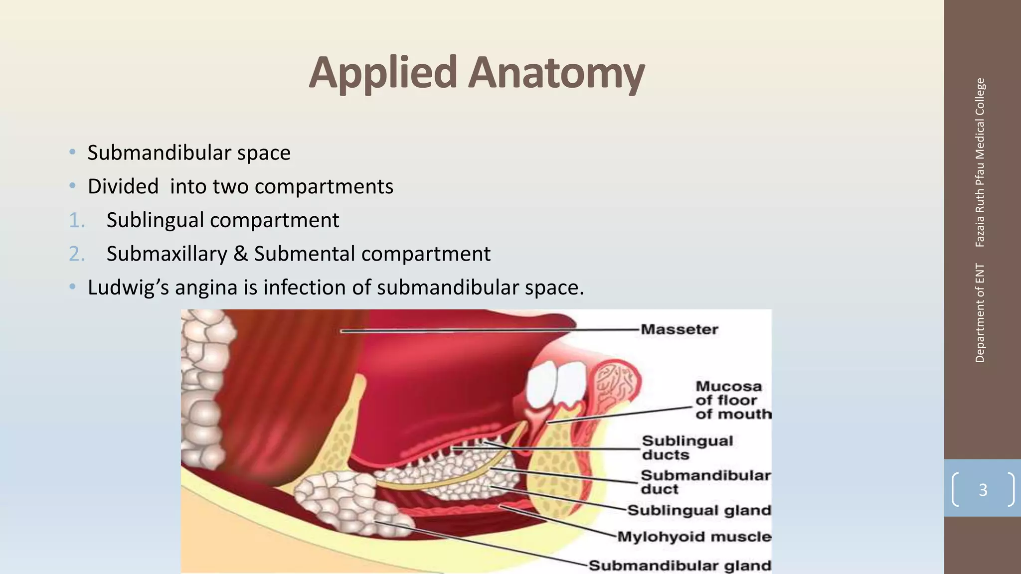


The document outlines Ludwig's angina, an infection of the submandibular space characterized by bilateral swelling and potential airway obstruction. It discusses its etiology, clinical features, differential diagnoses, and management strategies, emphasizing the necessity for airway protection and prompt treatment. Complications of Ludwig's angina include septicemia and aspiration pneumonia, highlighting the urgency of addressing this condition in a clinical setting.


![Down syndrome (2)[1].pptx pediatric lecture](https://cdn.slidesharecdn.com/ss_thumbnails/downsyndrome21-240709094926-fcdd02d9-thumbnail.jpg?width=640&height=640&fit=bounds)


![ABDOMINAL EXAMINATION Presentation[1].pptx](https://cdn.slidesharecdn.com/ss_thumbnails/abdominalexaminationpresentation1-240105120242-b6318479-thumbnail.jpg?width=640&height=640&fit=bounds)


