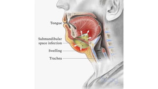
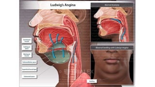
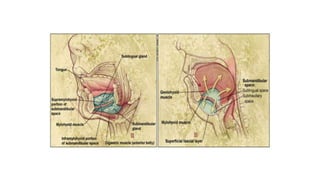
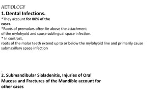
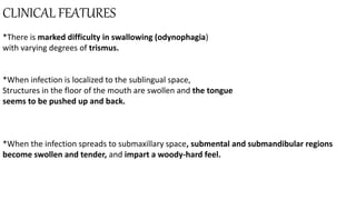
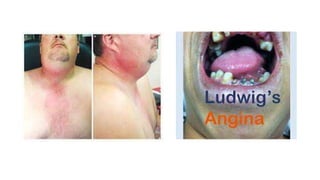
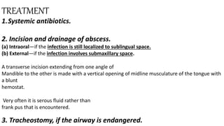
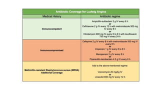
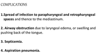
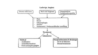
2. *Ludwig angina is life-threatening cellulitis of the soft tissue
involving the floor of the mouth and neck.
*Ludwig angina involves 3 compartments of the floor of the
mouth: sublingual, submental, and submandibular.
*The infection is rapidly progressive, leading to potential airway
obstruction.
3. CLINICAL ANATOMY:
*Submandibular space lies between the mucous membrane of
The floor of the mouth and tongue on one side and the superficial
layer of deep cervical fascia extending between the hyoid
bone and mandible on the other.
4. It is divided into two compartments by the
mylohyoid muscle:
1. Sublingual compartment (above the mylohyoid).
2. Submaxillary and submental compartment (below the
mylohyoid).
The two compartments are continuous around
the posterior border of the mylohyoid muscle.
Ludwig’s angina is an infection of the submandibular space.
5.
6.
7.
8. AETIOLOGY
1.Dental Infections.
*They account for 80% of the
cases.
*Roots of premolars often lie above the attachment
of the mylohyoid and cause sublingual space infection.
* In contrast,
roots of the molar teeth extend up to or below the mylohyoid line and primarily cause
submaxillary space infection
.
2. Submandibular Sialadenitis, Injuries of Oral
Mucosa and Fractures of the Mandible account for
other cases
9. 3.BACTERIOLOGY
*Mixed infections involving both aerobes and anaerobes are
common.
*Alpha-haemolytic Streptococci, Staphylococci
and Bacteroides groups are common.
*Rarely Haemophilus
Influenzae, Escherichia coli, and Pseudomonas are seen
10. CLINICAL FEATURES
*There is marked difficulty in swallowing (odynophagia)
with varying degrees of trismus.
*When infection is localized to the sublingual space,
Structures in the floor of the mouth are swollen and the tongue
seems to be pushed up and back.
*When the infection spreads to submaxillary space, submental and submandibular regions
become swollen and tender, and impart a woody-hard feel.
11. *Usually, there is cellulitis of the tissues rather than frank
abscess.
*Tongue is
progressively pushed upwards and backward threatening
the airway.
*Laryngeal edema may appear
12.
13.
14. TREATMENT
1.Systemic antibiotics.
2. Incision and drainage of abscess.
(a) Intraoral—if the infection is still localized to sublingual space.
(b) External—if the infection involves submaxillary space.
A transverse incision extending from one angle of
Mandible to the other is made with a vertical opening of midline musculature of the tongue with
a blunt
hemostat.
Very often it is serous fluid rather than
frank pus that is encountered.
3. Tracheostomy, if the airway is endangered.
15.
16. COMPLICATIONS
1.Spread of infection to parapharyngeal and retropharyngeal
spaces and thence to the mediastinum.
2. Airway obstruction due to laryngeal edema, or swelling and
pushing back of the tongue.
3. Septicemia.
4. Aspiration pneumonia.