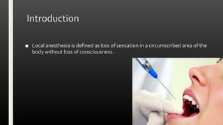
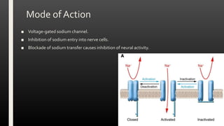
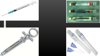
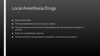
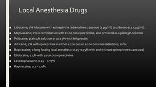
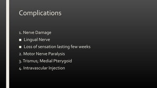
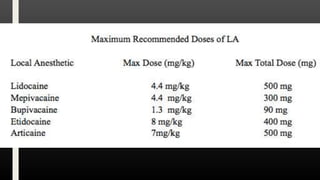
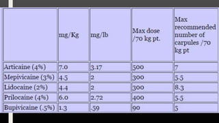
LOCAL ANESTHESIA
Dr. Hadi Munib
Oral and Maxillofacial Surgery Resident
Outline
■ Introduction
■ Mode of Action
■ Techniques
■ Drugs
■ Complications
■ References
Introduction
■ Local anesthesia is defined as loss of sensation in a circumscribed area of the
body without loss of consciousness.
Mode of Action
■ Voltage-gated sodium channel.
■ Inhibition of sodium entry into nerve cells.
■ Blockade of sodium transfer causes inhibition of neural activity.
Techniques for LocalAnesthesia
■ Topical Anesthesia
■ Infiltration Anesthesia
■ Regional Block Anesthesia
■ SupplementalTechniques
Topical Anesthesia
■ To lessen the discomfort of needle penetration.
■ No evidence that topical application lessens the discomfort of regional block techniques.
■ Creams, ointments and sprays.
■ Lidocaine and Benzocaine.
■ Oraqix®; a combination of lidocaine and Prilocaine.
Infiltration Anesthesia
■ Maxilla for dental pulpal anesthesia and Mandible of children for anesthesia of the
deciduous dentition.
■ Partly opened mouth; 1.0–2.0 ml of solution is deposited at a rate of 30 s/ml.
■ Supraperiosteal; BoneTouch?
■ 45 minutes of anesthesia of the dental pulps when a vasoconstrictor is used
■ The patient may have subjective anesthesia of the soft tissues for 1.5–2 hours.
Posterior Superior Alveolar Nerve Block
■ Maxillary Molar teeth, associated bone and buccal gingivae.
■ The first molar will not be satisfactorily anesthetized in some individuals as the Mesiobuccal root is
supplied by MSA
■ 20 mm penetration
Middle Superior Alveolar Nerve Block
■ Premolar pulps as well as the mesio-buccal pulp of the maxillary first permanent molar tooth.
■ Anterior Superior Alveolar Nerve Block; Maxillary incisors and canine teeth. It can be anesthetized
either by an infraorbital nerve block or by a buccal infiltration in the region of the apex of the canine.
Regional Blocks – ID Block
■ InferiorAlveolar Nerve block (Halstead technique)
■ Pterygo-temporal space on the medial aspect of the mandibular ramus.
■ Teeth, Bone and the soft tissues of the lower lip to the midline as well as the reflected and attached
gingivae from the premolar teeth to the midline.
■ Two common problems with this method are either contacting bone too soon or failure to touch
bone.
■ Management
■ Facial Paresis
■ Lidocaine with epinephrine will anesthetize the hard tissues including the teeth for around 45
minutes; subjective soft tissue numbness may be apparent for up to 3 hours.
■ When the so-called long-acting solutions are employed anesthesia of the teeth can last for 6–8
hours.
Gow-GatesTechnique
■ Lingual, Long buccal, Mylohyoid andAuriculotemporal nerves.
■ More superiorly with more nerves involved
■ The target is the mandibular condyle.
■ Plane parallel to a line visualized between the corner of the mouth and the intertragal
notch.
■ Syringe introduction into the mouth across the maxillary canine tooth of the opposite side.
■ The needle is advanced in this direction until the bone of the condyle is contacted.
AkinosiVaziraniTechnique
■ Administered with the patient’s mouth closed.
■ No bony end-point for needle insertion.
■ Buccal sulcus along a plane level with the mucogingival junction of the maxillary mucosa.
■ Posterior aspect of the maxilla until the hub of the needle is adjacent to the distal surface of the
maxillary second molar.
■ If the anterior aspect of the mandibular ramus is contacted then either a more lateral movement or
the patient instructed to move their lower jaw over to the side being injected.
■ This method anesthetizes the inferior alveolar nerve, lingual nerve, nerve to mylohyoid, and
occasionally the long buccal nerve.
Mental Nerve Block
■ Premolars anteriorly as well as the soft tissues of the lower lip and chin to the midline on one side.
■ Depth of the buccal sulcus between the premolar teeth and advanced to a zone below the premolar
apices.
Long Buccal Nerve Block
■ Buccal gingivae and mucosa and part of the cheek in the mandibular molar region.
■ The nerve can be anesthetized by either buccal infiltration in the zone of interest or by a regional
block
■ Coronoid notch of the mandible (the point at which the thumb rests during the Halstead technique).
Maxillary Nerve Block
■ Intra-Oral Approach;Tuberosity and Greater Palatine Foramen
■ Extra-Oral Approach
Intra-OralApproach
■ Tuberosity approach; high in the buccal sulcus in the plane of the distal surface of the maxillary
second molar tooth.The needle is advanced at an angle of 45 superiorly, posteriorly and medially at
a 30 mm depth
■ Greater palatine foramen approach; Pterygopalatine fossa via the greater palatine foramen.The
needle is inserted into the greater palatine foramen and advanced at an angle of 45o superiorly and
posteriorly to a depth of 30 mm.
Infraorbital Nerve Block
■ Intraoral or Extraoral.
■ The intraoral approach; buccal sulcus between the premolar teeth and towards the infraorbital
foramen, which is being palpated extra-orally by the operator’s non-syringe hand.
■ Second premolar to the central incisor, the gingivae adjacent to these teeth and the mucosal and skin
surfaces of one half of the upper lip and part of the skin on the lateral aspect of the nose.
Greater Palatine Nerve Block
■ Soft tissues of the palate from the foramen anteriorly to the canine
region.
■ Palatal Infiltration.
■ Infiltration and blocks provide around 45 minutes of soft tissue
anesthesia.
Nasopalatine nerve block;Anesthesia to the hard palate adjacent to the incisor teeth
bilaterally.
SupplementalAnesthesiaTechniques
■ Intraosseous anesthesia
■ Buccal Infiltrations
■ A perforation through soft tissue and cortical bone is made using a perforator matched to the
needle (these are provided in customized systems) or with a small round dental bur.
■ The point of penetration is 2 mm below the intersection of two imaginary lines.
1. Line joining the lowest part of the buccal gingival margin of the tooth of interest and its posterior
neighbor.
2. Line at 90º to the former line that bisects the interdental papilla.
■ Once the perforation has reached cancellous bone, Anesthesia is given.
SupplementalAnesthesiaTechniques
■ Intraligamentary (periodontal ligament) anesthesia
■ Specified systems or conventional.
■ Delivery of solution (0.2 ml per root) is performed slowly with controlled pressure.
■ Not very successful in mandibular incisors
IntrapulpalAnesthesia
Local Anesthesia Drugs
■ Esters and Amides
■ The local anesthetics in current use are amides.
■ The ester procaine is only used in those patients who are proven to be allergic to
amides.
■ Esters are metabolized in plasma.
■ Amides primarily undergo hepatic metabolism; Articaine is an exception.
Local Anesthesia Drugs
■ Lidocaine; 2% lidocaine with epinephrine (adrenaline) 1:200 000 (5 μg/ml) to 1:80 000 (12.5 μg/ml).
■ Mepivacaine; 2% in combination with 1:100 000 epinephrine, also provided as a plain 3% solution
■ Prilocaine; plain 4% solution or as a 3% with felypressin
■ Articaine; 4% with epinephrine in either 1:100 000 or 1:200 000 concentrations, safer.
■ Bupivacaine; a long-lasting local anesthetic; 0.25–0.75% with and without epinephrine (1:200 000)
■ Etidocaine; 1.5% with 1:100,000 epinephrine
■ Levobupivacaine; 0.25 – 0.75%
■ Ropivacaine; 0.2 – 1.0%
Complications
1. Nerve Damage
■ Lingual Nerve
■ Loss of sensation lasting few weeks
2. Motor Nerve Paralysis
3.Trismus; Medial Pterygoid
4. Intravascular Injection
Systemic Complications
■ Allergy
■ Infection
■ Toxicity
■ Drug Interactions
References
■ Chapter 5: Local Anesthesia
THANKYOU

Local Anesthesia

  • 1.
    LOCAL ANESTHESIA Dr. HadiMunib Oral and Maxillofacial Surgery Resident
  • 2.
    Outline ■ Introduction ■ Modeof Action ■ Techniques ■ Drugs ■ Complications ■ References
  • 3.
    Introduction ■ Local anesthesiais defined as loss of sensation in a circumscribed area of the body without loss of consciousness.
  • 4.
    Mode of Action ■Voltage-gated sodium channel. ■ Inhibition of sodium entry into nerve cells. ■ Blockade of sodium transfer causes inhibition of neural activity.
  • 8.
    Techniques for LocalAnesthesia ■Topical Anesthesia ■ Infiltration Anesthesia ■ Regional Block Anesthesia ■ SupplementalTechniques
  • 9.
    Topical Anesthesia ■ Tolessen the discomfort of needle penetration. ■ No evidence that topical application lessens the discomfort of regional block techniques. ■ Creams, ointments and sprays. ■ Lidocaine and Benzocaine. ■ Oraqix®; a combination of lidocaine and Prilocaine.
  • 10.
    Infiltration Anesthesia ■ Maxillafor dental pulpal anesthesia and Mandible of children for anesthesia of the deciduous dentition. ■ Partly opened mouth; 1.0–2.0 ml of solution is deposited at a rate of 30 s/ml. ■ Supraperiosteal; BoneTouch? ■ 45 minutes of anesthesia of the dental pulps when a vasoconstrictor is used ■ The patient may have subjective anesthesia of the soft tissues for 1.5–2 hours.
  • 14.
    Posterior Superior AlveolarNerve Block ■ Maxillary Molar teeth, associated bone and buccal gingivae. ■ The first molar will not be satisfactorily anesthetized in some individuals as the Mesiobuccal root is supplied by MSA ■ 20 mm penetration
  • 15.
    Middle Superior AlveolarNerve Block ■ Premolar pulps as well as the mesio-buccal pulp of the maxillary first permanent molar tooth. ■ Anterior Superior Alveolar Nerve Block; Maxillary incisors and canine teeth. It can be anesthetized either by an infraorbital nerve block or by a buccal infiltration in the region of the apex of the canine.
  • 17.
    Regional Blocks –ID Block ■ InferiorAlveolar Nerve block (Halstead technique) ■ Pterygo-temporal space on the medial aspect of the mandibular ramus. ■ Teeth, Bone and the soft tissues of the lower lip to the midline as well as the reflected and attached gingivae from the premolar teeth to the midline. ■ Two common problems with this method are either contacting bone too soon or failure to touch bone. ■ Management ■ Facial Paresis ■ Lidocaine with epinephrine will anesthetize the hard tissues including the teeth for around 45 minutes; subjective soft tissue numbness may be apparent for up to 3 hours. ■ When the so-called long-acting solutions are employed anesthesia of the teeth can last for 6–8 hours.
  • 21.
    Gow-GatesTechnique ■ Lingual, Longbuccal, Mylohyoid andAuriculotemporal nerves. ■ More superiorly with more nerves involved ■ The target is the mandibular condyle. ■ Plane parallel to a line visualized between the corner of the mouth and the intertragal notch. ■ Syringe introduction into the mouth across the maxillary canine tooth of the opposite side. ■ The needle is advanced in this direction until the bone of the condyle is contacted.
  • 24.
    AkinosiVaziraniTechnique ■ Administered withthe patient’s mouth closed. ■ No bony end-point for needle insertion. ■ Buccal sulcus along a plane level with the mucogingival junction of the maxillary mucosa. ■ Posterior aspect of the maxilla until the hub of the needle is adjacent to the distal surface of the maxillary second molar. ■ If the anterior aspect of the mandibular ramus is contacted then either a more lateral movement or the patient instructed to move their lower jaw over to the side being injected. ■ This method anesthetizes the inferior alveolar nerve, lingual nerve, nerve to mylohyoid, and occasionally the long buccal nerve.
  • 26.
    Mental Nerve Block ■Premolars anteriorly as well as the soft tissues of the lower lip and chin to the midline on one side. ■ Depth of the buccal sulcus between the premolar teeth and advanced to a zone below the premolar apices.
  • 28.
    Long Buccal NerveBlock ■ Buccal gingivae and mucosa and part of the cheek in the mandibular molar region. ■ The nerve can be anesthetized by either buccal infiltration in the zone of interest or by a regional block ■ Coronoid notch of the mandible (the point at which the thumb rests during the Halstead technique).
  • 31.
    Maxillary Nerve Block ■Intra-Oral Approach;Tuberosity and Greater Palatine Foramen ■ Extra-Oral Approach
  • 32.
    Intra-OralApproach ■ Tuberosity approach;high in the buccal sulcus in the plane of the distal surface of the maxillary second molar tooth.The needle is advanced at an angle of 45 superiorly, posteriorly and medially at a 30 mm depth ■ Greater palatine foramen approach; Pterygopalatine fossa via the greater palatine foramen.The needle is inserted into the greater palatine foramen and advanced at an angle of 45o superiorly and posteriorly to a depth of 30 mm.
  • 34.
    Infraorbital Nerve Block ■Intraoral or Extraoral. ■ The intraoral approach; buccal sulcus between the premolar teeth and towards the infraorbital foramen, which is being palpated extra-orally by the operator’s non-syringe hand. ■ Second premolar to the central incisor, the gingivae adjacent to these teeth and the mucosal and skin surfaces of one half of the upper lip and part of the skin on the lateral aspect of the nose.
  • 35.
    Greater Palatine NerveBlock ■ Soft tissues of the palate from the foramen anteriorly to the canine region. ■ Palatal Infiltration. ■ Infiltration and blocks provide around 45 minutes of soft tissue anesthesia.
  • 38.
    Nasopalatine nerve block;Anesthesiato the hard palate adjacent to the incisor teeth bilaterally.
  • 39.
    SupplementalAnesthesiaTechniques ■ Intraosseous anesthesia ■Buccal Infiltrations ■ A perforation through soft tissue and cortical bone is made using a perforator matched to the needle (these are provided in customized systems) or with a small round dental bur. ■ The point of penetration is 2 mm below the intersection of two imaginary lines. 1. Line joining the lowest part of the buccal gingival margin of the tooth of interest and its posterior neighbor. 2. Line at 90º to the former line that bisects the interdental papilla. ■ Once the perforation has reached cancellous bone, Anesthesia is given.
  • 41.
    SupplementalAnesthesiaTechniques ■ Intraligamentary (periodontalligament) anesthesia ■ Specified systems or conventional. ■ Delivery of solution (0.2 ml per root) is performed slowly with controlled pressure. ■ Not very successful in mandibular incisors
  • 42.
  • 43.
    Local Anesthesia Drugs ■Esters and Amides ■ The local anesthetics in current use are amides. ■ The ester procaine is only used in those patients who are proven to be allergic to amides. ■ Esters are metabolized in plasma. ■ Amides primarily undergo hepatic metabolism; Articaine is an exception.
  • 44.
    Local Anesthesia Drugs ■Lidocaine; 2% lidocaine with epinephrine (adrenaline) 1:200 000 (5 μg/ml) to 1:80 000 (12.5 μg/ml). ■ Mepivacaine; 2% in combination with 1:100 000 epinephrine, also provided as a plain 3% solution ■ Prilocaine; plain 4% solution or as a 3% with felypressin ■ Articaine; 4% with epinephrine in either 1:100 000 or 1:200 000 concentrations, safer. ■ Bupivacaine; a long-lasting local anesthetic; 0.25–0.75% with and without epinephrine (1:200 000) ■ Etidocaine; 1.5% with 1:100,000 epinephrine ■ Levobupivacaine; 0.25 – 0.75% ■ Ropivacaine; 0.2 – 1.0%
  • 45.
    Complications 1. Nerve Damage ■Lingual Nerve ■ Loss of sensation lasting few weeks 2. Motor Nerve Paralysis 3.Trismus; Medial Pterygoid 4. Intravascular Injection
  • 46.
    Systemic Complications ■ Allergy ■Infection ■ Toxicity ■ Drug Interactions
  • 49.
    References ■ Chapter 5:Local Anesthesia
  • 50.