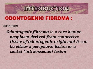
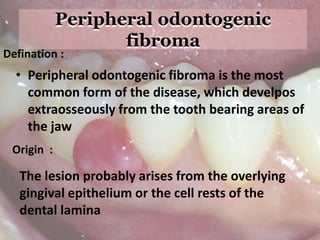
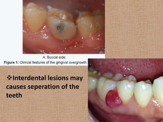
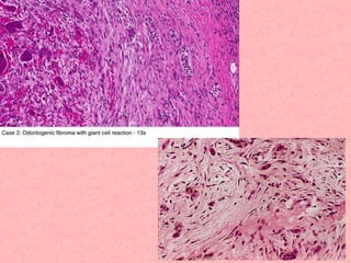
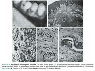
PERIPHERAL
ODONTOGENIC FIBROMA
Presentation by:
SWALIHA ALTHAF
Content
• INTRODUCTION
• CLASSIFICATION
• PERPHERAL ODONTOGENIC FIBROMA
• CLINICAL FEATURES
• RADIOLOGHIC FEATURES
• HISTOPATHOLOGY
• DIFFERENTIAL DIAGNOSIS
• TREATMENT
Odontogenic fibroma is a rare benign
neoplasm derived from connective
tissue of odontogenic origin and it can
be either a peripheral lesion or a
cental (intraosseous) lesion
ODONTOGENIC FIBROMA :
DEFINITION :
Odontogenic tumours
(classification)
A. Benign
I. Odontogenic epithelium :
• Ameloblastoma
• Squamous odontogenic tumour
• Calcifying epithelial odontogenic tumour
(pindborg tumour)
• Adenomatoid odontogenic tumour
• Keratocystic odontogenic tumour
II. Odontogenic epithelium with
odontogenic ectomesenchyme
• Ameloblastic fibroma
• Ameloblastic fibrodentinoma
• Ameloblastic fibro-odontoma
• Complex odontoma
• Compound odontoma
• Odontoameloblastoma
• Calcifying cystic odontogenic tumour
• Dentinogenic ghost cell tumour
III. Odontogenic ectomesenchyme :
• Odontogenic fibroma
• Myxoma (myxofibroma)
• Cementoblastoma (benign cementoblatoma,true
cementoma)
B.Malignant
I. Odontogenic carcinomas :
• Malignant ameloblastoma
• Ameloblastoma carcinoma
• Primary intraosseous carcinoma
• Clear cell odontogenic carcinoma
• Ghost cell odontogenic carcinoma
II. Odontogenic sarcomas :
• Ameloblastoma fibrosarcoma
• Ameloblastoma fibrodentinosarcoma
• Ameloblastoma fibro-odontosarcoma
• Peripheral odontogenic fibroma is the most
common form of the disease, which develpos
extraosseously from the tooth bearing areas of
the jaw
Defination :
Origin :
The lesion probably arises from the overlying
gingival epithelium or the cell rests of the
dental lamina
Clinical Features
• There was no gender predilection, while
the ages of the patients ranged from 5 to
65 years.
• Clinically appears as a show enlarging,
exophytic well-circumscribed ,sessile
growth of the gingiva.
Occurs in the mandible than the maxilla.
The lesion is usually firm in consistency it is painless and
the overlying epithelium is of normal colour.
Most of the lesion occur on the facial gingiva and their
size ranges between 0.5 to 1.5 centimeter in diameter
• More than one lesion can occur in the oral
cavity but rarely .
• There can be erythematous changes or even
ulcerations on the surface epithelium which
result from trauma.
• Consistency of the lesion varies,since lesions
may occur with ossification or without
ossification
Interdental lesions may
causes seperation of the
teeth
• Since peripheral odontogenic fibromas are small
lesions and they occur extraosseously within the
gingiva,radiographic changes in the bone are not
apparently found
• In certain saucerization of the cortical bone or
widening of the periodontal ligament space at the
cervical region may be seen
• Numerous foci of small radioopaque masses are
sometimes found within few lesions of peripheral
odontogenic fibroma,which indicate calcification
within indicate calcifcations within the tumor.
Radiographic Features :
Odontogenic fibroma producing a large irregular radiolucent
area containing numerous radiopaque foci
HISTOPATHOLOGY
• The peripheral odontogenic fibroma consists of a
markedly cellular fibrous connective tissue
parenchyma with scanty or numerous non-
neoplastic islands,strands and cords of columnar
or cuboidal,sometimes vacuolated odontogenic
epithelium.
• When numerous,the peripheral odontogenic
fibroma has been occasionally mistaken for an
epithelial neoplasma such as a peripheral
ameloblastoma
• This epithelium is usually deep in the
lesions,away from the surface epitheluim and is
the lesion,away from the surface epithelium and
is sometimes found ‘cuffing’ calcifications.
• Calcified tissue may or may not be present in the
peripheral odontogenic fibroma
• If found ,it may resemble trabeculae of bone or
osteoid,dentin or osteodentin (sometimes
described as dysplastic dentin ) or cementum
like material.
• Mature fibrous connective tissue stroma is
present and is sometimes highly
vascularized,particularly in the less cellular
areas.
• Myxomatous changes may also be found in the
stroma and the presence of inflammation is
variable.
Peripheral ossifying fibroma
Peripheral giant cell granuloma
True fibroma
Neurofibroma
Fibroepithelial polyp
• The lesion is treated by surgical excision
• Recurrence is not common.
Peripheral odontogenic fibroma

Peripheral odontogenic fibroma

  • 1.
  • 2.
    Content • INTRODUCTION • CLASSIFICATION •PERPHERAL ODONTOGENIC FIBROMA • CLINICAL FEATURES • RADIOLOGHIC FEATURES • HISTOPATHOLOGY • DIFFERENTIAL DIAGNOSIS • TREATMENT
  • 3.
    Odontogenic fibroma isa rare benign neoplasm derived from connective tissue of odontogenic origin and it can be either a peripheral lesion or a cental (intraosseous) lesion ODONTOGENIC FIBROMA : DEFINITION :
  • 4.
    Odontogenic tumours (classification) A. Benign I.Odontogenic epithelium : • Ameloblastoma • Squamous odontogenic tumour • Calcifying epithelial odontogenic tumour (pindborg tumour) • Adenomatoid odontogenic tumour • Keratocystic odontogenic tumour
  • 5.
    II. Odontogenic epitheliumwith odontogenic ectomesenchyme • Ameloblastic fibroma • Ameloblastic fibrodentinoma • Ameloblastic fibro-odontoma • Complex odontoma • Compound odontoma • Odontoameloblastoma • Calcifying cystic odontogenic tumour • Dentinogenic ghost cell tumour
  • 6.
    III. Odontogenic ectomesenchyme: • Odontogenic fibroma • Myxoma (myxofibroma) • Cementoblastoma (benign cementoblatoma,true cementoma) B.Malignant I. Odontogenic carcinomas : • Malignant ameloblastoma • Ameloblastoma carcinoma • Primary intraosseous carcinoma
  • 7.
    • Clear cellodontogenic carcinoma • Ghost cell odontogenic carcinoma II. Odontogenic sarcomas : • Ameloblastoma fibrosarcoma • Ameloblastoma fibrodentinosarcoma • Ameloblastoma fibro-odontosarcoma
  • 8.
    • Peripheral odontogenicfibroma is the most common form of the disease, which develpos extraosseously from the tooth bearing areas of the jaw Defination : Origin : The lesion probably arises from the overlying gingival epithelium or the cell rests of the dental lamina
  • 9.
    Clinical Features • Therewas no gender predilection, while the ages of the patients ranged from 5 to 65 years. • Clinically appears as a show enlarging, exophytic well-circumscribed ,sessile growth of the gingiva.
  • 10.
    Occurs in themandible than the maxilla. The lesion is usually firm in consistency it is painless and the overlying epithelium is of normal colour. Most of the lesion occur on the facial gingiva and their size ranges between 0.5 to 1.5 centimeter in diameter
  • 11.
    • More thanone lesion can occur in the oral cavity but rarely . • There can be erythematous changes or even ulcerations on the surface epithelium which result from trauma. • Consistency of the lesion varies,since lesions may occur with ossification or without ossification
  • 12.
    Interdental lesions may causesseperation of the teeth
  • 13.
    • Since peripheralodontogenic fibromas are small lesions and they occur extraosseously within the gingiva,radiographic changes in the bone are not apparently found • In certain saucerization of the cortical bone or widening of the periodontal ligament space at the cervical region may be seen • Numerous foci of small radioopaque masses are sometimes found within few lesions of peripheral odontogenic fibroma,which indicate calcification within indicate calcifcations within the tumor. Radiographic Features :
  • 14.
    Odontogenic fibroma producinga large irregular radiolucent area containing numerous radiopaque foci
  • 15.
    HISTOPATHOLOGY • The peripheralodontogenic fibroma consists of a markedly cellular fibrous connective tissue parenchyma with scanty or numerous non- neoplastic islands,strands and cords of columnar or cuboidal,sometimes vacuolated odontogenic epithelium. • When numerous,the peripheral odontogenic fibroma has been occasionally mistaken for an epithelial neoplasma such as a peripheral ameloblastoma
  • 17.
    • This epitheliumis usually deep in the lesions,away from the surface epitheluim and is the lesion,away from the surface epithelium and is sometimes found ‘cuffing’ calcifications. • Calcified tissue may or may not be present in the peripheral odontogenic fibroma • If found ,it may resemble trabeculae of bone or osteoid,dentin or osteodentin (sometimes described as dysplastic dentin ) or cementum like material.
  • 18.
    • Mature fibrousconnective tissue stroma is present and is sometimes highly vascularized,particularly in the less cellular areas. • Myxomatous changes may also be found in the stroma and the presence of inflammation is variable.
  • 20.
    Peripheral ossifying fibroma Peripheralgiant cell granuloma True fibroma Neurofibroma Fibroepithelial polyp
  • 21.
    • The lesionis treated by surgical excision • Recurrence is not common.