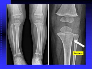
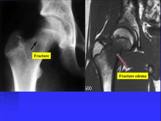
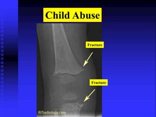
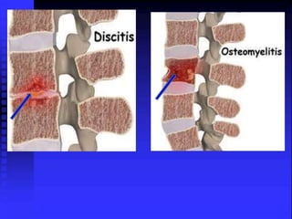
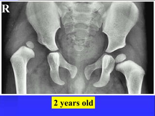
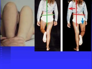
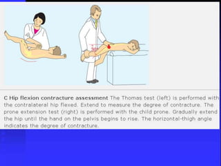
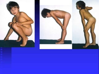
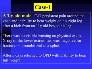
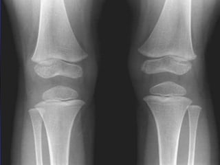
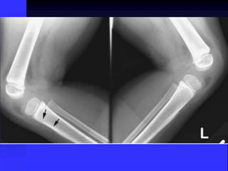
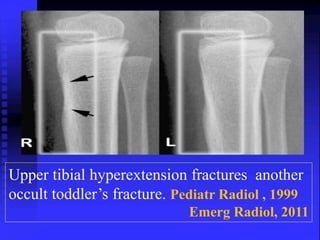
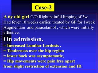
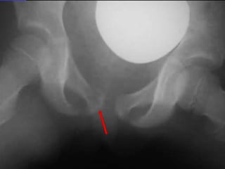
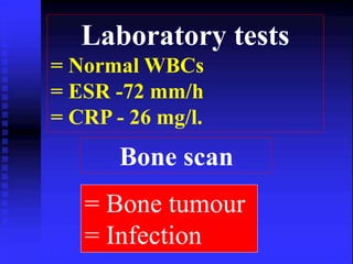
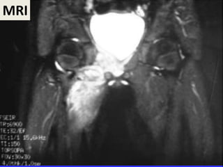
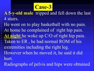
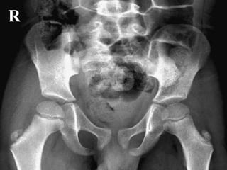
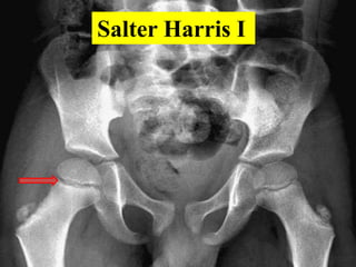
The Limping Child
Normal gait 
Stance Phase (weight-bearing phase) 
Heel-strike. 
Plantarflexion (foot flat) 
Toe-off 
Swing Phase 
begins with toe-off and ends with the heel-strike. 
+forward rotation and tilting of the pelvis, 
+stability of the lumbar spine
60% 40% 
Fully mature gait is attained by 4 years
1= Antalgic gait (anti-pain) 
Less time is spent in stance phase. 
2= A Trendelenburg gait 
In swing-phase drop of the of the pelvis on the weak 
side. 
3= Waddling gait 
Bilateral hip involvement or neurologic disease. 
4= Stiff-legged gait 
Knee extension and circumduction with pelvic 
elevation on the affected side.
5-Toe walking 
= Habitual = Muscle contractures 
= Puncture wound in heel. = LLD. 
6- Steppage gait 
Failed active dorsiflexion the foot, with exaggerated 
hip and knee flexion during the swing phase -- NM 
diseases. 
7- Stooped gait - pelvic or abdominal pathology
The commonest site 
= Hip (34%) 
= Knee (19%) 
= Leg (18%) 
= Spine (fewer than 2%) . 
Pain from the spine may be referred to the 
thigh or the abdomen 
Hip pain is referred to the thigh or the 
knee.
Evaluation of the Child With a Limp 
If the right questions are asked a 
provisional DX can be made in most 
cases at the primary examination. 
= Proper hx and physical exam 
= Specific features of the disease 
= Proper investigation
50 causes of limping child
Causes as any pathology 
= Musculoskeletal Trauma 
= Inflammation 
= Infection 
= Developmental or Congenital 
= Neoplasm 
= Autoimmune 
= Osteochondrosis 
= Neurological
Musculoskeletal Trauma 
Physeal Fracture 
Toddler’s Fracture 
Stress Fracture 
Child Abuse 
Soft tissue injury
Physeal Fracture
Early stage of ossification Another variation of normal
Normal 
Fracture
Toddler’s Fractures 
Typical nondisplaced spiral fracture of tibia 
with no fibular fracture. 
Initial x-ray: 
often normal, diagnosis by F/U films with 
lucent line or periosteal reaction 
Or by ultrasound
• No specific injury notable most of the time 
• Any child refuses to bear weight on leg 
Examine (hip, thigh , knee , Leg and foot)
Fracture 
Fracture Callus
Fracture
Buckle fracture—bunk-bed fracture. 
3-year-old male. 
Follow-up radiograph shows healing with sclerosis at 
the fracture site 
Fracture
Stress fracture 
Callus due to Fracture
Fracture 
Fracture edema
Child Abuse 
Fracture 
Fracture
Inflammation 
Transient Synovitis 
Juvenile Idiopathic Arthritis 
Viral Myositis
• Sudden hip pain or knee pain 
• Afebrile/low-grade fever (<38.5) 
• Able to ambulate with a limp 
–Antalgic gait 
• Hip: mildly decreased ROM
• Laboratory Evaluation 
–WBC count <12,000 
–ESR (<40); CRP (<5mg/L) 
• X-Ray 
• Joint space widening 
• >2mm on affected side. 
• Ultrasound: 
• Joint effusion and/or synovial swelling 
Kocher, Etal JBJS -1999. 
JBJS -2006
3-year-old
R 
>2mm
Treatment 
– Self-limited after 2-7 days 
– Bed rest 
–Ibuprofen 
• Decreased pain by 2.5 days Vs Placebo 
• Mean duration of pain 
–ibuprofen: 2 days 
–placebo: 4.5 days 
Annals of Emergency Medicine 2002
Prognosis 
–?? association with increased risk of 
Perthes disease (1%) 
(Clinical Pediatrics, 1985) 
–Recurrance rate in 4-15%
Septic Arthritis Vs Transient Synovitis 
= No child with a temperature >38.5 was 
found to have transient synovitis 
= CRP > 5mg/dL was the only independent risk 
factor strongly associated with Septic arthritis 
+++++++++++++++++++++++++++++++++++++ 
– 86% of patients with ESR < 40 mm/hr had 
transient synovitis 
– 71% of patients with CRP < 5mg/ or WBC 
< 12,000/mm3 had transient synovitis 
J Bone Joint Surg. 2006 
Orthop Clin N Am (2006)
Septic arthritis 
• Predicting of Septic arthritis 
–Fever >38.5 
–Cannot weight bear 
–ESR >40 in 1st hr 
–WBCs >12 
– CRP >5mg/L 
JBJS-Am. 1999.
Roll test 
The Dx of transient synovitis is more likely if an 
arc of 30 degrees or more of hip rotation is 
without pain.
Myositis 
= Viral myositis causing leg pain may be 
seen during influenza season 
= More common in older children
Infection 
Osteomyelitis 
Septic Arthritis 
Psoas abscess 
Discitis 
Children with septic arthritis or 
osteomylitis usually appear acutely ill 
than those with transient synovitis.
Culture the Tumour 
Biopsy the Infection. 
Osteomylitis   
Tumour
ANTIBIOTICS 
Guidelines 
 Central Line. 
 Large doses. 
 Drug level. 
 KFT. 
Duration 4-6 weeks 
* I.V : 
* Oral :
1- Afebrile for 24hr. 
2- Known org. 
3- Minimal symptoms 
4- Reliable parents 
5- ESR/CRP  Down.
Longer treatment required 
=Pelvis 
=Vertebrae 
=Diskitis 
=Calcaneum
Remember 
Tuberculosis is still around 
Arthroscopic drainge is dangerous
FABER test
Psoas Abscess Vs. Septic Arthritis 
Psoas sign. 
(Flexing the hip relieves the pain and allows painless 
IR+ER of the hip) 
Pain when the hip is passively extended or 
actively flexed against resistance. 
= Scoliosis, and Femoral N neuropathy 
Septic arthritis ROM is painfully limited in all directions. 
CT or MRI Diagnostic. 
American Family Physician-2009 Am J Orthop. 2008
Diskitis Vs. Vertebral osteomylitis 
Vertebral Osteomylitis. 
= Toxic appearance. 
= Radiographs :localized rarefaction of one vertebral body 
and bony destruction 
Diskitis: Lumbar disk space narrowing and destruction of 
adjacent vertebral end plates 
=MRI is the diagnostic study of choice. 
American Family Physician-2009
Don’t forget! 
Intra-abdominal pathology and 
testicular torsion may present 
simply as a limp – 
Examine abdomen and testicles 
in boys!!
Bony Deformities of the Hip 
DDH 
SCFE 
Perthes Disease 
Coxa Vara
R 
2 years old
= Femoral neck displaces ant. producing an 
apparent varus, the head is posterior 
= Occurs through Zone of hypertrophy
It is important to determine 
1. Stable 
Walking with or 
without crutches 
2. Unstable 
Non walker
Hip flexion 
Ext rotation 
deformity
The goal of treatment for SCFE is 
to prevent further slippage and to 
stabilize the epiphysis 
Screw advancement until 
FIVE 
threads engage the epiphysis
Complications 
1. Avascular necrosis. 
2. Chondrolysis. 
3. Osteoarthritis. 
4. Coxa vara 
NSA less than 120 degrees. 
5. Slipping of the 
opposite hip ≈ 20% of 
cases
Legg-Calvé-Perthes disease
Symptoms of Perthes usually 
have been present for weeks. 
• Hip, groin, thigh or knee pain 
• Limp :painless and intermittent. 
• Limitation of internal rotation.
* Slight widening of the left hip joint 
* Small joint effusion 
R
* Decrease epiphyseal hight 
R
R 
= The F.H smaller denser on the left 
= Joint widening can also be 2ry to 
hypertrophy of the cartilage.
Head-at-risk signs 
= Extrusion- subluxation (red arrow), 
= Metaphyseal reaction (yellow arrow), 
= Lateral rarifaction or Gage sign 
(white arrow)
Treatment Goals 
1-Relief of symptoms 
2-Restoration of ROM 
3-Containment
Management according to 
Lat. Pillar 
(Skeletal Age) 
* Age < 6y at any stage - Conservative. 
* Group A any age - Conservative. 
===================== 
* Group B 6>8Y --- Containment 
* Group C > 6Y --- Surgery.
=Bisphosphonates 
=Drilling of the head 
J Orthop Res 2005
10 Years 
Perthes
Perthes-11 years old
Developmental Coxa Vara 
Hilgenreiner’s Epiphyseal angle 
200-250
Surgery is indicated in 
= H.E. angle > 45 degrees 
= NS angle < 90-100 degrees 
= Trendelenburg gait 
= Limping
Idiopathic Chondrolysis of the Hip 
Autoimmune response in susceptible patient !!! 
= Female > male 5:1 
= Adolescent 
= Insidious onset of pain 
= Limp 
= Decreased ROM in all planes
Pathology 
= Thick fibrotic capsule 
= Dry joint 
= Thin synovium 
= Thin cartilage
Plain radiography 
N joint space 3.5-5 mm 
= < 3 mm joint space 
= Osteopenia
Pelvic tilt to right with medial hip joint 
space narrowing
Focus of abnormal signal intensity in middle 
one third of proximal femoral epiphysis.
Natural History 
= Acute phase: 6-16 months 
(inflammatory) 
= Chronic phase: 
# painful fibrous ankylosis 
# improvement 
50-60 % have favourable long 
term outcome
Treatment 
= Physiotherapy 
= NSAIDS, 
= Protected weight bearing 
= Etanercept (TNF) 
Orthopedics. 2009
Osteochondroses 
Köhler Disease 
Freiberg Disease 
Sever Disease 
Sinding-Larsen- Johansson Syndrome 
Osgood-Schlatter Disease 
Osteochondritis Dessicans
Tumors 
Bone cyst 
Osteoid Osteoma 
Osteosarcoma 
Ewing’s Sarcoma 
Acute Lymphoblatic Leukemia
Bone cyst
Osteoid Osteoma
Osteoid Osteoma
Ewing’s Sarcoma
Lucent metaphyseal bands (arrows), a finding 
present in 90% of patients with leukemia.
Case-1 
A 3-y-old male , C/O persistent pain around the 
knee and inability to bear weight on his right leg 
after a kick from an 11y old boy to his leg. 
There was no visible bruising on physical exam. 
X-ray of the lower extremities was negative for 
fracture --- immobilized in a splint. 
After 5 days returned to OPD with inability to bear 
full weight.
Upper tibial hyperextension fractures another 
occult toddler’s fracture. Pediatr Radiol , 1999 
Emerg Radiol, 2011
Case-2 
A 6y old girl C/O Right painful limping of 3w. 
Had fever 10 weeks earlier, treated by GP for 1week 
Augmentain and paracetamol , which were initially 
effective. 
On admission, 
= Increased Lumbar Lordosis . 
= Tenderness over the hip region 
= lower back was asymptomatic, 
= Hip movements were pain free apart 
from slight restriction of extension and IR.
Laboratory tests 
= Normal WBCs 
= ESR -72 mm/h 
= CRP - 26 mg/l. 
Bone scan 
= Bone tumour 
= Infection
MRI
Biopsy 
Pelvic osteomylitis 
She was treated with I.V AB for a 
week followed by oral AB for 10 w. 
J Pediatr Orthop B. 2010 
J Pediatr Surg. 2007
Case-3 
A 5-y-old male tripped and fell down the last 
4 stairs. 
He went on to play basketball with no pain. 
At home he complained of right hip pain. 
At night he woke up C/O of right hip pain. 
Taken to ER , he had normal ROM of his 
extremities including the right leg. 
However when he moved it, he said it did 
hurt. 
Radiographs of pelvis and hips were obtained
R
Salter Harris I
Case-4 
A 9-y-old boy presented with right hip 
pain and fever of 4 days duration. 
There was no history of trauma 
His Temp.was 39.9°C. 
O/E: pain on flexion and IR of the right 
hip. 
ESR= 58 mm CRP= 149 
mmol/l 
WBCs = Normal
# X-ray and ultrasonography of 
the hip were normal. 
# Blood cultures --Staph. Aureus 
# Creatine kinase :636 U/l 
On 3rd day found to have diffuse 
swelling in the hip region
= IV antibiotic therapy was continued. 
He left the hospital in good condition 
after 18 days with oral AB continued for 
4 weeks. 
Pyomyositis 
Eur J Pediatr. 2007 
J Pediatr Orthop B., 2004, 2008
Case-5 
A 3-y-old boy presented to ER with a 10-day 
history of limp. with low-grade pyrexia and 
hx of URTI. no history of trauma. 
O/E: 
Reluctant to weight bear. Temp.: 38.5°C, 
Good ROM was observed in both hips. 
ESR:50mm, CRP: 35, normal WBCs. 
X-rays were normal
Bone scan 
Multifocal lesions in the left femur and 
other areas of the skeleton
BM aspiration and trephine biopsy 
showed metastatic neuroblastoma. 
(CT Scan ) showed a large soft tissue 
mass of mixed density 
Neuroblastoma adrenal 
gland.
Case-6 
A 12 y old female patient, had hx of 
chronic pain left knee, limping of 14 m . 
She used to wake up at night from the pain, 
she became a well known case to one of the 
hospital as pethidine addict patient. 
O/E: mild wasting of thigh muscles, full 
ROM both lower limbs with no focal 
tenderness.
12 years old
Case-7 
A 9-y-old male , C/O: severe pain left hip and 
limping of 4 days duration, has been given NSAIDS 
He had history of fall a week before. 
O/E: Marked limitation of left hip ROM 
Temp. 39.5, ESR: 120mm, CRP: 98 
Hip USS– effusion—aspiration--- pus 
Arthrotomy – pus, drained and irrigation+ drain. 
After 72 hrs of IV AB, no significant drop of Temp, 
CRP: 130.
Pelvic abscess 
J Pediatr Orthop B, 2003
Case-8 
A 10 y old male patient presented to the 
Ped. Dept, as swollen left leg. 
There was no signs of infection 
Diagnosed as DVT and given treateemnt 
for one week without response. 
Started to be septic transferred to ICU 
Consulted us
DVT associated with osteomyelitis. Indian Pediatr. 2008 
J Bone Joint Surg A. 2007
Case-9 
A 4-y-old boy presented to ER for pain in 
the Rt knee and limping. 
Hx of fall 2 days. X-rays Rt knee -- N. 
Dx: simple contusion. 
Back after 2 weeks with no improvement 
The pain was more localized in medial tibial 
plateau 
No fever ,WBC,CRP were normal, as 
were the new x-rays
An ultrasound of the knee -- a small 
collection at the posterior side of the 
lateral tibial plateau appeared 
hypoechogenic. 
MRI revealed a slight hypersignal in the 
periphery, suggesting a subperiosteal 
collection in upper right tibial cortex 
The bone biopsy demonstrated no acute 
inflammation or tumoral lesion
2 weeks after being hospitalized 
=Thrombocytopenia (136 g/l ) 
= Anemia(11.4 g/l) 
= Neutropenia 
= No circulating blast cells were visible. 
Dx: Pre-B type ALL. 
(extramedullary diseases) 
Am J Hematol. 2010 
Orthopaedics & Traumatology Surgery (2009)
Conclusion 
= Is a chalange for the treating doctor. 
= Might be simple or major problem. 
= Careful history and physical exam. 
= Basic investigations 
= Septic arthritis Dx 
= Follow the basic D.Dx. 
= Team approach in Odd cases
  العرج عند الاطفال - Limping child - البروفيسور فريح ابوحسان - استشاري جراحة العظام وعظام الاطفال

العرج عند الاطفال - Limping child - البروفيسور فريح ابوحسان - استشاري جراحة العظام وعظام الاطفال

  • 1.
  • 2.
    Normal gait StancePhase (weight-bearing phase) Heel-strike. Plantarflexion (foot flat) Toe-off Swing Phase begins with toe-off and ends with the heel-strike. +forward rotation and tilting of the pelvis, +stability of the lumbar spine
  • 3.
    60% 40% Fullymature gait is attained by 4 years
  • 4.
    1= Antalgic gait(anti-pain) Less time is spent in stance phase. 2= A Trendelenburg gait In swing-phase drop of the of the pelvis on the weak side. 3= Waddling gait Bilateral hip involvement or neurologic disease. 4= Stiff-legged gait Knee extension and circumduction with pelvic elevation on the affected side.
  • 5.
    5-Toe walking =Habitual = Muscle contractures = Puncture wound in heel. = LLD. 6- Steppage gait Failed active dorsiflexion the foot, with exaggerated hip and knee flexion during the swing phase -- NM diseases. 7- Stooped gait - pelvic or abdominal pathology
  • 6.
    The commonest site = Hip (34%) = Knee (19%) = Leg (18%) = Spine (fewer than 2%) . Pain from the spine may be referred to the thigh or the abdomen Hip pain is referred to the thigh or the knee.
  • 7.
    Evaluation of theChild With a Limp If the right questions are asked a provisional DX can be made in most cases at the primary examination. = Proper hx and physical exam = Specific features of the disease = Proper investigation
  • 8.
    50 causes oflimping child
  • 9.
    Causes as anypathology = Musculoskeletal Trauma = Inflammation = Infection = Developmental or Congenital = Neoplasm = Autoimmune = Osteochondrosis = Neurological
  • 10.
    Musculoskeletal Trauma PhysealFracture Toddler’s Fracture Stress Fracture Child Abuse Soft tissue injury
  • 11.
  • 12.
    Early stage ofossification Another variation of normal
  • 13.
  • 15.
    Toddler’s Fractures Typicalnondisplaced spiral fracture of tibia with no fibular fracture. Initial x-ray: often normal, diagnosis by F/U films with lucent line or periosteal reaction Or by ultrasound
  • 16.
    • No specificinjury notable most of the time • Any child refuses to bear weight on leg Examine (hip, thigh , knee , Leg and foot)
  • 17.
  • 18.
  • 19.
    Buckle fracture—bunk-bed fracture. 3-year-old male. Follow-up radiograph shows healing with sclerosis at the fracture site Fracture
  • 20.
    Stress fracture Callusdue to Fracture
  • 21.
  • 22.
  • 23.
    Inflammation Transient Synovitis Juvenile Idiopathic Arthritis Viral Myositis
  • 25.
    • Sudden hippain or knee pain • Afebrile/low-grade fever (<38.5) • Able to ambulate with a limp –Antalgic gait • Hip: mildly decreased ROM
  • 26.
    • Laboratory Evaluation –WBC count <12,000 –ESR (<40); CRP (<5mg/L) • X-Ray • Joint space widening • >2mm on affected side. • Ultrasound: • Joint effusion and/or synovial swelling Kocher, Etal JBJS -1999. JBJS -2006
  • 27.
  • 28.
  • 29.
    Treatment – Self-limitedafter 2-7 days – Bed rest –Ibuprofen • Decreased pain by 2.5 days Vs Placebo • Mean duration of pain –ibuprofen: 2 days –placebo: 4.5 days Annals of Emergency Medicine 2002
  • 30.
    Prognosis –?? associationwith increased risk of Perthes disease (1%) (Clinical Pediatrics, 1985) –Recurrance rate in 4-15%
  • 31.
    Septic Arthritis VsTransient Synovitis = No child with a temperature >38.5 was found to have transient synovitis = CRP > 5mg/dL was the only independent risk factor strongly associated with Septic arthritis +++++++++++++++++++++++++++++++++++++ – 86% of patients with ESR < 40 mm/hr had transient synovitis – 71% of patients with CRP < 5mg/ or WBC < 12,000/mm3 had transient synovitis J Bone Joint Surg. 2006 Orthop Clin N Am (2006)
  • 32.
    Septic arthritis •Predicting of Septic arthritis –Fever >38.5 –Cannot weight bear –ESR >40 in 1st hr –WBCs >12 – CRP >5mg/L JBJS-Am. 1999.
  • 33.
    Roll test TheDx of transient synovitis is more likely if an arc of 30 degrees or more of hip rotation is without pain.
  • 34.
    Myositis = Viralmyositis causing leg pain may be seen during influenza season = More common in older children
  • 35.
    Infection Osteomyelitis SepticArthritis Psoas abscess Discitis Children with septic arthritis or osteomylitis usually appear acutely ill than those with transient synovitis.
  • 38.
    Culture the Tumour Biopsy the Infection. Osteomylitis   Tumour
  • 39.
    ANTIBIOTICS Guidelines Central Line.  Large doses.  Drug level.  KFT. Duration 4-6 weeks * I.V : * Oral :
  • 40.
    1- Afebrile for24hr. 2- Known org. 3- Minimal symptoms 4- Reliable parents 5- ESR/CRP  Down.
  • 41.
    Longer treatment required =Pelvis =Vertebrae =Diskitis =Calcaneum
  • 42.
    Remember Tuberculosis isstill around Arthroscopic drainge is dangerous
  • 43.
  • 44.
    Psoas Abscess Vs.Septic Arthritis Psoas sign. (Flexing the hip relieves the pain and allows painless IR+ER of the hip) Pain when the hip is passively extended or actively flexed against resistance. = Scoliosis, and Femoral N neuropathy Septic arthritis ROM is painfully limited in all directions. CT or MRI Diagnostic. American Family Physician-2009 Am J Orthop. 2008
  • 45.
    Diskitis Vs. Vertebralosteomylitis Vertebral Osteomylitis. = Toxic appearance. = Radiographs :localized rarefaction of one vertebral body and bony destruction Diskitis: Lumbar disk space narrowing and destruction of adjacent vertebral end plates =MRI is the diagnostic study of choice. American Family Physician-2009
  • 47.
    Don’t forget! Intra-abdominalpathology and testicular torsion may present simply as a limp – Examine abdomen and testicles in boys!!
  • 48.
    Bony Deformities ofthe Hip DDH SCFE Perthes Disease Coxa Vara
  • 49.
  • 52.
    = Femoral neckdisplaces ant. producing an apparent varus, the head is posterior = Occurs through Zone of hypertrophy
  • 54.
    It is importantto determine 1. Stable Walking with or without crutches 2. Unstable Non walker
  • 55.
    Hip flexion Extrotation deformity
  • 56.
    The goal oftreatment for SCFE is to prevent further slippage and to stabilize the epiphysis Screw advancement until FIVE threads engage the epiphysis
  • 58.
    Complications 1. Avascularnecrosis. 2. Chondrolysis. 3. Osteoarthritis. 4. Coxa vara NSA less than 120 degrees. 5. Slipping of the opposite hip ≈ 20% of cases
  • 59.
  • 61.
    Symptoms of Perthesusually have been present for weeks. • Hip, groin, thigh or knee pain • Limp :painless and intermittent. • Limitation of internal rotation.
  • 63.
    * Slight wideningof the left hip joint * Small joint effusion R
  • 64.
  • 65.
    R = TheF.H smaller denser on the left = Joint widening can also be 2ry to hypertrophy of the cartilage.
  • 66.
    Head-at-risk signs =Extrusion- subluxation (red arrow), = Metaphyseal reaction (yellow arrow), = Lateral rarifaction or Gage sign (white arrow)
  • 67.
    Treatment Goals 1-Reliefof symptoms 2-Restoration of ROM 3-Containment
  • 68.
    Management according to Lat. Pillar (Skeletal Age) * Age < 6y at any stage - Conservative. * Group A any age - Conservative. ===================== * Group B 6>8Y --- Containment * Group C > 6Y --- Surgery.
  • 69.
    =Bisphosphonates =Drilling ofthe head J Orthop Res 2005
  • 70.
  • 71.
  • 75.
    Developmental Coxa Vara Hilgenreiner’s Epiphyseal angle 200-250
  • 77.
    Surgery is indicatedin = H.E. angle > 45 degrees = NS angle < 90-100 degrees = Trendelenburg gait = Limping
  • 78.
    Idiopathic Chondrolysis ofthe Hip Autoimmune response in susceptible patient !!! = Female > male 5:1 = Adolescent = Insidious onset of pain = Limp = Decreased ROM in all planes
  • 79.
    Pathology = Thickfibrotic capsule = Dry joint = Thin synovium = Thin cartilage
  • 80.
    Plain radiography Njoint space 3.5-5 mm = < 3 mm joint space = Osteopenia
  • 81.
    Pelvic tilt toright with medial hip joint space narrowing
  • 82.
    Focus of abnormalsignal intensity in middle one third of proximal femoral epiphysis.
  • 83.
    Natural History =Acute phase: 6-16 months (inflammatory) = Chronic phase: # painful fibrous ankylosis # improvement 50-60 % have favourable long term outcome
  • 84.
    Treatment = Physiotherapy = NSAIDS, = Protected weight bearing = Etanercept (TNF) Orthopedics. 2009
  • 85.
    Osteochondroses Köhler Disease Freiberg Disease Sever Disease Sinding-Larsen- Johansson Syndrome Osgood-Schlatter Disease Osteochondritis Dessicans
  • 86.
    Tumors Bone cyst Osteoid Osteoma Osteosarcoma Ewing’s Sarcoma Acute Lymphoblatic Leukemia
  • 87.
  • 88.
  • 89.
  • 90.
  • 91.
    Lucent metaphyseal bands(arrows), a finding present in 90% of patients with leukemia.
  • 95.
    Case-1 A 3-y-oldmale , C/O persistent pain around the knee and inability to bear weight on his right leg after a kick from an 11y old boy to his leg. There was no visible bruising on physical exam. X-ray of the lower extremities was negative for fracture --- immobilized in a splint. After 5 days returned to OPD with inability to bear full weight.
  • 98.
    Upper tibial hyperextensionfractures another occult toddler’s fracture. Pediatr Radiol , 1999 Emerg Radiol, 2011
  • 99.
    Case-2 A 6yold girl C/O Right painful limping of 3w. Had fever 10 weeks earlier, treated by GP for 1week Augmentain and paracetamol , which were initially effective. On admission, = Increased Lumbar Lordosis . = Tenderness over the hip region = lower back was asymptomatic, = Hip movements were pain free apart from slight restriction of extension and IR.
  • 101.
    Laboratory tests =Normal WBCs = ESR -72 mm/h = CRP - 26 mg/l. Bone scan = Bone tumour = Infection
  • 102.
  • 103.
    Biopsy Pelvic osteomylitis She was treated with I.V AB for a week followed by oral AB for 10 w. J Pediatr Orthop B. 2010 J Pediatr Surg. 2007
  • 104.
    Case-3 A 5-y-oldmale tripped and fell down the last 4 stairs. He went on to play basketball with no pain. At home he complained of right hip pain. At night he woke up C/O of right hip pain. Taken to ER , he had normal ROM of his extremities including the right leg. However when he moved it, he said it did hurt. Radiographs of pelvis and hips were obtained
  • 105.
  • 106.
  • 108.
    Case-4 A 9-y-oldboy presented with right hip pain and fever of 4 days duration. There was no history of trauma His Temp.was 39.9°C. O/E: pain on flexion and IR of the right hip. ESR= 58 mm CRP= 149 mmol/l WBCs = Normal
  • 109.
    # X-ray andultrasonography of the hip were normal. # Blood cultures --Staph. Aureus # Creatine kinase :636 U/l On 3rd day found to have diffuse swelling in the hip region
  • 111.
    = IV antibiotictherapy was continued. He left the hospital in good condition after 18 days with oral AB continued for 4 weeks. Pyomyositis Eur J Pediatr. 2007 J Pediatr Orthop B., 2004, 2008
  • 112.
    Case-5 A 3-y-oldboy presented to ER with a 10-day history of limp. with low-grade pyrexia and hx of URTI. no history of trauma. O/E: Reluctant to weight bear. Temp.: 38.5°C, Good ROM was observed in both hips. ESR:50mm, CRP: 35, normal WBCs. X-rays were normal
  • 113.
    Bone scan Multifocallesions in the left femur and other areas of the skeleton
  • 114.
    BM aspiration andtrephine biopsy showed metastatic neuroblastoma. (CT Scan ) showed a large soft tissue mass of mixed density Neuroblastoma adrenal gland.
  • 115.
    Case-6 A 12y old female patient, had hx of chronic pain left knee, limping of 14 m . She used to wake up at night from the pain, she became a well known case to one of the hospital as pethidine addict patient. O/E: mild wasting of thigh muscles, full ROM both lower limbs with no focal tenderness.
  • 116.
  • 118.
    Case-7 A 9-y-oldmale , C/O: severe pain left hip and limping of 4 days duration, has been given NSAIDS He had history of fall a week before. O/E: Marked limitation of left hip ROM Temp. 39.5, ESR: 120mm, CRP: 98 Hip USS– effusion—aspiration--- pus Arthrotomy – pus, drained and irrigation+ drain. After 72 hrs of IV AB, no significant drop of Temp, CRP: 130.
  • 120.
    Pelvic abscess JPediatr Orthop B, 2003
  • 121.
    Case-8 A 10y old male patient presented to the Ped. Dept, as swollen left leg. There was no signs of infection Diagnosed as DVT and given treateemnt for one week without response. Started to be septic transferred to ICU Consulted us
  • 123.
    DVT associated withosteomyelitis. Indian Pediatr. 2008 J Bone Joint Surg A. 2007
  • 124.
    Case-9 A 4-y-oldboy presented to ER for pain in the Rt knee and limping. Hx of fall 2 days. X-rays Rt knee -- N. Dx: simple contusion. Back after 2 weeks with no improvement The pain was more localized in medial tibial plateau No fever ,WBC,CRP were normal, as were the new x-rays
  • 126.
    An ultrasound ofthe knee -- a small collection at the posterior side of the lateral tibial plateau appeared hypoechogenic. MRI revealed a slight hypersignal in the periphery, suggesting a subperiosteal collection in upper right tibial cortex The bone biopsy demonstrated no acute inflammation or tumoral lesion
  • 129.
    2 weeks afterbeing hospitalized =Thrombocytopenia (136 g/l ) = Anemia(11.4 g/l) = Neutropenia = No circulating blast cells were visible. Dx: Pre-B type ALL. (extramedullary diseases) Am J Hematol. 2010 Orthopaedics & Traumatology Surgery (2009)
  • 130.
    Conclusion = Isa chalange for the treating doctor. = Might be simple or major problem. = Careful history and physical exam. = Basic investigations = Septic arthritis Dx = Follow the basic D.Dx. = Team approach in Odd cases