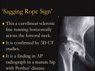
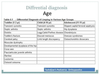
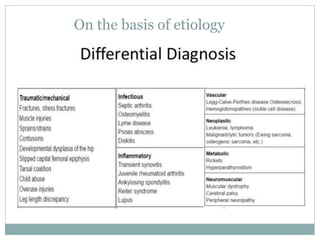
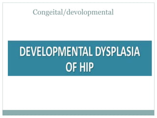
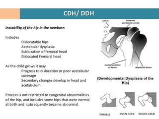
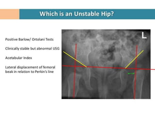
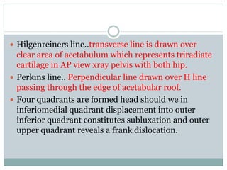
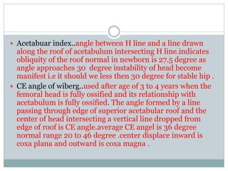
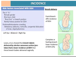
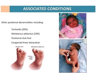
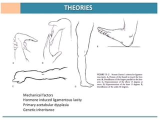
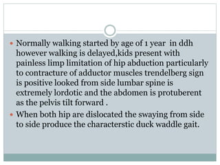
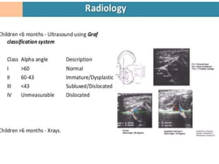
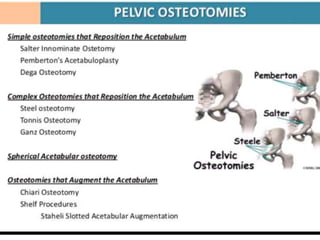
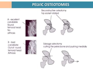
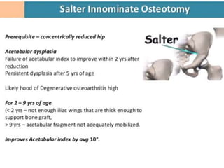
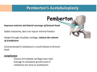
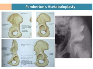
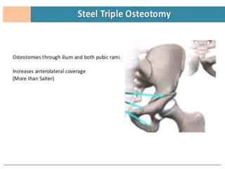
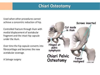
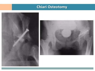
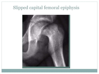
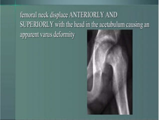
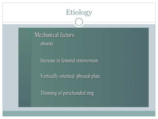
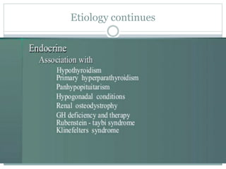
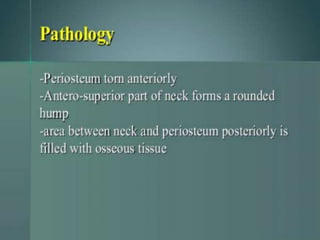
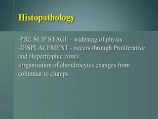
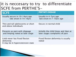
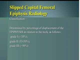
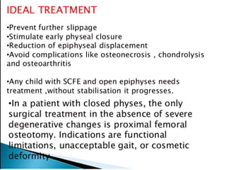
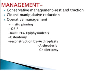
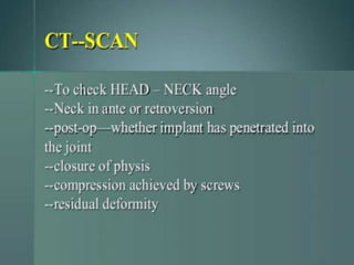
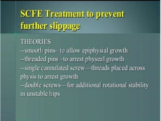
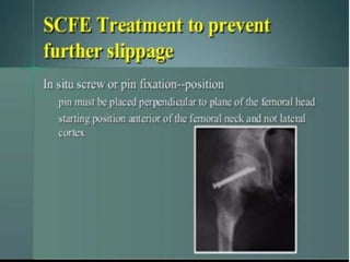
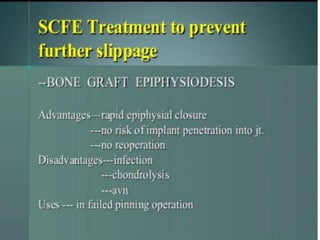
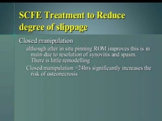
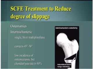
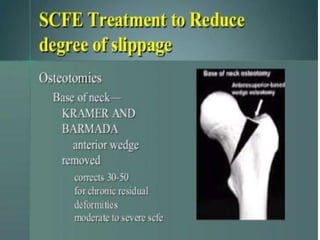
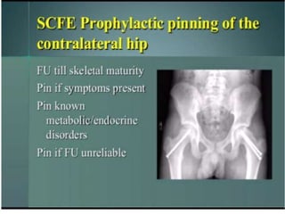
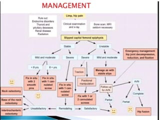
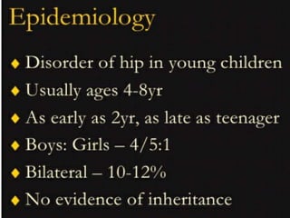
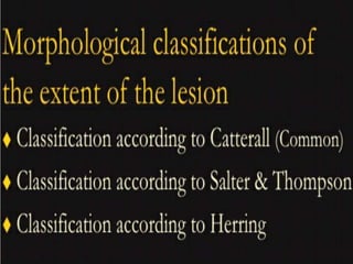
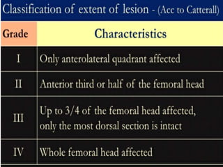
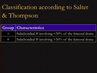
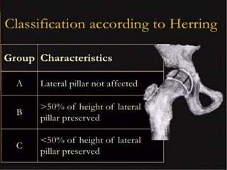
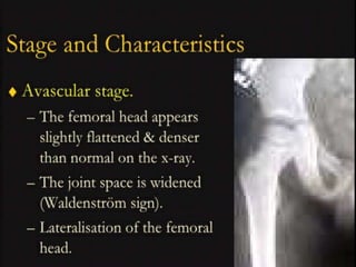
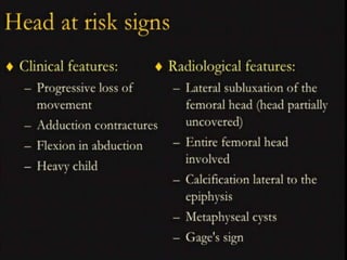
This document discusses differential diagnoses for a limping child based on etiology. It covers congenital/developmental causes like developmental dysplasia of the hip (DDH), providing details on diagnostic imaging techniques like the Hilgenreiners line and acetabular index. The pathoanatomy of DDH is described, from changes in newborns to changes in older walking children. Clinical presentation of DDH is outlined, noting delayed walking and characteristic gait abnormalities. Other causes discussed briefly include slipped capital femoral epiphysis and Perthes disease.
7. Hilgenreiners line..transverse line is drawn over
clear area of acetabulum which represents triradiate
cartilage in AP view xray pelvis with both hip.
Perkins line.. Perpendicular line drawn over H line
passing through the edge of acetabular roof.
Four quadrants are formed head should we in
inferiomedial quadrant displacement into outer
inferior quadrant constitutes subluxation and outer
upper quadrant reveals a frank dislocation.
8. Acetabuar index..angle between H line and a line drawn
along the roof of acetabulum intersecting H line.indicates
obliquity of the roof normal in newborn is 27.5 degree as
angle approaches 30 degree instability of head become
manifest i.e it should we less then 30 degree for stable hip .
CE angle of wiberg..used after age of 3 to 4 years when the
femoral head is fully ossified and its relationship with
acetabulum is fully ossified. The angle formed by a line
passing through edge of superior acetabular roof and the
center of head intersecting a vertical line dropped from
edge of roof is CE angle.average CE angel is 36 degree
normal range 20 to 46 degree .center displace inward is
coxa plana and outward is coxa magna .
9.
10.
11.
12. Pathoanatomy
Depend on age and duration since the hip is remain dislocated or
subluxated .
Change in newborn ..acetabular fossa is shallow and small roof of
superior portion is oblique no resistance to upward glid of head by
muscle pull.neolimbus- presence of hypertrophied acetabular labrum
,femoral head is enlarged and cannot adapted for socket, capsule is
thickened ,ligamentum teres may be hypertrophied degenrated or even
absent,inferior central acetabular fossa filled with fibrofatty tissue.
Change in walking child..exaggregation of changes seen in newborn
femoral head is completely displace out of acetabulum and comes to
rest against lateral wall of illium depression in bone producing a false
acetabulum a pseudo joint is formed between the iliac bone and
femoral head which rests against it.femoral neck is anteverted with
significant valgus deformity .
13.
14.
15.
16.
17.
18.
19.
20. Normally walking started by age of 1 year in ddh
however walking is delayed,kids present with
painless limp limitation of hip abduction particularly
to contracture of adductor muscles trendelberg sign
is positive looked from side lumbar spine is
extremely lordotic and the abdomen is protuberent
as the pelvis tilt forward .
When both hip are dislocated the swaying from side
to side produce the characterstic duck waddle gait.