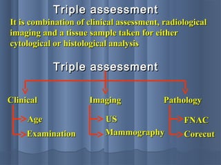
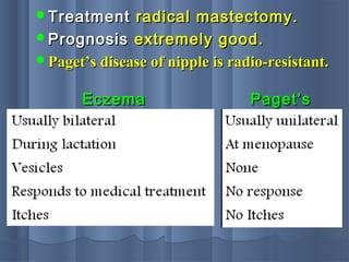
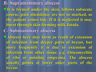
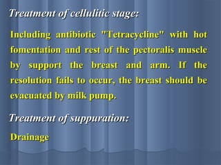
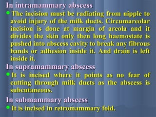
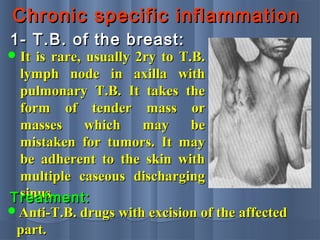
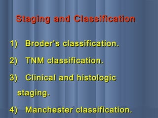
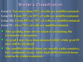
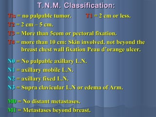
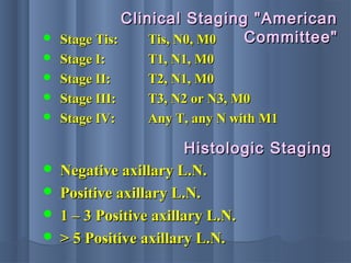
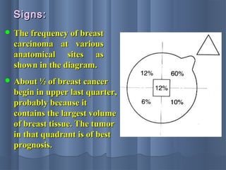
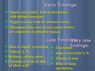
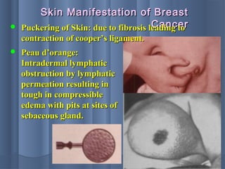
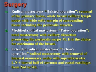
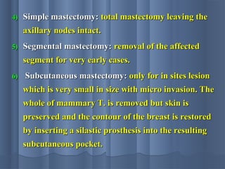
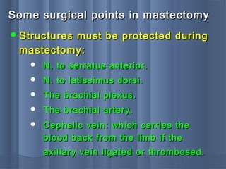
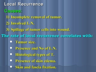
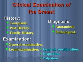
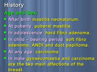
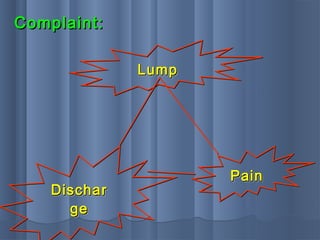
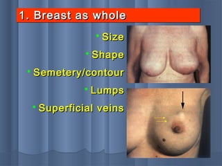
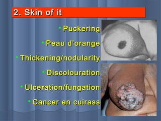
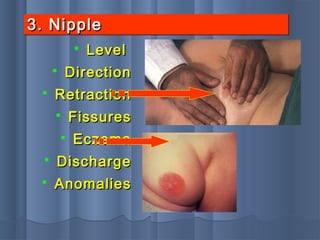
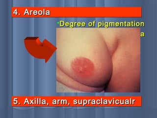
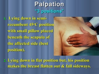
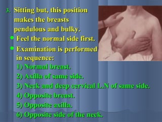
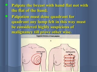
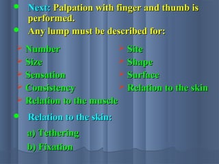
THETHE
BREASTBREAST
ByBy
Prof. Dr. Mohamad YahiaProf. Dr. Mohamad Yahia
Professor of General SurgeryProfessor of General Surgery
Zagazig Faculty of MedicineZagazig Faculty of Medicine
SURGICAL ANATOMYSURGICAL ANATOMY
The breast consists of followingThe breast consists of following
parts:parts:
1- The mammary gland.1- The mammary gland.
2- The superficial fascia.2- The superficial fascia.
3- The overlying skin with areola and3- The overlying skin with areola and
nipple.nipple.
The mammary glandThe mammary gland lies in the superficiallies in the superficial
fascia, it is modified skin gland differ fromfascia, it is modified skin gland differ from
other glands in the body being it doesn't haveother glands in the body being it doesn't have
a capsule and not enclosed in a sheath ofa capsule and not enclosed in a sheath of
fascia. It is formed of 16-20 lobes each lobe isfascia. It is formed of 16-20 lobes each lobe is
divided into lobules, the lobules are separateddivided into lobules, the lobules are separated
by septa of fibrous tissue attached from theby septa of fibrous tissue attached from the
fascia of the chest wall to the subcutaneousfascia of the chest wall to the subcutaneous
tissue "tissue "Cooper`sCooper`s LigLig".".
Thus, the breast is divided into number ofThus, the breast is divided into number of
fascial compartment; each lobe pours its secretionfascial compartment; each lobe pours its secretion
into lactiferous duct. All the lactiferous ductsinto lactiferous duct. All the lactiferous ducts
converge upon the nipple and under the areola,converge upon the nipple and under the areola,
every lactiferous duct enlarges to form lactiferousevery lactiferous duct enlarges to form lactiferous
sinus and then becomes narrow again to open onsinus and then becomes narrow again to open on
summit of the nipple. The whole gland extendssummit of the nipple. The whole gland extends
upwards to 2ry rib, downwards to six costalupwards to 2ry rib, downwards to six costal
cartilage medially to edge of the sternum, laterallycartilage medially to edge of the sternum, laterally
to mid-axillary line. The nipple is a conicalto mid-axillary line. The nipple is a conical
projection placed usually over 4th intercostal space.projection placed usually over 4th intercostal space.
It is surrounded by areola which is circular area ofIt is surrounded by areola which is circular area of
pigmented skin.pigmented skin.
Blood supply of the breast:Blood supply of the breast:
A.A. Arterial supplyArterial supply
1)1) Superior thoracic artery "from the first partSuperior thoracic artery "from the first part
of axillary artery".of axillary artery".
2)2) Lateral and acromiothoracic arteries "fromLateral and acromiothoracic arteries "from
the second part of axillary artery".the second part of axillary artery".
These two vessels supply the lateral aspect of the breast.These two vessels supply the lateral aspect of the breast.
3)3) Perforating branches of intercostal arteries.Perforating branches of intercostal arteries.
4)4) Perforating branch of internal mammaryPerforating branch of internal mammary
artery.artery.
These two vessels supply the medial aspect of the breast.These two vessels supply the medial aspect of the breast.
MedialLateral
B.B. Venous supply:Venous supply: Venous return simplyVenous return simply
follow the arteries.follow the arteries.
The lymph drainage of the breast, as with anyThe lymph drainage of the breast, as with any
other organ follows pathway of its blood supplyother organ follows pathway of its blood supply
and therefore it travels:and therefore it travels:
C.C. Lymphatic drainageLymphatic drainage
1-Along tributaries of the axillary vessels to1-Along tributaries of the axillary vessels to
axillary L.N.S.axillary L.N.S.
2-Along tributaries of internal thoracic vessels2-Along tributaries of internal thoracic vessels
piercing pectoralis major to traverse eachpiercing pectoralis major to traverse each
intercostal space to L.N.S. along the internalintercostal space to L.N.S. along the internal
mammary chain.mammary chain.
There are two main lymphatic plexuses:There are two main lymphatic plexuses:
Lymphatic plexusLymphatic plexus
 Sub-areolar plexus of Sappy, for superficialSub-areolar plexus of Sappy, for superficial
parts of the breast.parts of the breast.
 Pectoral plexus on the pectoral fascia forPectoral plexus on the pectoral fascia for
deep parts of the breast.deep parts of the breast.
Lymphatics of the breast do not cross theLymphatics of the breast do not cross the
midline but cross the diaphragm, so lymphaticmidline but cross the diaphragm, so lymphatic
spread from the lower medial part of thespread from the lower medial part of the
breast can spread through lymphatics of thebreast can spread through lymphatics of the
liver into falciform ligament forming 2ryliver into falciform ligament forming 2ry
umbilical nodule which can be occur alsoumbilical nodule which can be occur also
from lymphatics though the posterior rectusfrom lymphatics though the posterior rectus
sheath.sheath.
1.1. Anterior group" Pectoral": along the lower border ofAnterior group" Pectoral": along the lower border of
pectoralis M. It drains the whole chest wall, upperpectoralis M. It drains the whole chest wall, upper
abdomen down to umbilicus.abdomen down to umbilicus.
2.2. Posterior group" Subscapular": closely related toPosterior group" Subscapular": closely related to
subscapular V. It drains the back to iliac crest.subscapular V. It drains the back to iliac crest.
3.3. Lateral group" humeral": along side the axillaryLateral group" humeral": along side the axillary
vein. It drains the upper limb.vein. It drains the upper limb.
4.4. Central group: In floor of the axilla. It drains theCentral group: In floor of the axilla. It drains the
three upper groupsthree upper groups
5.5. Apical group" infraclavicular": immediately behindApical group" infraclavicular": immediately behind
the clavicle.the clavicle.
6.6. It drains the center group.It drains the center group.
Axillary lymph notesAxillary lymph notes
1.1. Abscess of the breast should be open by a radialAbscess of the breast should be open by a radial
incision to avoid cutting across the lactiferous ducts.incision to avoid cutting across the lactiferous ducts.
2.2. Retraction in relation to cancer of the breast:Retraction in relation to cancer of the breast:
Some surgical clinical important pointsSome surgical clinical important points
 Retraction of the skin "dimpling" due to invasion ofRetraction of the skin "dimpling" due to invasion of
Cooper’s ligaments.Cooper’s ligaments.
 Retraction of the nipple due to extension of theRetraction of the nipple due to extension of the
growth along the main milk ducts with fibrosisgrowth along the main milk ducts with fibrosis
leading to indrawing of the nipple.leading to indrawing of the nipple.
 Peau d`orange: The pits of hair follicles appear to bePeau d`orange: The pits of hair follicles appear to be
retracted beneath the level of the surrounding skin.retracted beneath the level of the surrounding skin.
It is due to blockage of lymphatics draining the skinIt is due to blockage of lymphatics draining the skin
leading to edema of the skin.leading to edema of the skin.
1.1. MammographyMammography::
Investigation of the breastInvestigation of the breast
 Soft tissue mammography.Soft tissue mammography.
 Xero mammography.Xero mammography.
Mammography is the only reliable meansMammography is the only reliable means
of detecting breast cancer before a massof detecting breast cancer before a mass
can be palpated.can be palpated.
The principle of soft tissue mammography isThe principle of soft tissue mammography is
based on detecting a difference in radiographicbased on detecting a difference in radiographic
densities between the normal beast and cancerdensities between the normal beast and cancer
containing area. Carcinoma tends to be densercontaining area. Carcinoma tends to be denser
than normal tissues and show irregular outlinesthan normal tissues and show irregular outlines
with fine calcified spots.with fine calcified spots.
Indications of mammographyIndications of mammography
1.1. To evaluate the opposite breast if carcinomaTo evaluate the opposite breast if carcinoma
is diagnosed in one breast.is diagnosed in one breast.
2.2. To evaluate doubtful masses in fatty breast.To evaluate doubtful masses in fatty breast.
3.3. To screen a selected group of women withTo screen a selected group of women with
high risk factor for developing cancer breast.high risk factor for developing cancer breast.
BUTBUT mammography is inappropriate formammography is inappropriate for
patient under the age of 35 years old for twopatient under the age of 35 years old for two
reasons:reasons:
1.1. Incidence of breast cancer is low.Incidence of breast cancer is low.
2.2. The normal breast is too dense in youngThe normal breast is too dense in young
female to permit sufficient diagnostic details.female to permit sufficient diagnostic details.
UltrasonographyUltrasonography
It is useful in young women with denseIt is useful in young women with dense
breasts in whom mammograms are difficult tobreasts in whom mammograms are difficult to
interpret. It is helpful in differentiating solidinterpret. It is helpful in differentiating solid
from cystic swelling. It can also used to locatefrom cystic swelling. It can also used to locate
impalpable breast lumps.impalpable breast lumps.
GalactographyGalactography
Soft tissue X-rays combined with injectionSoft tissue X-rays combined with injection
of radio-opaque material into a major duct,of radio-opaque material into a major duct,
specially used in demonstration of duct tumor.specially used in demonstration of duct tumor.
ThermographyThermography
A technique by which the heat emissionA technique by which the heat emission
from the surface of the breast in form of infra-from the surface of the breast in form of infra-
red radiation can be recorded, as the skin overred radiation can be recorded, as the skin over
malignant tumor of breast is usually warmermalignant tumor of breast is usually warmer
than surrounding, but infection may give falsethan surrounding, but infection may give false
+ve.+ve.
Magnetic resonance imagingMagnetic resonance imaging
It can be useful to distinguish scar fromIt can be useful to distinguish scar from
recurrence in women who have hadrecurrence in women who have had
previous breast conservation therapy forprevious breast conservation therapy for
cancer. It is gold standard for imaging thecancer. It is gold standard for imaging the
breasts of women with implants.breasts of women with implants.
Biopsy and cytologyBiopsy and cytology
A-A- Trucut orTrucut or corecutcorecut biopsy may be usedbiopsy may be used
under local anesthesia.under local anesthesia.
B-B- Fine needle aspiration cytology "Fine needle aspiration cytology "FNACFNAC""
is the least invasive and more accurate ifis the least invasive and more accurate if
both operator and cytologist areboth operator and cytologist are
experienced.experienced.
C-C- Open biopsy (excisional or incisional).Open biopsy (excisional or incisional).
Triple assessmentTriple assessment
It is combination of clinical assessment, radiologicalIt is combination of clinical assessment, radiological
imaging and a tissue sample taken for eitherimaging and a tissue sample taken for either
cytological or histological analysiscytological or histological analysis
ClinicalClinical ImagingImaging PathologyPathology
AgeAge
ExaminationExamination
USUS
MammographyMammography
FNACFNAC
CorecutCorecut
Triple assessmentTriple assessment
THE NIPPLETHE NIPPLE
1.1. Athelia:Athelia: absence of nipple "rare".absence of nipple "rare".
2.2. Polythelia:Polythelia: multiple nipples along linemultiple nipples along line
extended from ant. fold of axilla to groin “extended from ant. fold of axilla to groin “
milklinemilkline”.”.
3.3. Fissure:Fissure: it is cracked nipple, occurs duringit is cracked nipple, occurs during
lactation must probably due to lack oflactation must probably due to lack of
cleanliness which lead to maceration andcleanliness which lead to maceration and
falling the superficial layers epithelium andfalling the superficial layers epithelium and
sucking becomes painful.sucking becomes painful.
Treatment: Cleaning with boric acid lotion keeping
it dry. Evacuate the milk with breast pump.
4.4. RetractionRetraction::
 Congenital retraction.Congenital retraction.
 Remote simple retractionRemote simple retraction occurs atoccurs at
puberty, it is simple inversion,puberty, it is simple inversion,
unknown etiology, bilateral in 25% ofunknown etiology, bilateral in 25% of
cases.cases. Treatment:Treatment: drawing out thedrawing out the
nipple between finger and thumbnipple between finger and thumb
daily for three weeks during anddaily for three weeks during and
sonly often puberty.sonly often puberty.
 Recent retraction:Recent retraction: UsuallyUsually
accompanied with scirrhousaccompanied with scirrhous
carcinoma occurs at womanhood.carcinoma occurs at womanhood.
5- Nipple discharge5- Nipple discharge
a- Clear, serous dischargea- Clear, serous discharge
Physiological "early pregnancy".Physiological "early pregnancy".
Mammary dysplasia.Mammary dysplasia.
b-b- Blood stained dischargeBlood stained discharge
Intra ductal papilloma.Intra ductal papilloma.
Intra ductal carcinoma.Intra ductal carcinoma.
Duct ectasia.Duct ectasia.
c-c- Black or brownish greenBlack or brownish green
Mammary dysplasia with cyst.Mammary dysplasia with cyst.
Duct ectasia.Duct ectasia.
d-d- MilkMilk
Following lactation.Following lactation.
Galactocele.Galactocele.
Increase in secretion of pituitary prolactin.Increase in secretion of pituitary prolactin.
Contraceptive pills.Contraceptive pills.
e-e- PurulentPurulent
Breast abscess.Breast abscess.
Fistula.Fistula.
The most common cases are:The most common cases are:
1.1. Duct papilloma.Duct papilloma.
2.2. Mammary fistula.Mammary fistula.
3.3. Duct ectasia.Duct ectasia.
4.4. Mammary dysplasia.Mammary dysplasia.
5.5. Paget’s disease.Paget’s disease.
AssociatedAssociated
with lumpwith lump
LumpectomyLumpectomy
++
biopsybiopsy
No lumpNo lump
Treatment of abnormal dischargeTreatment of abnormal discharge
From oneFrom one
ductduct
MicrodochectomyMicrodochectomy
From manyFrom many
ductsducts
HB +veHB +ve
Over 40Over 40
HB –veHB –ve
under 40under 40
More than one ductMore than one duct
Observe and repeatObserve and repeat
occult blood testoccult blood test
periodicallyperiodically
Simple mastectomySimple mastectomy
+ biopsy+ biopsy
1.1. Discharge disappearsDischarge disappears
2.2. Localized to one duct:Localized to one duct:
Microdochectomy.Microdochectomy.
3.3. Lump appears: lumpectomy.Lump appears: lumpectomy.
PAGET'S DISEASEPAGET'S DISEASE
It is persistent eczema likeIt is persistent eczema like
condition usually occurs in patientcondition usually occurs in patient
over 50 years and, doesn't respond toover 50 years and, doesn't respond to
treatment. The disease started astreatment. The disease started as
eczema of nipple which is followed byeczema of nipple which is followed by
mass in underlying tissue, themass in underlying tissue, the
eczematous area looks bright red, iteczematous area looks bright red, it
may moist or dry and scaly.may moist or dry and scaly.
The mass develops 2-10 years afterThe mass develops 2-10 years after
appearance of eczema and it proves to beappearance of eczema and it proves to be
carcinoma of the breast, accepted theory iscarcinoma of the breast, accepted theory is
that the disease started as intraductalthat the disease started as intraductal
carcinoma which spread in two directions: onecarcinoma which spread in two directions: one
to the skin of nipple → eczema and other to theto the skin of nipple → eczema and other to the
breast tissue → mass.breast tissue → mass.
Microscopic features includes: hypertrophyMicroscopic features includes: hypertrophy
of epidermis which becomes thick Paget cellsof epidermis which becomes thick Paget cells
which are large, clear and vacuolated andwhich are large, clear and vacuolated and
round, plasma cells in sub epidermisround, plasma cells in sub epidermis
TreatmentTreatment radical mastectomy.radical mastectomy.
PrognosisPrognosis extremely good.extremely good.
Paget’s disease of nipple is radio-resistant.Paget’s disease of nipple is radio-resistant.
EczemaEczema Paget’sPaget’s
Duct EctesiaDuct Ectesia
"plasma cell mastitis""plasma cell mastitis"
It is primary dilatation of major ducts of the breastIt is primary dilatation of major ducts of the breast
which possible may be due to relaxant effects ofwhich possible may be due to relaxant effects of
progesterone or an auto-immune reaction. It isprogesterone or an auto-immune reaction. It is
commonly associated with discharging nipple incommonly associated with discharging nipple in
middle aged woman. The initial manifestation ismiddle aged woman. The initial manifestation is
worm-like swelling accompanied with local pain,worm-like swelling accompanied with local pain,
tenderness, hardness of the mass with nippletenderness, hardness of the mass with nipple
retraction.retraction.
It is benign lesion, but may be mistaken for eitherIt is benign lesion, but may be mistaken for either
cancer or abscess, so it is also called "Plasma cellcancer or abscess, so it is also called "Plasma cell
mastitis".mastitis".
Treatment:Treatment: Total excision is performed.Total excision is performed.
Mammary fistulaMammary fistula
It is due to subacute recurrentIt is due to subacute recurrent
abscess which ruptures usuallyabscess which ruptures usually
followed by supra mammary type orfollowed by supra mammary type or
due to congenital abnormality withdue to congenital abnormality with
abscess formationabscess formation
Treatment:Treatment: The track is laid open andThe track is laid open and
saucerized or excision of the fistulasaucerized or excision of the fistula
with its related duct.with its related duct.
Duct PapillomaDuct Papilloma
It arises from epithelium of one or more ofIt arises from epithelium of one or more of
main lactiferous duct. It may be single ormain lactiferous duct. It may be single or
multiple and usually pedunculated withmultiple and usually pedunculated with
narrow basenarrow base
Clinically:Clinically:
Female patient of 40 years old with bloody, painless,Female patient of 40 years old with bloody, painless,
profuse discharge from nipple. Cystic swelling mayprofuse discharge from nipple. Cystic swelling may
felt under areola due to obstruction of the duct withfelt under areola due to obstruction of the duct with
cystic formation. It shows tendency to malignantcystic formation. It shows tendency to malignant
change so it should be removed early.change so it should be removed early.
The bleeding nipple is more than in duct carcinomaThe bleeding nipple is more than in duct carcinoma
due to less fibrous tissue.due to less fibrous tissue.
Localization of the papilloma by:Localization of the papilloma by:
Investigation:Investigation:
1-1- Differential pressure.Differential pressure.
2-2- Duct mammography.Duct mammography.
3-3- Soft tissue mammography.Soft tissue mammography.
4-4- Cytology.Cytology.
Treatment:Treatment:
Discharging from the duct:Discharging from the duct:
microdochectomy is performed.microdochectomy is performed.
Multiple masses: simple mastectomy.Multiple masses: simple mastectomy.
Congenital abnormalitiesCongenital abnormalities
of the breastof the breast
1-1- AmaziaAmazia
Absence of breast (Absence of breast ( unilateralunilateral oror
bilateral).bilateral).
May associate with absence of theMay associate with absence of the
sternal portion of the pectoralissternal portion of the pectoralis
major "Poland’s syndrome"major "Poland’s syndrome"
Common in male.Common in male.
2- Polymazia2- Polymazia
Presence ofPresence of one or moreone or more..
May be found in axilla "most common site"May be found in axilla "most common site"
groin or lateral side of the thigh.groin or lateral side of the thigh.
Usually functionless but it may beUsually functionless but it may be
functionally during lactation.functionally during lactation.
3- Diffuse hypertrophy of the breasts3- Diffuse hypertrophy of the breasts
Usually in healthy girls at puberty and lessUsually in healthy girls at puberty and less
often during the first pregnancy due tooften during the first pregnancy due to
hypertrophyhypertrophy of stroma and fatof stroma and fat
Treatment:Treatment: Reduction mammoplasty.Reduction mammoplasty.
Injuries of the BreastInjuries of the Breast
HaematomaHaematoma
It is cystic lump with past history of trauma andIt is cystic lump with past history of trauma and
aspiration is diagnostic.aspiration is diagnostic.
Traumatic fat necrosisTraumatic fat necrosis
It is due to trauma (a blow or indirect violence)It is due to trauma (a blow or indirect violence)
prolonged pressure or needle.prolonged pressure or needle.
Biopsies of benign conditions. An area of fat becomesBiopsies of benign conditions. An area of fat becomes
devitalized and liberates fatty acids which react withdevitalized and liberates fatty acids which react with
calcium to form calcium soaps which becomecalcium to form calcium soaps which become
surrounded by fibrosis forming an irregular hardsurrounded by fibrosis forming an irregular hard
painless lump which may be mistaken for carcinoma,painless lump which may be mistaken for carcinoma,
so excision and frozen section are needed forso excision and frozen section are needed for
differentiation. History of trauma is not diagnostic.differentiation. History of trauma is not diagnostic.
Acute mastitis and acuteAcute mastitis and acute
breast abscessbreast abscess
1.1. Mastitis of infants.Mastitis of infants.
2.2. Mastitis of puberty.Mastitis of puberty.
3.3. Mastitis of Mumps.Mastitis of Mumps.
4.4. Mastitis of local irritation.Mastitis of local irritation.
5.5. Mastitis during lactation.Mastitis during lactation.
6.6. Mastitis due to bacterialMastitis due to bacterial
invasion.invasion.
1- Mastitis of infants1- Mastitis of infants
 It is the effect of female hormonalIt is the effect of female hormonal
stimulation through placenta. Thestimulation through placenta. The
breast becomes swollen and tenderbreast becomes swollen and tender
on the 3rd or 4th day of life. If theon the 3rd or 4th day of life. If the
breast of the infant is pressed lightlybreast of the infant is pressed lightly
drop of colourless fluid can bedrop of colourless fluid can be
expressed, few days later, milkyexpressed, few days later, milky
secretion from nipple, that is willsecretion from nipple, that is will
disappear during 3rd week anddisappear during 3rd week and
known as "witch’s milk".known as "witch’s milk".
2- Mastitis of puberty2- Mastitis of puberty
 Male breast may becomeMale breast may become
enlarged, firm tender duringenlarged, firm tender during
puberty, the condition subsidespuberty, the condition subsides
spontaneously and needs nospontaneously and needs no
special therapy. Suppurationspecial therapy. Suppuration
never occur.never occur.
3- Mastitis of mumps3- Mastitis of mumps
 Unilateral in femaleUnilateral in female
4- Mastitis due to local4- Mastitis due to local
irritationirritation
 It is due to too tight elasticIt is due to too tight elastic
brassiere, rare in male rare inbrassiere, rare in male rare in
suppuration.suppuration.
5- Mastitis due to5- Mastitis due to
lactationlactation
 It is due to milk engorgement, whenIt is due to milk engorgement, when
one of the duct becomes blockedone of the duct becomes blocked
with epithelial debris. The breastwith epithelial debris. The breast
becomes painful and tender.becomes painful and tender.
6- Mastitis due to bacterial6- Mastitis due to bacterial
invasion with abscessinvasion with abscess
formationformation
It is the most common variety ofIt is the most common variety of
mastitis. 32% of mastitis in female ismastitis. 32% of mastitis in female is
due to bacterial mastitis withoutdue to bacterial mastitis without
lactation probably may due tolactation probably may due to
infected haematoma. the commoninfected haematoma. the common
infected organism is "infected organism is " Staph aureusStaph aureus""
which is penicillin resistant.which is penicillin resistant.
Pathology:Pathology:
The organism reaches the breast throughThe organism reaches the breast through
milk ducts or through fissures in nipples butmilk ducts or through fissures in nipples but
blood borne infection is rare. The breast atblood borne infection is rare. The breast at
first congested and later suppurated.first congested and later suppurated.
Clinical features:Clinical features:
General features of Toxemia are usuallyGeneral features of Toxemia are usually
marked especially in commonmarked especially in common
intramammary abscess, it may be masked byintramammary abscess, it may be masked by
ill-advise use of antibiotics leading toill-advise use of antibiotics leading to
"antibioma". In case of supramammary"antibioma". In case of supramammary
abscess it usually has subacute course.abscess it usually has subacute course.
Local features:Local features:
Suppuration:Suppuration: A- Intramammary abscess.A- Intramammary abscess.
B- Supramammary abscess.B- Supramammary abscess.
C- Submammary abscess.C- Submammary abscess.
A- Intramammary abscessA- Intramammary abscess
 It may reach big size with lactation the breastIt may reach big size with lactation the breast
is swollen, tender with marked pain andis swollen, tender with marked pain and
dilated veins on the surface. When pus isdilated veins on the surface. When pus is
formed the pain becomes throbbing andformed the pain becomes throbbing and
temperature becomes hectic edema increases.temperature becomes hectic edema increases.
The fluctuation is not dependable sign so don'tThe fluctuation is not dependable sign so don't
wait for fluctuation.wait for fluctuation.
B- Supramammary abscessB- Supramammary abscess
It is formed under the skin, follows subacuteIt is formed under the skin, follows subacute
course, pain attendance are not so marked, socourse, pain attendance are not so marked, so
the patient comes late. If it is neglected it maythe patient comes late. If it is neglected it may
burst through skin forming milk fistula.burst through skin forming milk fistula.
C- Submammary abscessC- Submammary abscess
Abscess here may form as result of extensionAbscess here may form as result of extension
of infection from deeper parts of breast, butof infection from deeper parts of breast, but
more frequently it is due to extension ofmore frequently it is due to extension of
infection from other tissue, e.g. Osteomyelitisinfection from other tissue, e.g. Osteomyelitis
of ribs or pointing empyema. The abscessof ribs or pointing empyema. The abscess
usually points at lower outer parts of theusually points at lower outer parts of the
breastbreast..
Treatment of cellulitic stage:Treatment of cellulitic stage:
Including antibiotic "Tetracycline" with hotIncluding antibiotic "Tetracycline" with hot
fomentation and rest of the pectoralis musclefomentation and rest of the pectoralis muscle
by support the breast and arm. If theby support the breast and arm. If the
resolution fails to occur, the breast should beresolution fails to occur, the breast should be
evacuated by milk pump.evacuated by milk pump.
Treatment of suppuration:Treatment of suppuration:
DrainageDrainage
In intramammary abscessIn intramammary abscess
The incision must be radiating from nipple toThe incision must be radiating from nipple to
avoid injury of the milk ducts. Circumareolaravoid injury of the milk ducts. Circumareolar
incision is done at margin of areola and itincision is done at margin of areola and it
divides the skin only then long haemostate isdivides the skin only then long haemostate is
pushed into abscess cavity to break any fibrouspushed into abscess cavity to break any fibrous
bands or adhesion inside it. And drain is leftbands or adhesion inside it. And drain is left
inside it.inside it.
In supramammary abscessIn supramammary abscess
It is incised where it points as no fear ofIt is incised where it points as no fear of
cutting through milk ducts as the abscess iscutting through milk ducts as the abscess is
subcutaneous.subcutaneous.
In submammary abscessIn submammary abscess
It is incised in retromammary fold.It is incised in retromammary fold.
Chronic specific inflammationChronic specific inflammation
1- T.B. of the breast:1- T.B. of the breast:
 It is rare, usually 2ry to T.B.It is rare, usually 2ry to T.B.
lymph node in axilla withlymph node in axilla with
pulmonary T.B. It takes thepulmonary T.B. It takes the
form of tender mass orform of tender mass or
masses which may bemasses which may be
mistaken for tumors. It maymistaken for tumors. It may
be adherent to the skin withbe adherent to the skin with
multiple caseous dischargingmultiple caseous discharging
sinus.sinus.Treatment:Treatment:
Anti-T.B. drugs with excision of the affectedAnti-T.B. drugs with excision of the affected
part.part.
2- Syphilis of the breast:2- Syphilis of the breast:
It occurs from nursing of syphilic child.It occurs from nursing of syphilic child.
It extremely rare.It extremely rare.
Treatment:Treatment: Anti-syphilitic drugsAnti-syphilitic drugs
3- Actinomycosis of the breast:3- Actinomycosis of the breast:
 Extremely rare, 2ry to involved lung,Extremely rare, 2ry to involved lung,
presented with indurated mass with sulphurpresented with indurated mass with sulphur
discharging sinus.discharging sinus.
Treatment:Treatment: Penicillin "large doses" withPenicillin "large doses" with
excision of the affected part.excision of the affected part.
Chronic non-specificChronic non-specific
inflammationinflammation
Chronic abscess:Chronic abscess:
It is usually from in adequate use of antibiotics forIt is usually from in adequate use of antibiotics for
acute mastitis and it is very difficult to differentiateacute mastitis and it is very difficult to differentiate
between it and “scirrhous carcinoma” of the breast.between it and “scirrhous carcinoma” of the breast.
For differentiation:For differentiation:
1- History of acute mastitis.1- History of acute mastitis.
2- It is less resistant in center than at periphery2- It is less resistant in center than at periphery
3- No nipple retraction3- No nipple retraction
4- Rounded posterior surface.4- Rounded posterior surface.
5- Lymph node is small, firm, tender and mobile.5- Lymph node is small, firm, tender and mobile.
 In doubtful cases, frozen biopsy is recommended toIn doubtful cases, frozen biopsy is recommended to
confirm or exclude malignancy.confirm or exclude malignancy.
MondorsMondors
disease:disease: It is thrombophlebitis of superficialIt is thrombophlebitis of superficial
veins of the breast and anterior chestveins of the breast and anterior chest
wall in absence of injury orwall in absence of injury or
inflammation due to unknown causes.inflammation due to unknown causes.
 Clinically:Clinically: presented with induratedpresented with indurated
subcutaneous cord like structure. Itsubcutaneous cord like structure. It
may be diagnosed as lymphaticmay be diagnosed as lymphatic
permeation of an occult carcinoma.permeation of an occult carcinoma.
 Treatment:Treatment: restriction of armrestriction of arm
movements.movements.
Benign Breast DiseasesBenign Breast Diseases
1. ANDI “fibroadenosis / cystic hyperplasia”1. ANDI “fibroadenosis / cystic hyperplasia”
2. Fibroadenoma (hard & soft)2. Fibroadenoma (hard & soft)
3. Cysts3. Cysts
4. Duct papilloma4. Duct papilloma 5. Duct ectasia5. Duct ectasia
6. Fat necrosis6. Fat necrosis 7. Monder’s disease7. Monder’s disease
8. Pregnancy related disease (galactocele & puerperal8. Pregnancy related disease (galactocele & puerperal
abscess)abscess)
9. Congenital disorders (inverted nipple and9. Congenital disorders (inverted nipple and
supramammary breast)supramammary breast)
10. Non breast disorders (Tietze’s disease, sebaceous10. Non breast disorders (Tietze’s disease, sebaceous
cysts, other skin lesions)cysts, other skin lesions)
Fibroadenosis/Cystic hyperplasiaFibroadenosis/Cystic hyperplasia
“Aberrations of normal development“Aberrations of normal development
and involution” (ANDI)and involution” (ANDI)
““MammaryMammary dysplesiadysplesia””
Aetiology:Aetiology:
It is an aberration of normal physiological changesIt is an aberration of normal physiological changes
related to ovarian activity. The blood hormonerelated to ovarian activity. The blood hormone
levels are usually within normal limits and it maylevels are usually within normal limits and it may
be that the disease is caused by an abnormalbe that the disease is caused by an abnormal
breast tissue response rather than an abnormalbreast tissue response rather than an abnormal
stimulus.stimulus.
Pathology:Pathology:
Site:Site: Bilateral common but may be unilateral.Bilateral common but may be unilateral.
Diffuse or localized type.Diffuse or localized type.
Macroscopic:Macroscopic: When sectioned with knife, the affectedWhen sectioned with knife, the affected
area in the breast are white or yellow and of India-rubberarea in the breast are white or yellow and of India-rubber
consistency but never present gritty sensation ofconsistency but never present gritty sensation of
carcinoma.carcinoma.
Microscopically:Microscopically:
1) Cystic formation: solitary or multiple.1) Cystic formation: solitary or multiple.
2) Adenosis2) Adenosis
3) Fibrosis: the fat and elastic tissue replaced by dense3) Fibrosis: the fat and elastic tissue replaced by dense
white fibrous tissue.white fibrous tissue.
4) Epitheliosis: hyperplasia of epithelium.4) Epitheliosis: hyperplasia of epithelium.
5) Papillomatosis: Epithelial hyperplasia may be so5) Papillomatosis: Epithelial hyperplasia may be so
extensive that it results in papillomatous formation.extensive that it results in papillomatous formation.
Five features with variationFive features with variation
Clinical Features:Clinical Features:
 Mild degree of the condition are very common beingMild degree of the condition are very common being
usually a tender or painful area of breast tissue ofusually a tender or painful area of breast tissue of
increased density.increased density.
 The pain and swelling may be mainly premenstrualThe pain and swelling may be mainly premenstrual
and caused by vascular engorgement.and caused by vascular engorgement.
 By for it is the commonest disease of female breastBy for it is the commonest disease of female breast
(25 – 45 years).(25 – 45 years).
 The main complain is painful or tender mass orThe main complain is painful or tender mass or
masses in the breast.masses in the breast.
 The pain increases during or before the menstruationThe pain increases during or before the menstruation
and relieved by pregnancy and lactation.and relieved by pregnancy and lactation.
 The pain may mistake for angina as it is shooting toThe pain may mistake for angina as it is shooting to
the arm.the arm.
 The nodules felt between the thumb and fingers andThe nodules felt between the thumb and fingers and
vaguely by flat the of the hand. It is not adherent tovaguely by flat the of the hand. It is not adherent to
the skin ofthe skin of Pectoralis fasciaPectoralis fascia.. N.B: The mass which isN.B: The mass which is
felt by flat of the hand inside the breast:felt by flat of the hand inside the breast:
* Cyst formation* Cyst formation * Chronic abscess* Chronic abscess * Malignancy* Malignancy
 There may be discharge from nipple (serous, green-There may be discharge from nipple (serous, green-
brown).brown).
 L.N. in axilla may be tender but never hard.L.N. in axilla may be tender but never hard.
 The localized type “sector type” of the disease mayThe localized type “sector type” of the disease may
simulate carcinoma.simulate carcinoma.
The relationship of mammary dysplasiaThe relationship of mammary dysplasia
to carcinoma:to carcinoma:
 The mater is not yet settled.The mater is not yet settled.
 It is considered as not precancerous but, theIt is considered as not precancerous but, the
incidence of carcinoma in female within cysticincidence of carcinoma in female within cystic
disease has been reported to be about 3 or 5disease has been reported to be about 3 or 5
times that of general female population.times that of general female population.
Others report prominent epithelium hyperplasia asOthers report prominent epithelium hyperplasia as
finding that has a significant frequency relationship tofinding that has a significant frequency relationship to
subsequent carcinoma of the breast.subsequent carcinoma of the breast.
Treatment:Treatment:
1)1)Reassurance is very important.Reassurance is very important.
2)2)Support the breast.Support the breast.
3)3)Testosterone 5 mg/3 times daily for 2 monthsTestosterone 5 mg/3 times daily for 2 months
but not recommended in young female.but not recommended in young female.
4)4)Multiple cysts: Surgical treatment.Multiple cysts: Surgical treatment.
5)5)Localized mass: excisional biopsy.Localized mass: excisional biopsy.
6)6)Iodine in milk may cause improvement ofIodine in milk may cause improvement of
fibrosis.fibrosis.
7)7)For severe pain: Danazol synthetic androgenFor severe pain: Danazol synthetic androgen
may be used.may be used.
CYSTS OF THE BREASTCYSTS OF THE BREAST
1) Cysts connected to big ducts (1) Cysts connected to big ducts (galactocelegalactocele
&& Simple retention cystSimple retention cyst).).
2) Cysts connected to small ducts (Cysts of2) Cysts connected to small ducts (Cysts of
mammary dysplasia).mammary dysplasia).
3) Cysts connected to the tumors (Duct3) Cysts connected to the tumors (Duct
papiloma, serocystic disease,papiloma, serocystic disease,
cystadenoma, degenerated cyst incystadenoma, degenerated cyst in
malignancy).malignancy).
4) Cysts found in the stroma (Lymphatic4) Cysts found in the stroma (Lymphatic
cyst, blood cyst, hydatid cyst).cyst, blood cyst, hydatid cyst).
Galactocele:Galactocele:
Rare lesion, due to obstruction of main milk ductRare lesion, due to obstruction of main milk duct
by inspirated milk or fibrosis. It is painless cysticby inspirated milk or fibrosis. It is painless cystic
swelling behind nipple and areola since lactationswelling behind nipple and areola since lactation
and milk expressed from areola by pressure overand milk expressed from areola by pressure over
the cyst.the cyst.
Treatment:Treatment:
Excision or repeated aspiration.Excision or repeated aspiration.
Aspiration of any cyst is not safe if:Aspiration of any cyst is not safe if: 4R4R
 RRefilling of the cyst.efilling of the cyst.
 RRed aspirated fluid.ed aspirated fluid.
 RResidual lump after aspiration.esidual lump after aspiration.
 RRevealing malignant cells by cystological examinationevealing malignant cells by cystological examination
FibroadenomaFibroadenoma
Two types:Two types: hard pericanalicular typehard pericanalicular type wherewhere
the ducts are surrounded by dense C.T.the ducts are surrounded by dense C.T. softsoft
intracanalicular typeintracanalicular type where the ducts arewhere the ducts are
surrounded by loose C.T.surrounded by loose C.T.
Pathology:Pathology:
Hard fibroadenoma:Hard fibroadenoma:
 15 – 35 y, commoner, slow rate of growth small in size.15 – 35 y, commoner, slow rate of growth small in size.
 Cut section:Cut section: smooth, not gritty, bulges on cutting.smooth, not gritty, bulges on cutting.
 Clinically:Clinically: painless small, firm well defined freelypainless small, firm well defined freely
mobile “breast mouse” mass no axillary L.N.mobile “breast mouse” mass no axillary L.N.
 Complication:Complication: rarely turns malignant.rarely turns malignant.
 Treatment:Treatment: Excisional biopsy.Excisional biopsy.
Soft fibroadenoma:Soft fibroadenoma:
 30 – 50 y, less common, more rapid rate of30 – 50 y, less common, more rapid rate of
growth, huge size, may burst through the skin.growth, huge size, may burst through the skin.
 Clinically:Clinically: painless, large, soft swelling withpainless, large, soft swelling with
more rapid rate of growth.more rapid rate of growth.
 Complication:Complication: cystadenoma, cystosarcomacystadenoma, cystosarcoma
phyllodes more liable to be malignantphyllodes more liable to be malignant
transformation.transformation.
Treatment:Treatment:
 If small: decisional biopsy.If small: decisional biopsy.
 If larger: simple mastectomy + biopsy.If larger: simple mastectomy + biopsy.
Cystosarcoma PhyllodesCystosarcoma Phyllodes
(Serocystic Disease of Brodie)(Serocystic Disease of Brodie)
““PhylloidsPhylloids TumorsTumors””
 It is a giant soft fibro adenoma.It is a giant soft fibro adenoma.
 Not sarcomaNot sarcoma
 Not cysticNot cystic
 It grows rapidly causing pressure necrosis of theIt grows rapidly causing pressure necrosis of the
overlying skin without infiltrating it and the tumoroverlying skin without infiltrating it and the tumor
fungates from it “Probe Test” is used to differentiate itfungates from it “Probe Test” is used to differentiate it
from fungating malignant tissue.from fungating malignant tissue.
 The surface is bosselated with area of softening.The surface is bosselated with area of softening.
 Dilated veins may be seen in skin over it.Dilated veins may be seen in skin over it.
 Neither attached to the skin nor to deeper structures.Neither attached to the skin nor to deeper structures.
 No axillary L.N unless secondary infection occurs.No axillary L.N unless secondary infection occurs.
MastalgiaMastalgia
1) Cyclic mastalgia: It occurs in mammary dysplesia1) Cyclic mastalgia: It occurs in mammary dysplesia
tenderness and heaviness in the breast immediatelytenderness and heaviness in the breast immediately
before each period.before each period.
2) Non–cyclic mastalgia: as in duct ectasia, trauma or2) Non–cyclic mastalgia: as in duct ectasia, trauma or
idiopathic.idiopathic.
3) Acute inflammation: lactational mastitis, abscess.3) Acute inflammation: lactational mastitis, abscess.
4) Cancer: only in"5 – 10"of cases.4) Cancer: only in"5 – 10"of cases.
5) Extramammary cause:5) Extramammary cause:
 Tietze’s disease: condritis of costal cartilage.Tietze’s disease: condritis of costal cartilage.
 Biliary diseases.Biliary diseases.
 PleurisyPleurisy
It is a pain in the breastIt is a pain in the breast
1)1) Exclude cancer.Exclude cancer.
2)2) Reassurance.Reassurance.
3)3) Pain chart.Pain chart.
4)4) Treatment the specific cause.Treatment the specific cause.
Treatment of mastalgia:Treatment of mastalgia:
Breast CancerBreast Cancer
Age:Age: It may occur at any age but theIt may occur at any age but the
commonest age is over fifty.commonest age is over fifty.
Race:Race: In general, the rates reported fromIn general, the rates reported from
developing countries are low, where asdeveloping countries are low, where as
rates are high in developed countriesrates are high in developed countries
except in Japan.except in Japan.
Etiology and PathogenesisEtiology and Pathogenesis
Family history:Family history: Women whose mother orWomen whose mother or
sisters had breast cancer are 2 or 3sisters had breast cancer are 2 or 3
times more likely to develop the diseasetimes more likely to develop the disease
than controls.than controls.
 Mammary dysplasia complained by proliferativeMammary dysplasia complained by proliferative
changes papillomatosis or solid hyperplasia ischanges papillomatosis or solid hyperplasia is
associated with an increased incidence of cancer.associated with an increased incidence of cancer.
 A woman who has had cancer in one breast is atA woman who has had cancer in one breast is at
increased risk of developing cancer in other breast.increased risk of developing cancer in other breast.
 A woman with uterine cancer has a higher risk ofA woman with uterine cancer has a higher risk of
breast cancer.breast cancer.
Previous Medical History:Previous Medical History:
 Early menarche (under age 12) and late menopauseEarly menarche (under age 12) and late menopause
(of the age 50) are associated with increase in risk of(of the age 50) are associated with increase in risk of
developing breast cancer.developing breast cancer.
Menstrual History:Menstrual History:
 Unmarried woman or married woman with feverUnmarried woman or married woman with fever
pregnancy or married woman with first children ofpregnancy or married woman with first children of
the age 30 has a higher incidence of breast cancer.the age 30 has a higher incidence of breast cancer.
Marital History:Marital History:
 There is some evidence that continuousThere is some evidence that continuous
administration of estrogens toadministration of estrogens to
postmenopausal women may result in anpostmenopausal women may result in an
increased risk of breast cancer after 10 –increased risk of breast cancer after 10 –
12 years.12 years.
 It should be noted that the adrenal glandIt should be noted that the adrenal gland
is a major source of estrogen andis a major source of estrogen and
androgen in postmenopausal women.androgen in postmenopausal women.
 There is no correlation between thyroidThere is no correlation between thyroid
mass and breast cancer, but somemass and breast cancer, but some
reported higher incidence of breast cancerreported higher incidence of breast cancer
in patient with hypothyroidism.in patient with hypothyroidism.
Hormonal Factor:Hormonal Factor:
1)1) Old age.Old age.
2)2) High socioeconomic factor.High socioeconomic factor.
3)3) Early menarche.Early menarche.
4)4) Late menopause.Late menopause.
5)5) Never married.Never married.
6)6) Fewer Pregnancies.Fewer Pregnancies.
7)7) Female who has had 1st children of the 30 years old.Female who has had 1st children of the 30 years old.
8)8) Female who is non-lactating.Female who is non-lactating.
9)9) History of benign breast disease (BBD).History of benign breast disease (BBD).
10) Continuous administration of estrogen to10) Continuous administration of estrogen to
postmenopausal women.postmenopausal women.
11) Family history of cancer breast.11) Family history of cancer breast.
Risk Factors for Female Breast:Risk Factors for Female Breast:
As an infective agent carried from theAs an infective agent carried from the
mother to baby in the milk "Bitter milkmother to baby in the milk "Bitter milk
factor” but, no evidence to support it infactor” but, no evidence to support it in
human body although it is effective in rats.human body although it is effective in rats.
Milk Factor:Milk Factor:
It has a role in animal but, no evidence inIt has a role in animal but, no evidence in
human.human.
Role of Virus:Role of Virus:
1)1) Broder’s classification.Broder’s classification.
2)2) TNM classification.TNM classification.
3)3) Clinical and histologicClinical and histologic
staging.staging.
4)4) Manchester classification.Manchester classification.
Staging and ClassificationStaging and Classification
Grad I:Grad I: Not more than 25% of cells are undifferentiated.Not more than 25% of cells are undifferentiated.
Grad II:Grad II: From 25% to 50% of cells are undifferentiated.From 25% to 50% of cells are undifferentiated.
Grad III:Grad III: From 50% to 75% of cells are undifferentiated.From 50% to 75% of cells are undifferentiated.
Grad IV:Grad IV: Over 75% of cells are undifferentiated.Over 75% of cells are undifferentiated.
Broder’s Classification:Broder’s Classification:
 This grading must not be taken in estimating theThis grading must not be taken in estimating the
treatment or prognosis.treatment or prognosis.
 As grad I may have disseminated wildly while grad IVAs grad I may have disseminated wildly while grad IV
may still be localized.may still be localized.
 The undifferentiated tissue are usually radio sensitive,The undifferentiated tissue are usually radio sensitive,
but, commonly recur, while high differentiated tissuebut, commonly recur, while high differentiated tissue
tend to be (radio resistant).tend to be (radio resistant).
TisTis = no palpable tumor.= no palpable tumor. T1T1 = 2 cm or less.= 2 cm or less.
T2T2 = 2 cm – 5 cm.= 2 cm – 5 cm.
T3T3 = More than 5com or pectoral fixation.= More than 5com or pectoral fixation.
T4T4 = more than 10 cm: Skin involved, not beyond the= more than 10 cm: Skin involved, not beyond the
breast chest wall fixation Peau d’orange ulcer.breast chest wall fixation Peau d’orange ulcer.
N0N0 = No palpable axillary L.N.= No palpable axillary L.N.
N1N1 = axillary mobile L.N.= axillary mobile L.N.
N2N2 = axillary fixed L.N.= axillary fixed L.N.
N3N3 = Supra clavicular L.N or edema of Arm.= Supra clavicular L.N or edema of Arm.
M0M0 = No distant metastases.= No distant metastases.
M1M1 = Metastases beyond breast.= Metastases beyond breast.
T.N.M. Classification:T.N.M. Classification:
 Stage Tis:Stage Tis: Tis, N0, M0Tis, N0, M0
 Stage I:Stage I: T1, N1, M0T1, N1, M0
 Stage II:Stage II: T2, N1, M0T2, N1, M0
 Stage III:Stage III: T3, N2 or N3, M0T3, N2 or N3, M0
 Stage IV:Stage IV: Any T, any N with M1Any T, any N with M1
Clinical Staging "AmericanClinical Staging "American
Committee"Committee"
 Negative axillary L.N.Negative axillary L.N.
 Positive axillary L.N.Positive axillary L.N.
 1 – 3 Positive axillary L.N.1 – 3 Positive axillary L.N.
 > 5 Positive axillary L.N.> 5 Positive axillary L.N.
Histologic StagingHistologic Staging
 Stage I:Stage I: Mobile lump without axillary L.N.Mobile lump without axillary L.N.
 Stage II:Stage II: Mobile Lump with mobile L.N.Mobile Lump with mobile L.N.
 Stage III:Stage III: Fixed lump with fixed L.N.Fixed lump with fixed L.N.
 Stage IV:Stage IV: Distant metastases.Distant metastases.
Manchester StagingManchester Staging
 In about 70% of cases the presenting complaint isIn about 70% of cases the presenting complaint is
painless lump.painless lump.
 Less frequent symptoms are breast pain, nippleLess frequent symptoms are breast pain, nipple
discharge, erosion, retraction and redness withdischarge, erosion, retraction and redness with
hardness.hardness.
 Rarely, an axillary mass or bone pain may be 1stRarely, an axillary mass or bone pain may be 1st
symptom.symptom.
Clinical featuresClinical features
Symptoms:Symptoms:
 The frequency of breastThe frequency of breast
carcinoma at variouscarcinoma at various
anatomical sites asanatomical sites as
shown in the diagram.shown in the diagram.
Signs:Signs:
 About ½ of breast cancerAbout ½ of breast cancer
begin in upper last quarter,begin in upper last quarter,
probably because itprobably because it
contains the largest volumecontains the largest volume
of breast tissue. The tumorof breast tissue. The tumor
in that quadrant is of bestin that quadrant is of best
prognosis.prognosis.
 Single, non tender, firm to hard massSingle, non tender, firm to hard mass
with defined margins.with defined margins.
 Nipple erosion with or without a mass.Nipple erosion with or without a mass.
 Mammography may detect cancer beforeMammography may detect cancer before
development of palpable masses.development of palpable masses.
Early Findings:Early Findings:
 Skin or nipple retraction.Skin or nipple retraction.
 Axillary L.N.Axillary L.N.
 Redness, edema, pain.Redness, edema, pain.
 Fixation of mass to skinFixation of mass to skin
or chest wall.or chest wall.
Late Findings:Late Findings:
 Ulceration.Ulceration.
 Supra clavicular L.N.Supra clavicular L.N.
 Edema of arm.Edema of arm.
 Bone or lungBone or lung
metastases.metastases.
Very lateVery late
findings:findings:
Skin Manifestation of BreastSkin Manifestation of Breast
CancerCancer
 Peau d’orange:Peau d’orange:
Intradermal lymphaticIntradermal lymphatic
obstruction by lymphaticobstruction by lymphatic
permeation resulting inpermeation resulting in
tough in compressibletough in compressible
edema with pits at sites ofedema with pits at sites of
sebaceous gland.sebaceous gland.
 Puckering of Skin: due to fibrosis leading toPuckering of Skin: due to fibrosis leading to
contraction of cooper’s ligament.contraction of cooper’s ligament.
 Cancer en Cuirass: with progressive lymphaticCancer en Cuirass: with progressive lymphatic
obstruction the entire skin of the breast andobstruction the entire skin of the breast and
sometimes the chest wall becomes deeply pigmentedsometimes the chest wall becomes deeply pigmented
hard, thick and rigid tethering.hard, thick and rigid tethering.
 Skin Nodule: due to lymphatic permeationSkin Nodule: due to lymphatic permeation
occurring at skin of the breast or around umbilicus.occurring at skin of the breast or around umbilicus.
 Ulceration and fungation.Ulceration and fungation.
 Edema of the arm.Edema of the arm.
1)1) Peau d’orange.Peau d’orange.
2)2) Cancer en cuirasse.Cancer en cuirasse.
3)3) Edema of the arm.Edema of the arm.
4)4) Lymphangio sarcoma.Lymphangio sarcoma.
5)5) Serous effusion into peritoneal or pleural cavities.Serous effusion into peritoneal or pleural cavities.
Phenomena resulting four lymphaticPhenomena resulting four lymphatic
obstruction in cases of cancer breastobstruction in cases of cancer breast
LymphangiosarcomaLymphangiosarcoma
It is post mastectomy lymphadema it may beIt is post mastectomy lymphadema it may be
confused with recurrent carcinoma of the breast.confused with recurrent carcinoma of the breast.
A.A. Early post operative:Early post operative: within matter of days thewithin matter of days the
radical mastectomy may be related to axillary veinradical mastectomy may be related to axillary vein
thrombosis but, it is wise to regard it as infective inthrombosis but, it is wise to regard it as infective in
origin and treatment with full course of antibiotics.origin and treatment with full course of antibiotics.
B.B. Late edema:Late edema: from several months to many yearsfrom several months to many years
after operation it may be associated with localafter operation it may be associated with local
infection, so it is treated vigorously with antibioticsinfection, so it is treated vigorously with antibiotics
while the only treatment of value is elevation withwhile the only treatment of value is elevation with
elastic bandage from wrist to the axillary.elastic bandage from wrist to the axillary.
C.C. Brawny Arm:Brawny Arm: can result from advanced neoplasticcan result from advanced neoplastic
infiltration of unarmored or in completely removedinfiltration of unarmored or in completely removed
axillary or supra clavicular L.N. edema is persistentaxillary or supra clavicular L.N. edema is persistent
brawny (does not pit).brawny (does not pit).
Edema of the ArmEdema of the Arm
1)1) Duct papilloma.Duct papilloma.
2)2) Huge soft fibro adenoma.Huge soft fibro adenoma.
3)3) Epithelial type of ANDI.Epithelial type of ANDI.
Precancerous Breast LesionsPrecancerous Breast Lesions
Easley Detection of BreastEasley Detection of Breast
carcinomacarcinoma1)1) Mass Examination:Mass Examination: clinical examination of womenclinical examination of women
in the cancer age in special clinics annually inin the cancer age in special clinics annually in
certain area.certain area.
2)2) Self Examination: all women above 30 years agoSelf Examination: all women above 30 years ago
should be palpate and examine her breast by herselfshould be palpate and examine her breast by herself
monthly in front of a mirror.monthly in front of a mirror.
3)3) Mammography, Ultrasonography:Mammography, Ultrasonography: as a screeningas a screening
program.program.
Pathology of CarcinomaPathology of Carcinoma
of the Breast:of the Breast:
All types of carcinoma of the breastAll types of carcinoma of the breast
arise in epithelium of the duct, witharise in epithelium of the duct, with
variation in the power of infiltration ofvariation in the power of infiltration of
the breast tissue.the breast tissue.
1)1) Site of affection.Site of affection.
2)2) Pathological classification.Pathological classification.
It is a duct carcinoma with slowIt is a duct carcinoma with slow
progress an good prognosis.progress an good prognosis.
1.1. Columnar cell carcinomaColumnar cell carcinoma
It is a duct carcinoma withIt is a duct carcinoma with infiltrationinfiltration
and invasion of breast tissueand invasion of breast tissue
resulting in variable degree ofresulting in variable degree of
cellular and fibrous tissues.cellular and fibrous tissues.
2.2. Spheroidal cell carcinomaSpheroidal cell carcinoma
A.A. Atrophic scirrhous carcinoma:Atrophic scirrhous carcinoma: there is markedthere is marked
fibrous with minimal cellular tissue. The progress isfibrous with minimal cellular tissue. The progress is
slow with small rounded nodule of dense whitishslow with small rounded nodule of dense whitish
tissue with central yellowish area.tissue with central yellowish area.
B.B. Scirrhous carcinoma:Scirrhous carcinoma: there is marked fibrous withthere is marked fibrous with
more cellular tissue, presented with hard mass, whichmore cellular tissue, presented with hard mass, which
on cutting with knife the cut surface is concave, grayon cutting with knife the cut surface is concave, gray
in color with small granular whitish streaks radiatingin color with small granular whitish streaks radiating
into surrounding tissues with no capsule.into surrounding tissues with no capsule.
C.C. Encephaloid carcinoma:Encephaloid carcinoma: the cells are more active,the cells are more active,
spread rapidly, with minimal fibrous tissue. The massspread rapidly, with minimal fibrous tissue. The mass
is soft, gray and homogeneous with early presentation.is soft, gray and homogeneous with early presentation.
D.D. Mastitis carcinoma:Mastitis carcinoma: highly malignant, occurs duringhighly malignant, occurs during
pregnancy and lactation. The cellular activity ispregnancy and lactation. The cellular activity is
higher than the fibrous reaction.higher than the fibrous reaction.
3.3. Paget’s disease.Paget’s disease.
The cells are distended with colloidThe cells are distended with colloid
material and the nucleus is pushed tomaterial and the nucleus is pushed to
one side "signet ring”. It is bulkyone side "signet ring”. It is bulky
tumor not highly malignant as intumor not highly malignant as in
colloid type of carcinoma of stomachcolloid type of carcinoma of stomach
or colon.or colon.
4.4. Colloid carcinomaColloid carcinoma
1. Local:1. Local: into breast tissue and later tointo breast tissue and later to
pectoral M. and chest wall.pectoral M. and chest wall.
3)3) SpreadSpread
 Axillary and internal mammary lymph nodesAxillary and internal mammary lymph nodes
may be affected early.may be affected early.
 Other breast, supra circular lymph nodes andOther breast, supra circular lymph nodes and
mediastinum may be affected later.mediastinum may be affected later.
 2ry carcinoma in umbilicus via lymphatic of2ry carcinoma in umbilicus via lymphatic of
falciform ligament.falciform ligament.
 Pathway of lymphatic spread are eitherPathway of lymphatic spread are either
embolization or permeation.embolization or permeation.
 In embolization, the malignant cells are carriedIn embolization, the malignant cells are carried
as emboli in lymph stream to the lymph node.as emboli in lymph stream to the lymph node.
2.2. Lymphatic:Lymphatic:
 In permeation, the malignant cells grow asIn permeation, the malignant cells grow as
malignant column in the lymphatic vessel. Thismalignant column in the lymphatic vessel. This
may reach the lymph nodes or may be arrested atmay reach the lymph nodes or may be arrested at
some point giving rise to nodules away from thesome point giving rise to nodules away from the
primary growth in the breast tissue, in the skin, inprimary growth in the breast tissue, in the skin, in
the other breast.the other breast.
 To the bones, liver or brain. It usually affect theTo the bones, liver or brain. It usually affect the
ribs and vertebrae with osteolytic activity leadingribs and vertebrae with osteolytic activity leading
to pathological fractures due to these secondaries.to pathological fractures due to these secondaries.
3. Blood3. Blood
Special Clinical from ofSpecial Clinical from of
Carcinoma of BreastCarcinoma of Breast
1) Mastitis carcinoma1) Mastitis carcinoma mostmost
malignant.malignant.
2) Scirrhous carcinoma2) Scirrhous carcinoma most common.most common.
3) Atrophic scirrhous carcinoma.3) Atrophic scirrhous carcinoma.
4) Encephaloid or medullary carcinoma4) Encephaloid or medullary carcinoma
most big.most big.
5) Duct carcinoma5) Duct carcinoma most bloody.most bloody.
1. Mastitis1. Mastitis
carcinomatosacarcinomatosaThis is the most of carcinoma andThis is the most of carcinoma and
the most difficult to diagnose as itthe most difficult to diagnose as it
easily mistaken for acute mastitis oreasily mistaken for acute mastitis or
breast abscess for the followingbreast abscess for the following
reasons:reasons:
i.i. The course is rapid, and the tumor is painful (inThe course is rapid, and the tumor is painful (in
contrast to common carcinoma of the breast whichcontrast to common carcinoma of the breast which
is usually painless).is usually painless).
ii.ii. Redness, hotness and edema are marked features.Redness, hotness and edema are marked features.
iii.iii. The axillary lymph nodes are usually enlarged.The axillary lymph nodes are usually enlarged.
Retraction of nipple may he found. Diagnosis mayRetraction of nipple may he found. Diagnosis may
be helped by noticing that:be helped by noticing that:
1.1. The edema is marked and more extensive.The edema is marked and more extensive.
2.2. Pyrexia and leucocytosis are usually absent.Pyrexia and leucocytosis are usually absent.
3.3. No response to antibiotics. The Prognosis is alwaysNo response to antibiotics. The Prognosis is always
bad and patients usually die within few months ofbad and patients usually die within few months of
onset. Hormonal therapy, termination of pregnancyonset. Hormonal therapy, termination of pregnancy
and ovariectomy may give some improvement.and ovariectomy may give some improvement.
Radical mastectomyRadical mastectomy contraindicated.contraindicated.
Fortunately this is a rare type of carcinoma.Fortunately this is a rare type of carcinoma.
2. Scirrhous carcinoma2. Scirrhous carcinoma
(65%)(65%) This is a commonest carcinoma ofThis is a commonest carcinoma of
the breastthe breast
 95 % of cases the patients present95 % of cases the patients present
with a painless lump discoveredwith a painless lump discovered
accidentally during washingaccidentally during washing
 The breast may be smaller than theThe breast may be smaller than the
other breast with higher level.other breast with higher level.
 The nipple is retracted with evidenceThe nipple is retracted with evidence
of malignant skin features.of malignant skin features.
 The mass is hard and may be fixedThe mass is hard and may be fixed
to the muscle or to the skin.to the muscle or to the skin.
 It occurs in old women with very slow progress.It occurs in old women with very slow progress.
a.a. Atrophic scirrhous carcinoma (5Atrophic scirrhous carcinoma (5
%)%)
 It occurs in younger age (25-35 years), the mass isIt occurs in younger age (25-35 years), the mass is
bigger and softer, the prognosis is not so bad likebigger and softer, the prognosis is not so bad like
scirrhous carcinoma as the women usuallyscirrhous carcinoma as the women usually
presented with an early big mass in the breast.presented with an early big mass in the breast.
b.b. Encephaloid or medullaryEncephaloid or medullary
carcinoma (15 %)carcinoma (15 %)
 It is painless bloody charged tumor with massIt is painless bloody charged tumor with mass
under the areola. The prognosis is good due to earlyunder the areola. The prognosis is good due to early
bloody presentation and late lymphatic spread.bloody presentation and late lymphatic spread.
c.c. Duct carcinomaDuct carcinoma
Treatment of Breast CancerTreatment of Breast Cancer
A.A. Early breast cancer:Early breast cancer:
No evidence of disease beyondNo evidence of disease beyond T2T2
N1.N1.
The aim of treatment:The aim of treatment: curative.curative.
B.B. Advanced breast cancer:Advanced breast cancer:
Disease beyondDisease beyond T2 N1.T2 N1.
The aim of the treatment:The aim of the treatment: palliative.palliative.
A- Treatment of early breast cancerA- Treatment of early breast cancer
Although the spread of breastAlthough the spread of breast
cancer is centrifugal (i.e. both tocancer is centrifugal (i.e. both to
axillary and to internal mammaryaxillary and to internal mammary
nodes), removal of the growth, thenodes), removal of the growth, the
affected breast and either removal ofaffected breast and either removal of
axillary nodes or their treatment byaxillary nodes or their treatment by
irradiation constitute effective localirradiation constitute effective local
treatment.treatment.
SurgerySurgery
1)1) Radical mastectomyRadical mastectomy ““Halsted operationHalsted operation”: removal”: removal
of the primary lesion, whole breast axillary lymphof the primary lesion, whole breast axillary lymph
nodes with wide softy margin of surroundingnodes with wide softy margin of surrounding
tissue including the pectoralis muscles.tissue including the pectoralis muscles.
2)2) Modified radical mastectomyModified radical mastectomy ““Patey operationPatey operation”:”:
total mastectomy with axillary dissectiontotal mastectomy with axillary dissection
preserving the pectoralis major M. It is the choicepreserving the pectoralis major M. It is the choice
for carcinoma of the breast.for carcinoma of the breast.
3)3) Extended radical mastectomyExtended radical mastectomy ““Urban’sUrban’s
operationoperation” radical mastectomy with removal of” radical mastectomy with removal of
internal mammary nodes with supraclavicularinternal mammary nodes with supraclavicular
L.N + lateral half of sternum and costal cartilagesL.N + lateral half of sternum and costal cartilages
from 2nd to 5th.from 2nd to 5th.
4)4) Simple mastectomy:Simple mastectomy: total mastectomy leaving thetotal mastectomy leaving the
axillary nodes intact.axillary nodes intact.
5)5) Segmental mastectomy:Segmental mastectomy: removal of the affectedremoval of the affected
segment for very early cases.segment for very early cases.
6)6) Subcutaneous mastectomy:Subcutaneous mastectomy: only for in sites lesiononly for in sites lesion
which is very small in size with micro invasion. Thewhich is very small in size with micro invasion. The
whole of mammary T. is removed but skin iswhole of mammary T. is removed but skin is
preserved and the contour of the breast is restoredpreserved and the contour of the breast is restored
by inserting a silastic prosthesis into the resultingby inserting a silastic prosthesis into the resulting
subcutaneous pocket.subcutaneous pocket.
Some surgical points in mastectomySome surgical points in mastectomy
 Structures must be protected duringStructures must be protected during
mastectomy:mastectomy:
 N. to serratus anterior.N. to serratus anterior.
 N. to latissimus dorsi.N. to latissimus dorsi.
 The brachial plexus.The brachial plexus.
 The brachial artery.The brachial artery.
 Cephalic vein: which carries theCephalic vein: which carries the
blood back from the limb if theblood back from the limb if the
axillary vein ligated or thrombosed.axillary vein ligated or thrombosed.
 Dissection begins from axilla forDissection begins from axilla for
the following reasons:the following reasons:
 To allow early exploration of axilla.To allow early exploration of axilla.
 To avoid lymphatic dissemination ifTo avoid lymphatic dissemination if
the tumor is mobilized first.the tumor is mobilized first.
 The chest wall remains covered byThe chest wall remains covered by
the breast most of the time ofthe breast most of the time of
operation. This will minimizesoperation. This will minimizes
shock and pulmonary complication.shock and pulmonary complication.
 Radical mastectomy may be requiredRadical mastectomy may be required
for some cases of advanced localfor some cases of advanced local
disease of the tumor invade thedisease of the tumor invade the
muscle.muscle.
 Extended radical mastectomy couldExtended radical mastectomy could
be only for patients with medicalbe only for patients with medical
lesions, axillary L.N. involvement butlesions, axillary L.N. involvement but
no signs of distant metastases.no signs of distant metastases.
Complications of radicalComplications of radical
mastectomymastectomy
 HaemorrhageHaemorrhage
 HaemothoraxHaemothorax
 Injury of nerve to serratus anterior:Injury of nerve to serratus anterior:
winging of scapula.winging of scapula.
 Injury to nerve to latissimus dorsi: failureInjury to nerve to latissimus dorsi: failure
of adduction.of adduction.
 Early postoperative edema: due toEarly postoperative edema: due to
axillary vein thrombosis or infection, alsoaxillary vein thrombosis or infection, also
it may indicate complete clearance ofit may indicate complete clearance of
axilla.axilla.
 ShockShock
 PneumothoraxPneumothorax
IrradiationIrradiation
1.1. McWhirter’s methods “postoperativeMcWhirter’s methods “postoperative
irradiation”.irradiation”.
2.2. Preoperative irradiation.Preoperative irradiation.
3.3. QUART = Quadrantectomy + axillaryQUART = Quadrantectomy + axillary
dissection + radiotherapy.dissection + radiotherapy.
 Indications of post operativeIndications of post operative
radiotherapy:radiotherapy:
 When L.N.S are involved and have notWhen L.N.S are involved and have not
certainly been removed by mastectomy.certainly been removed by mastectomy.
 When a simple mastectomy policy has beenWhen a simple mastectomy policy has been
Indications of preoperative irradiation:Indications of preoperative irradiation:
1.1. Carcinoma of male breast.Carcinoma of male breast.
2.2. in small female breast with shortin small female breast with short
skin.skin.
 In these two cases, the skin graftingIn these two cases, the skin grafting
after radical mastectomy is usuallyafter radical mastectomy is usually
needed and if post operativeneeded and if post operative
irradiation is given the graft willirradiation is given the graft will
slough.slough.
Disadvantages of pre-operativeDisadvantages of pre-operative
irradiation:irradiation:
 It delays the time of operation.It delays the time of operation.
 It renders the tissue more vascular.It renders the tissue more vascular.
 The patient may refuse the operationThe patient may refuse the operation
if irradiation causes some reductionif irradiation causes some reduction
in size of tumor.in size of tumor.
However:However: recent studies show that prerecent studies show that pre
operative irradiation is better inoperative irradiation is better in
preventing post operative localpreventing post operative local
recurrence than post operative.recurrence than post operative.
It is now used as an adjuvant toIt is now used as an adjuvant to
primary therapy in stage I and stage IIprimary therapy in stage I and stage II
with positive axillary L.N. and have nowith positive axillary L.N. and have no
evidence of distant metastases andevidence of distant metastases and
treated for one by surgery or irradiationtreated for one by surgery or irradiation
or both. The aim of adjuvant therapy isor both. The aim of adjuvant therapy is
to eradicate occult distant metastasesto eradicate occult distant metastases
and residual tumor in chest wall. CMFand residual tumor in chest wall. CMF
program 12 monthly cycles is used.program 12 monthly cycles is used.
Adjuvant chemotherapy CMFAdjuvant chemotherapy CMF
A. Immediate care against wound complications:A. Immediate care against wound complications:
1)1) Fluid collection (seroma, hematoma) to avoidFluid collection (seroma, hematoma) to avoid
that, meticulous hemostasis pressure dressing,that, meticulous hemostasis pressure dressing,
suction drainage and immobility of arm for 3–4suction drainage and immobility of arm for 3–4
days.days.
2)2) Skin flap necrosis to avoid that, cut flaps mustSkin flap necrosis to avoid that, cut flaps must
be thick, gentle handling and closure withoutbe thick, gentle handling and closure without
tension.tension.
B.B. Follow up care:Follow up care: to detect recurrences and toto detect recurrences and to
observe other breast every 6 months till 5 yearsobserve other breast every 6 months till 5 years
then every 8 – 12 months.then every 8 – 12 months.
Postoperative care againstPostoperative care against
complication:complication:
A- Treatment of advanced breast cancerA- Treatment of advanced breast cancer
A. Local therapy:A. Local therapy:
1) Surgery.1) Surgery.
2) Radio therapy.2) Radio therapy.
3) Cytotoxic.3) Cytotoxic.
B. Systemic therapy:B. Systemic therapy:
1) Hormonal1) Hormonal
therapy.therapy.
2) Cytotoxic therapy.2) Cytotoxic therapy.
3) Management of3) Management of
hypercalcemia.hypercalcemia.
4) Immune therapy.4) Immune therapy.
A) Local therapyA) Local therapy
(1) Palliative surgery indications:(1) Palliative surgery indications:
a. Solitary skin secondaries.a. Solitary skin secondaries.
b. Local recurrence or chest wall.b. Local recurrence or chest wall.
c.c. Palliative simple mastectomy forPalliative simple mastectomy for
untreated fungating tissue.untreated fungating tissue.
d. Effects of distant metastases as:d. Effects of distant metastases as:
 Paraplegia from vertebral columnParaplegia from vertebral column
involvement: laminectomy is indicated.involvement: laminectomy is indicated.
 Pathological fracture: internal fixation isPathological fracture: internal fixation is
indicated.indicated.
(2) Radiotherapy:(2) Radiotherapy:
 Radical irradiation of the breast,Radical irradiation of the breast,
chest wall and axillary lymph nodeschest wall and axillary lymph nodes
for locally advanced inoperablefor locally advanced inoperable
lesions with no evidence of distantlesions with no evidence of distant
metastases.metastases.
 Palliative radiotherapy may be usedPalliative radiotherapy may be used
for locally advanced cases withfor locally advanced cases with
distant metastases in order to controldistant metastases in order to control
ulcer-ation, pain and otherulcer-ation, pain and other
manifestations of breast cancer.manifestations of breast cancer.
 Palliative radio therapy is useful inPalliative radio therapy is useful in
treatment of isolated bony
(3)(3) Local cytotoxic drugs:Local cytotoxic drugs:
 It is indicated in cases of malignantIt is indicated in cases of malignant
pleural effusion.pleural effusion.
 This effusion is best controlled byThis effusion is best controlled by
closed tube drainage of the chest andclosed tube drainage of the chest and
intra-pleural instillations of sclerosingintra-pleural instillations of sclerosing
agent through inter costal tube.agent through inter costal tube.
 So after water seal drainage andSo after water seal drainage and
removal of the fluid: 500mg ofremoval of the fluid: 500mg of
tetracycline dissolved in 30ml of salinetetracycline dissolved in 30ml of saline
are then infected into the pleural cavityare then infected into the pleural cavity
through the tube, which is clamped forthrough the tube, which is clamped for
6 hours.
 The patient’s position is clampedThe patient’s position is clamped
frequently to distribute thefrequently to distribute the
tetracycline within the pleural cavity.tetracycline within the pleural cavity.
This procedure is repeated for 5-6This procedure is repeated for 5-6
days, thus, if the sclerosing action ofdays, thus, if the sclerosing action of
tetracycline is effective adherence oftetracycline is effective adherence of
visceral to partial pleura is occurred.visceral to partial pleura is occurred.
 Other chemo therapeutic agent asOther chemo therapeutic agent as
thiotepa may cause nausea, vomitingthiotepa may cause nausea, vomiting
or bone marrow depression.or bone marrow depression.
B) Systemic therapyB) Systemic therapy
(1) Hormonal Therapy:(1) Hormonal Therapy:
A- Prediction of response toA- Prediction of response to
hormone manipulation.hormone manipulation.
1. The free interval:1. The free interval:
 It is the time from the primaryIt is the time from the primary
treatment to appearance oftreatment to appearance of
metastases.metastases.
 The chance of successThe chance of success
increases as the free intervalincreases as the free interval
lengthens.lengthens.
2) The site of the first2) The site of the first
metastases:metastases: Visceral metastases e.g. brain,Visceral metastases e.g. brain,
lung, liver are less regress thanlung, liver are less regress than
skeletal skin or L.N deposits.skeletal skin or L.N deposits.
3) The menopausal status of the host:3) The menopausal status of the host:
 Pre-menopausal women are betterPre-menopausal women are better
than others. Women within 5 yearsthan others. Women within 5 years
post menopausal are with badpost menopausal are with bad
prognosis with hormonal treatment.prognosis with hormonal treatment.
4) Pregnancy:4) Pregnancy:
 Tumors occur during pregnancyTumors occur during pregnancy
or lactation diagnosed lateor lactation diagnosed late
because physiologic changes inbecause physiologic changes in
the breast may obscure the truethe breast may obscure the true
nature of the lesion.nature of the lesion.
5) Extent of the disease:5) Extent of the disease:
 Jaundice ascites and cachexia,Jaundice ascites and cachexia,
indicates hopeless cases.indicates hopeless cases.
(2) Hormone Receptor Sites:(2) Hormone Receptor Sites:
The presence or absence ofThe presence or absence of
estrogen receptor on tumor cellsestrogen receptor on tumor cells
appears to be a major prognosticappears to be a major prognostic
factor and is of great importance infactor and is of great importance in
managing patients with recurrent ormanaging patients with recurrent or
metastatic disease.metastatic disease.
(3) Forms of Hormonal treatment:(3) Forms of Hormonal treatment:
1) Estrogen:1) Estrogen:
 Women more than 5 years postWomen more than 5 years post
menopausal.menopausal.
 Diethyl stillbesterol 5 mg 13 times 1 daily.Diethyl stillbesterol 5 mg 13 times 1 daily.
 Side effect: anorexia, nausea, vomiting,Side effect: anorexia, nausea, vomiting,
pigmentation, enlarged breasts, Na and Hpigmentation, enlarged breasts, Na and H22OO
retention.retention.
When estrogen therapy stopped:When estrogen therapy stopped:
uterine bleeding occurs in majority ofuterine bleeding occurs in majority of
post menopausal women.post menopausal women.
2) Anti estrogens:2) Anti estrogens:
 These are group of compounds, capable ofThese are group of compounds, capable of
decreasing the specific uptake of estrogen bydecreasing the specific uptake of estrogen by
various target tissues.various target tissues.
 Mode of action: either by competitiveMode of action: either by competitive
binding to estrogen receptor sites or bybinding to estrogen receptor sites or by
failure of anti estrogen complex when Transfailure of anti estrogen complex when Trans
located into nucleus to stimulate estrogenlocated into nucleus to stimulate estrogen
receptors.receptors.
 Dosage: tamoxifen citrate 10 mg/twice/daily/Dosage: tamoxifen citrate 10 mg/twice/daily/
rolly. It is the best treatment for estrogenrolly. It is the best treatment for estrogen
receptor +ve post menopausal women withreceptor +ve post menopausal women with
non visceral metastatic breast cancer.non visceral metastatic breast cancer.
3) Androgens:3) Androgens:
 For premenopausal women with advanced breastFor premenopausal women with advanced breast
cancer.cancer.
 It may be usefully added to castration in patientsIt may be usefully added to castration in patients
under age 35.under age 35.
 Dosage: testosterone propionate 100 mg I.M/ 3Dosage: testosterone propionate 100 mg I.M/ 3
times/weekly (Danazol)times/weekly (Danazol)
 Side effects: Increased libido and masculinizationSide effects: Increased libido and masculinization
e.ge.g (Hisutism, Hoarseness of voice, loss of scalp(Hisutism, Hoarseness of voice, loss of scalp
hair), fluid retention, anorexia, vomiting andhair), fluid retention, anorexia, vomiting and
hepatotoxicity, virilization in prolonged treatmenthepatotoxicity, virilization in prolonged treatment
for 6 months.for 6 months.
 Estrogen and androgen therapy are generally ofEstrogen and androgen therapy are generally of
limited value in patients with metastases to liver,limited value in patients with metastases to liver,
lung or brain.lung or brain.
4) Corticosteroids4) Corticosteroids
 Indications:Indications:
a)a) Patient with acute seriousPatient with acute serious
symptoms.symptoms.
b)b) Too ill patient unfit for majorToo ill patient unfit for major
endocrine ablation.endocrine ablation.
c)c) To improve hypercalcemia, brainTo improve hypercalcemia, brain
and lung metastases symptoms.and lung metastases symptoms.
 Dosage:Dosage: cortisone 150mg twice or 3cortisone 150mg twice or 3
times daily.times daily.
5) Endocrine ablation5) Endocrine ablation
A. Castration:A. Castration:
 Oophorectomy in pre menopausal women withOophorectomy in pre menopausal women with
advanced metastatic or recurrent cancer breastadvanced metastatic or recurrent cancer breast
with 50% regression of estrogen – receptors +vewith 50% regression of estrogen – receptors +ve
cases.cases.
 No value in post menopausal women.No value in post menopausal women.
 Castration can be performed by bilateralCastration can be performed by bilateral
oophorectomy or irradiation but, oophorectomyoophorectomy or irradiation but, oophorectomy
is preferable because it rules out the possibilityis preferable because it rules out the possibility
of residual ovarian function.of residual ovarian function.
B.B. Adrenalectomy or hypophysectomy:Adrenalectomy or hypophysectomy:
 Patients who respond to castration or toPatients who respond to castration or to
hormonal therapy are most likely to benefithormonal therapy are most likely to benefit
from removal of adrenals or pituitary.from removal of adrenals or pituitary.
 Adrenalectomy is better than hypophysectomyAdrenalectomy is better than hypophysectomy
due to its wider availability and greater ease ofdue to its wider availability and greater ease of
post operative endocrine management.post operative endocrine management.
 Recently chemical method of adrenalRecently chemical method of adrenal
suppression may be used amino glutethimidesuppression may be used amino glutethimide
1g/daily in a combination with hydrocortisone1g/daily in a combination with hydrocortisone
40 mg/daily it is simple, non toxic, effective.40 mg/daily it is simple, non toxic, effective.
Summery of EndocrineSummery of Endocrine
TherapyTherapy
A.A. Pre menopausal women:Pre menopausal women:
oophorectomy.oophorectomy.
B.B. Post menopausal women (5y):Post menopausal women (5y):
 Tamoxifen 10mg/twice/d.Tamoxifen 10mg/twice/d.
 Diethyl stiebesteral 15mg/d.Diethyl stiebesteral 15mg/d.
C.C. 2ry Endocrine Therapy:2ry Endocrine Therapy: may be indicatedmay be indicated
in women who had a good response toin women who had a good response to
primary endocrine manipulation and thenprimary endocrine manipulation and then
relapsed it includes:relapsed it includes:
1)1) Hypophysectomy.Hypophysectomy.
2)2) Adrenalectomy: surgically or withAdrenalectomy: surgically or with
aminoglutethmide 1g/daily.aminoglutethmide 1g/daily.
3)3) Androgens testosterone 100mgAndrogens testosterone 100mg
/I.M/3 times/weekly or recent drug/I.M/3 times/weekly or recent drug
(Danazol).(Danazol).
Systemic ChemotherapySystemic Chemotherapy
 It is commonly used as the 1st line of treatmentIt is commonly used as the 1st line of treatment
for advanced breast cancer because the responsefor advanced breast cancer because the response
are more rapid and the rate of response isare more rapid and the rate of response is
greater when drugs are used in combinationgreater when drugs are used in combination
than when drugs are used alone.than when drugs are used alone.
 The most useful single chemotherapeutic agent isThe most useful single chemotherapeutic agent is
"Adriamycin“ with rate of response 40 – 50%."Adriamycin“ with rate of response 40 – 50%.
 CAF program every 4 weeks.CAF program every 4 weeks.
 CMFP program every 4 weeks.CMFP program every 4 weeks.
C: cyclophosphamide.C: cyclophosphamide.
A: adriamycin.A: adriamycin.
F: fluorouracil.F: fluorouracil.
M: methotrexate.M: methotrexate.
Hypercalcemia with Breast CancerHypercalcemia with Breast Cancer
 Hypercalcemia is may be due to osteolytic sterolsHypercalcemia is may be due to osteolytic sterols
secreted by breast tumors.secreted by breast tumors.
 The symptoms include confusion, nausea, vomit-The symptoms include confusion, nausea, vomit-
ing constipation, dehydration and polyuria.ing constipation, dehydration and polyuria.
1)1) Hydration with isotonic saline, in addition toHydration with isotonic saline, in addition to
rapid mobilization of bedridden institution ofrapid mobilization of bedridden institution of
low calcium diet devoid of daily products.low calcium diet devoid of daily products.
2)2) If the patient is under androgen or estrogensIf the patient is under androgen or estrogens
therapy for breast cancer, with drown therapytherapy for breast cancer, with drown therapy
must be done.must be done.
TreatmentTreatment
3)3) Chelating agents as Na Citrate promote renalChelating agents as Na Citrate promote renal
excreting of cancer.excreting of cancer.
4)4) Potent diuretics as frusamide inhibitPotent diuretics as frusamide inhibit
readsorption of Ca by renal tubule.readsorption of Ca by renal tubule.
5)5) EDTA: but it’s effect is transient.EDTA: but it’s effect is transient.
 But these measures, may not benefit in patientsBut these measures, may not benefit in patients
with impaired renal function or congestivewith impaired renal function or congestive
failure, so other measures may used as:failure, so other measures may used as:
1) Predispose 60 – 100 mg/day to reduce1) Predispose 60 – 100 mg/day to reduce
resumption of cancer from bone.resumption of cancer from bone.
2)2) Oral phosphate.Oral phosphate.
3)3) Mithramycin 25 mg/kg/I.V is effective, drug ofMithramycin 25 mg/kg/I.V is effective, drug of
choice if hydration is not possible.choice if hydration is not possible.
Other causes of hypercalcemiaOther causes of hypercalcemia
1)1) Myeloma.Myeloma. 2) Lung carcinoma.2) Lung carcinoma.
3)3) Prostatic carcinoma.Prostatic carcinoma. 4) Lymphomas.4) Lymphomas.
5)5) LeukemiasLeukemias 6) Sarcoidosis.6) Sarcoidosis.
7)7) Drugs: vitamin D intoxication, estrogen therapyDrugs: vitamin D intoxication, estrogen therapy
for breast cancer, excess Ca intake in combina-for breast cancer, excess Ca intake in combina-
tion with antacids (milk alkali syndrome).tion with antacids (milk alkali syndrome).
8)8) Immobilization.Immobilization.
9)9) Acute osteoporosis.Acute osteoporosis.
10) Hyperthyroidism.10) Hyperthyroidism.
11) Primary hyperparathyroidism.11) Primary hyperparathyroidism.
Management of Breast Cancer DuringManagement of Breast Cancer During
Pregnancy and LactationPregnancy and Lactation
1)1) Carcinoma during 1st half of pregnancy:Carcinoma during 1st half of pregnancy: radicalradical
mastectomy without irradiation or interruptionmastectomy without irradiation or interruption
of pregnancy.of pregnancy.
2)2) Carcinoma during 2nd half of pregnancy:Carcinoma during 2nd half of pregnancy:
require more individual consideration smallrequire more individual consideration small
lesion not treatment until after delivery if rapidlesion not treatment until after delivery if rapid
growth occurs or the lesion is already stage IIIgrowth occurs or the lesion is already stage III
pregnancy must be terminated lactation ispregnancy must be terminated lactation is
suppressed by androgens and the lesion issuppressed by androgens and the lesion is
treated.treated.
3)3)Breast cancer during lactationBreast cancer during lactation are treatedare treated
in conventional manner after suppressionin conventional manner after suppression
of lactation.of lactation.
4)4)Breast cancer under 35 yearsBreast cancer under 35 years areare
encouraged to plan pregnancies after aencouraged to plan pregnancies after a
minimum one year following mastectomy.minimum one year following mastectomy.
Local RecurrenceLocal Recurrence
1)1) Incomplete removal of tumor.Incomplete removal of tumor.
2)2) Involved L.N.Involved L.N.
3)3) Spillage of tumor cells into wound.Spillage of tumor cells into wound.
The rate of local recurrence correlates with:The rate of local recurrence correlates with:
 Tumor size.Tumor size.
 Presence and No of L.N.Presence and No of L.N.
 Histological types of T.Histological types of T.
 Presence of skin edema.Presence of skin edema.
 Skin and fascia fixation.Skin and fascia fixation.
Causes:Causes:
Clinical Examination ofClinical Examination of
the Breastthe Breast
 Complaint.Complaint.
 Past History.Past History.
 Family History.Family History.
History:History:
General examination.General examination.
Local examination:Local examination:
Examination:Examination:
 General consideration.General consideration.
 Inspection.Inspection.
 Palpation.Palpation.
 Anatomical.Anatomical.
 Pathological.Pathological.
Diagnosis:Diagnosis:
HistoryHistory
 After birthAfter birth mastitis neonatorum.mastitis neonatorum.
 At pubertyAt puberty puberal mastitis.puberal mastitis.
 In adolescenceIn adolescence hard fibro adenoma.hard fibro adenoma.
 In child – bearing periodIn child – bearing period soft fibrosoft fibro
adenoma, ANDI and duct papilloma.adenoma, ANDI and duct papilloma.
 At any age:At any age: carcinomacarcinoma
 In maleIn male gynaecomastia and carcinomagynaecomastia and carcinoma
are the two main affections of theare the two main affections of the
breast.breast.
Age and Sex:Age and Sex:
LumpLumpLumpLump
Complaint:Complaint:
DischarDischar
gege
DischarDischar
gege
PainPainPainPain
A.A. Pain less lump:Pain less lump: carcinoma, fibro adenosis/cysticcarcinoma, fibro adenosis/cystic
hyperplasia, fibro adenoma, fat necrosis.hyperplasia, fibro adenoma, fat necrosis.
B.B. Painful lump:Painful lump: fibro adenosis hyperplasia, abscessfibro adenosis hyperplasia, abscess
fat necrosis, carcinoma.fat necrosis, carcinoma.
C.C. Pain and tenderness but no lump:Pain and tenderness but no lump: pregnancypregnancy
mastitis, mild fibro adenosis/cystic hyperplasia.mastitis, mild fibro adenosis/cystic hyperplasia.
D.D. Discharge:Discharge:
 Site.Site.
 Amount.Amount.
 Odour.Odour.
 Colour.Colour.
 Duct papilloma.Duct papilloma.
 Mammary fistula.Mammary fistula.
 Duct ectasia.Duct ectasia.
 Mammary dysplasia.Mammary dysplasia.
 Duct carcinoma.Duct carcinoma.
 Pregnancy and lactation.Pregnancy and lactation.
 Abscess.Abscess.
 Paget’s diseasePaget’s disease
The common discharging breastThe common discharging breast
diseases:diseases:
Past historyPast history
 Who has had cancer in one breast.Who has had cancer in one breast.
 With a cancer of uterus.With a cancer of uterus.
 Continuous administration of estrogenContinuous administration of estrogen
to post menopausal female.to post menopausal female.
Previous medical history:Previous medical history:
 Early menarrache and lateEarly menarrache and late
menopause.menopause.
Menstrual history:Menstrual history:
 Unmarried.Unmarried.
 Married but non-lactating.Married but non-lactating.
 Married without children.Married without children.
 Married with 1st child after 30 years old.Married with 1st child after 30 years old.
 Married with single or multipurpose.Married with single or multipurpose.
Marital history:Marital history:
 Abscess/inflammation.Abscess/inflammation.
 Fat necrosis.Fat necrosis.
Trauma:Trauma: Operation.Operation.
T.B.T.B.
 whose mothers or sisters had breast cancer.whose mothers or sisters had breast cancer.
Family History:Family History:
GENERALGENERALGENERALGENERAL
ExaminationExamination
LOCALLOCALLOCALLOCAL
 AbdomenAbdomen
 ChestChest
 Bones/spineBones/spine
ss
 UmbilicusUmbilicus
 P.V.P.V.
 GeneralGeneral
considerationconsideration
 InspectionInspection
 PalpationPalpation
 Any mass in the breast = carcinoma tillAny mass in the breast = carcinoma till
proved otherwise. It is a wrong statement,proved otherwise. It is a wrong statement,
but a correct management.but a correct management.
InspectionInspection
““3 positions”3 positions”
1.1. Standing orStanding or
sitting withsitting with
arm by thearm by the
side of bodyside of body ..
2.2. Sitting with raisingSitting with raising
the arms above thethe arms above the
head.head.
For accentuation ofFor accentuation of
lumps or dimples inlumps or dimples in
lower surface of thelower surface of the
breast.breast.
3.3. Bending forward:Bending forward:
For detection theFor detection the
degree of protrusiondegree of protrusion
of the breastof the breast
 The examiner must be stand inThe examiner must be stand in
front of the pat and look at bothfront of the pat and look at both
breasts.breasts.
 The two sides must be comparedThe two sides must be compared
starting with the normal side first.starting with the normal side first.
1. Breast as whole1. Breast as whole1. Breast as whole1. Breast as whole
 SizeSize
 ShapeShape
 Semetery/contourSemetery/contour
 LumpsLumps
 Superficial veinsSuperficial veins
2. Skin of it2. Skin of it2. Skin of it2. Skin of it
 PuckeringPuckering
 Peau d’orangePeau d’orange
 Thickening/nodularityThickening/nodularity
 DiscolourationDiscolouration
 Ulceration/fungationUlceration/fungation
 Cancer en cuirassCancer en cuirass
3. Nipple3. Nipple3. Nipple3. Nipple
 LevelLevel
 DirectionDirection
 RetractionRetraction
 FissuresFissures
 EczemaEczema
 DischargeDischarge
 AnomaliesAnomalies
4. Areola4. Areola4. Areola4. Areola
Degree of pigmentationDegree of pigmentation
EczemaEczema
5. Axilla, arm, supraclavicualr5. Axilla, arm, supraclavicualr5. Axilla, arm, supraclavicualr5. Axilla, arm, supraclavicualr
PalpationPalpation
““3 positions”3 positions”
1.1. Lying down in semi-Lying down in semi-
recumbent 45recumbent 45oo
C positionC position
with small pillow placedwith small pillow placed
beneath the scapula ofbeneath the scapula of
the affected side (bestthe affected side (best
position).position).
2.2. Lying down in flat position but, his positionLying down in flat position but, his position
makes the breast flatten out & fall sideways.makes the breast flatten out & fall sideways.
3.3. Sitting but, this positionSitting but, this position
makes the breastsmakes the breasts
pendulous and bulky.pendulous and bulky.
 Feel the normal side first.Feel the normal side first.
 Examination is performedExamination is performed
in sequence:in sequence:
1) Normal breast.1) Normal breast.
2) Axilla of same side.2) Axilla of same side.
3) Neck and deep cervical L.N of same side.3) Neck and deep cervical L.N of same side.
4) Opposite breast.4) Opposite breast.
5) Opposite axilla.5) Opposite axilla.
6) Opposite side of the neck.6) Opposite side of the neck.
 Palpate the breast with hand flat not withPalpate the breast with hand flat not with
the flat of the hand.the flat of the hand.
 Palpation must done quadrant forPalpation must done quadrant for
quadrant any lump felt in this way mustquadrant any lump felt in this way must
be considered highly suspicious ofbe considered highly suspicious of
malignancy till prove other wise.malignancy till prove other wise.
 Next:Next: Palpation with finger and thumb isPalpation with finger and thumb is
performed.performed.
 Any lump must be described for:Any lump must be described for:
 NumberNumber
 SizeSize
 SensationSensation
 ConsistencyConsistency
 Relation to the muscleRelation to the muscle
 SiteSite
 ShapeShape
 SurfaceSurface
 Relation to the skinRelation to the skin
 Relation to the skin:Relation to the skin:
a) Tetheringa) Tethering
b) Fixationb) Fixation
TetheringTethering
When malignant disease in the breastWhen malignant disease in the breast
begins to spread, it grows along thebegins to spread, it grows along the
cooper’s ligament infiltration of thatcooper’s ligament infiltration of that
ligament by tumor makes them shorterligament by tumor makes them shorter
and inelastic. This pulls the skin inand inelastic. This pulls the skin in
wordword  puckering of skin surface, butpuckering of skin surface, but
the under lying lump can still be movedthe under lying lump can still be moved
independently of the skin for a limitedindependently of the skin for a limited
distance so it is described as tethereddistance so it is described as tethered
to the skin.to the skin.
FixationFixation
 When a lump is fixed to the skin theWhen a lump is fixed to the skin the
two structures (lump and skin) cantwo structures (lump and skin) can
not be moved separately.not be moved separately.
 Fixity means that, there is directFixity means that, there is direct
continuous and widespreadcontinuous and widespread
infiltration of skin by underlyinginfiltration of skin by underlying
disease.disease.
 Relation to the muscle:Relation to the muscle: the samethe same
definitions (tethering and fixation)definitions (tethering and fixation)
apply to the deep attachments of aapply to the deep attachments of a
lump in the breast. But, it is morelump in the breast. But, it is more
difficult to distinguish betweendifficult to distinguish between
them because you can not seethem because you can not see
puckering or movement of thepuckering or movement of the
muscle.muscle.
 Examination of the lump while theExamination of the lump while the
hand of the pat pressing on her hiphand of the pat pressing on her hip
 Palpation of the nipplePalpation of the nipple
 If there isIf there is retractionretraction  tray to event ittray to event it
by gentle pressure on either side of it.by gentle pressure on either side of it.
 If there isIf there is dischargedischarge  try to find itstry to find its
source by gentle pressure on eachsource by gentle pressure on each
segment of the breast and areola. If it issegment of the breast and areola. If it is
visiblevisible  try to detect its nature fromtry to detect its nature from
its color.its color.
 Palpation of axilla: for 5 axillary L.N.Palpation of axilla: for 5 axillary L.N.
 Examination of the arm.Examination of the arm.
AnteriorPosterior Lateral
Apical
Central
 ExaminationExamination
of the armsof the arms
BREASTBREAST
BBreast Mass/breast as a wholereast Mass/breast as a whole
RRetractionetraction
EEdema, eczemadema, eczema
AAxillary involvedxillary involved
SSanguineousanguineous
TTendernessenderness
Questions of ExaminationQuestions of Examination
1)1) Management of abnormalManagement of abnormal
discharge from nipple.discharge from nipple.
2)2) Acute mastitis and breast abscess.Acute mastitis and breast abscess.
3)3) Begin breast mass.Begin breast mass.
4)4) Acute breast mass.Acute breast mass.
5)5) Chronic breast mass.Chronic breast mass.
6)6) Management of breast cancerManagement of breast cancer
(early – advanced).(early – advanced).
7)7) Cysts of the breast.Cysts of the breast.
D.D of acute breastD.D of acute breast
masses:masses:
1)1) Acute mastitis.Acute mastitis.
2)2) Acute abscess.Acute abscess.
3)3) Acute mastitis carcinomatosis.Acute mastitis carcinomatosis.
4)4) Milk engorgement.Milk engorgement.
D.D of Ch. Breast masses:D.D of Ch. Breast masses:
1)1) Carcinoma.Carcinoma.
2)2) Fibro adenoma.Fibro adenoma.
3)3) Sector type of mammarySector type of mammary
dysplasia.dysplasia.
4)4) Ch. Breast abscess.Ch. Breast abscess.
5)5) Traumatic fat necrosis.Traumatic fat necrosis.
D.D of cysticD.D of cystic
swellings:swellings:
1)1) Cysts connected to big ducts.Cysts connected to big ducts.
2)2) Cysts connected to small ducts.Cysts connected to small ducts.
3)3) Cysts connected to Tumors.Cysts connected to Tumors.
4)4) Cysts found in the stroma.Cysts found in the stroma.
D.D of massive swelling inD.D of massive swelling in
the breast (huge breast):the breast (huge breast):
1)1) Diffuse hypertrophy.Diffuse hypertrophy.
2)2) Soft fibro adenoma.Soft fibro adenoma.
3)3) Encephaloid carcinoma.Encephaloid carcinoma.
4)4) Cystosarcoma phyllodes.Cystosarcoma phyllodes.
5)5) Sarcoma.Sarcoma.
6)6) Filiarial elephantiasis.Filiarial elephantiasis.
7)7) Huge cystadenoma.Huge cystadenoma.
El-Shamy

Lecture of Breast

  • 2.
    THETHE BREASTBREAST ByBy Prof. Dr. MohamadYahiaProf. Dr. Mohamad Yahia Professor of General SurgeryProfessor of General Surgery Zagazig Faculty of MedicineZagazig Faculty of Medicine
  • 3.
    SURGICAL ANATOMYSURGICAL ANATOMY Thebreast consists of followingThe breast consists of following parts:parts: 1- The mammary gland.1- The mammary gland. 2- The superficial fascia.2- The superficial fascia. 3- The overlying skin with areola and3- The overlying skin with areola and nipple.nipple.
  • 4.
    The mammary glandThemammary gland lies in the superficiallies in the superficial fascia, it is modified skin gland differ fromfascia, it is modified skin gland differ from other glands in the body being it doesn't haveother glands in the body being it doesn't have a capsule and not enclosed in a sheath ofa capsule and not enclosed in a sheath of fascia. It is formed of 16-20 lobes each lobe isfascia. It is formed of 16-20 lobes each lobe is divided into lobules, the lobules are separateddivided into lobules, the lobules are separated by septa of fibrous tissue attached from theby septa of fibrous tissue attached from the fascia of the chest wall to the subcutaneousfascia of the chest wall to the subcutaneous tissue "tissue "Cooper`sCooper`s LigLig".".
  • 5.
    The mammary glandThemammary gland lies in the superficiallies in the superficial fascia, it is modified skin gland differ fromfascia, it is modified skin gland differ from other glands in the body being it doesn't haveother glands in the body being it doesn't have a capsule and not enclosed in a sheath ofa capsule and not enclosed in a sheath of fascia. It is formed of 16-20 lobes each lobe isfascia. It is formed of 16-20 lobes each lobe is divided into lobules, the lobules are separateddivided into lobules, the lobules are separated by septa of fibrous tissue attached from theby septa of fibrous tissue attached from the fascia of the chest wall to the subcutaneousfascia of the chest wall to the subcutaneous tissue "tissue "Cooper`sCooper`s LigLig".".
  • 6.
    Thus, the breastis divided into number ofThus, the breast is divided into number of fascial compartment; each lobe pours its secretionfascial compartment; each lobe pours its secretion into lactiferous duct. All the lactiferous ductsinto lactiferous duct. All the lactiferous ducts converge upon the nipple and under the areola,converge upon the nipple and under the areola, every lactiferous duct enlarges to form lactiferousevery lactiferous duct enlarges to form lactiferous sinus and then becomes narrow again to open onsinus and then becomes narrow again to open on summit of the nipple. The whole gland extendssummit of the nipple. The whole gland extends upwards to 2ry rib, downwards to six costalupwards to 2ry rib, downwards to six costal cartilage medially to edge of the sternum, laterallycartilage medially to edge of the sternum, laterally to mid-axillary line. The nipple is a conicalto mid-axillary line. The nipple is a conical projection placed usually over 4th intercostal space.projection placed usually over 4th intercostal space. It is surrounded by areola which is circular area ofIt is surrounded by areola which is circular area of pigmented skin.pigmented skin.
  • 7.
    Blood supply ofthe breast:Blood supply of the breast: A.A. Arterial supplyArterial supply 1)1) Superior thoracic artery "from the first partSuperior thoracic artery "from the first part of axillary artery".of axillary artery". 2)2) Lateral and acromiothoracic arteries "fromLateral and acromiothoracic arteries "from the second part of axillary artery".the second part of axillary artery". These two vessels supply the lateral aspect of the breast.These two vessels supply the lateral aspect of the breast. 3)3) Perforating branches of intercostal arteries.Perforating branches of intercostal arteries. 4)4) Perforating branch of internal mammaryPerforating branch of internal mammary artery.artery. These two vessels supply the medial aspect of the breast.These two vessels supply the medial aspect of the breast.
  • 8.
  • 9.
    B.B. Venous supply:Venoussupply: Venous return simplyVenous return simply follow the arteries.follow the arteries. The lymph drainage of the breast, as with anyThe lymph drainage of the breast, as with any other organ follows pathway of its blood supplyother organ follows pathway of its blood supply and therefore it travels:and therefore it travels: C.C. Lymphatic drainageLymphatic drainage 1-Along tributaries of the axillary vessels to1-Along tributaries of the axillary vessels to axillary L.N.S.axillary L.N.S. 2-Along tributaries of internal thoracic vessels2-Along tributaries of internal thoracic vessels piercing pectoralis major to traverse eachpiercing pectoralis major to traverse each intercostal space to L.N.S. along the internalintercostal space to L.N.S. along the internal mammary chain.mammary chain.
  • 10.
    There are twomain lymphatic plexuses:There are two main lymphatic plexuses: Lymphatic plexusLymphatic plexus  Sub-areolar plexus of Sappy, for superficialSub-areolar plexus of Sappy, for superficial parts of the breast.parts of the breast.  Pectoral plexus on the pectoral fascia forPectoral plexus on the pectoral fascia for deep parts of the breast.deep parts of the breast. Lymphatics of the breast do not cross theLymphatics of the breast do not cross the midline but cross the diaphragm, so lymphaticmidline but cross the diaphragm, so lymphatic spread from the lower medial part of thespread from the lower medial part of the breast can spread through lymphatics of thebreast can spread through lymphatics of the liver into falciform ligament forming 2ryliver into falciform ligament forming 2ry umbilical nodule which can be occur alsoumbilical nodule which can be occur also from lymphatics though the posterior rectusfrom lymphatics though the posterior rectus sheath.sheath.
  • 11.
    1.1. Anterior group"Pectoral": along the lower border ofAnterior group" Pectoral": along the lower border of pectoralis M. It drains the whole chest wall, upperpectoralis M. It drains the whole chest wall, upper abdomen down to umbilicus.abdomen down to umbilicus. 2.2. Posterior group" Subscapular": closely related toPosterior group" Subscapular": closely related to subscapular V. It drains the back to iliac crest.subscapular V. It drains the back to iliac crest. 3.3. Lateral group" humeral": along side the axillaryLateral group" humeral": along side the axillary vein. It drains the upper limb.vein. It drains the upper limb. 4.4. Central group: In floor of the axilla. It drains theCentral group: In floor of the axilla. It drains the three upper groupsthree upper groups 5.5. Apical group" infraclavicular": immediately behindApical group" infraclavicular": immediately behind the clavicle.the clavicle. 6.6. It drains the center group.It drains the center group. Axillary lymph notesAxillary lymph notes
  • 12.
    1.1. Abscess ofthe breast should be open by a radialAbscess of the breast should be open by a radial incision to avoid cutting across the lactiferous ducts.incision to avoid cutting across the lactiferous ducts. 2.2. Retraction in relation to cancer of the breast:Retraction in relation to cancer of the breast: Some surgical clinical important pointsSome surgical clinical important points  Retraction of the skin "dimpling" due to invasion ofRetraction of the skin "dimpling" due to invasion of Cooper’s ligaments.Cooper’s ligaments.  Retraction of the nipple due to extension of theRetraction of the nipple due to extension of the growth along the main milk ducts with fibrosisgrowth along the main milk ducts with fibrosis leading to indrawing of the nipple.leading to indrawing of the nipple.  Peau d`orange: The pits of hair follicles appear to bePeau d`orange: The pits of hair follicles appear to be retracted beneath the level of the surrounding skin.retracted beneath the level of the surrounding skin. It is due to blockage of lymphatics draining the skinIt is due to blockage of lymphatics draining the skin leading to edema of the skin.leading to edema of the skin.
  • 13.
    1.1. MammographyMammography:: Investigation ofthe breastInvestigation of the breast  Soft tissue mammography.Soft tissue mammography.  Xero mammography.Xero mammography. Mammography is the only reliable meansMammography is the only reliable means of detecting breast cancer before a massof detecting breast cancer before a mass can be palpated.can be palpated. The principle of soft tissue mammography isThe principle of soft tissue mammography is based on detecting a difference in radiographicbased on detecting a difference in radiographic densities between the normal beast and cancerdensities between the normal beast and cancer containing area. Carcinoma tends to be densercontaining area. Carcinoma tends to be denser than normal tissues and show irregular outlinesthan normal tissues and show irregular outlines with fine calcified spots.with fine calcified spots.
  • 14.
    Indications of mammographyIndicationsof mammography 1.1. To evaluate the opposite breast if carcinomaTo evaluate the opposite breast if carcinoma is diagnosed in one breast.is diagnosed in one breast. 2.2. To evaluate doubtful masses in fatty breast.To evaluate doubtful masses in fatty breast. 3.3. To screen a selected group of women withTo screen a selected group of women with high risk factor for developing cancer breast.high risk factor for developing cancer breast. BUTBUT mammography is inappropriate formammography is inappropriate for patient under the age of 35 years old for twopatient under the age of 35 years old for two reasons:reasons: 1.1. Incidence of breast cancer is low.Incidence of breast cancer is low. 2.2. The normal breast is too dense in youngThe normal breast is too dense in young female to permit sufficient diagnostic details.female to permit sufficient diagnostic details.
  • 15.
    UltrasonographyUltrasonography It is usefulin young women with denseIt is useful in young women with dense breasts in whom mammograms are difficult tobreasts in whom mammograms are difficult to interpret. It is helpful in differentiating solidinterpret. It is helpful in differentiating solid from cystic swelling. It can also used to locatefrom cystic swelling. It can also used to locate impalpable breast lumps.impalpable breast lumps.
  • 16.
    GalactographyGalactography Soft tissue X-rayscombined with injectionSoft tissue X-rays combined with injection of radio-opaque material into a major duct,of radio-opaque material into a major duct, specially used in demonstration of duct tumor.specially used in demonstration of duct tumor. ThermographyThermography A technique by which the heat emissionA technique by which the heat emission from the surface of the breast in form of infra-from the surface of the breast in form of infra- red radiation can be recorded, as the skin overred radiation can be recorded, as the skin over malignant tumor of breast is usually warmermalignant tumor of breast is usually warmer than surrounding, but infection may give falsethan surrounding, but infection may give false +ve.+ve.
  • 17.
    Magnetic resonance imagingMagneticresonance imaging It can be useful to distinguish scar fromIt can be useful to distinguish scar from recurrence in women who have hadrecurrence in women who have had previous breast conservation therapy forprevious breast conservation therapy for cancer. It is gold standard for imaging thecancer. It is gold standard for imaging the breasts of women with implants.breasts of women with implants.
  • 18.
    Biopsy and cytologyBiopsyand cytology A-A- Trucut orTrucut or corecutcorecut biopsy may be usedbiopsy may be used under local anesthesia.under local anesthesia. B-B- Fine needle aspiration cytology "Fine needle aspiration cytology "FNACFNAC"" is the least invasive and more accurate ifis the least invasive and more accurate if both operator and cytologist areboth operator and cytologist are experienced.experienced. C-C- Open biopsy (excisional or incisional).Open biopsy (excisional or incisional).
  • 19.
    Triple assessmentTriple assessment Itis combination of clinical assessment, radiologicalIt is combination of clinical assessment, radiological imaging and a tissue sample taken for eitherimaging and a tissue sample taken for either cytological or histological analysiscytological or histological analysis ClinicalClinical ImagingImaging PathologyPathology AgeAge ExaminationExamination USUS MammographyMammography FNACFNAC CorecutCorecut Triple assessmentTriple assessment
  • 20.
    THE NIPPLETHE NIPPLE 1.1.Athelia:Athelia: absence of nipple "rare".absence of nipple "rare". 2.2. Polythelia:Polythelia: multiple nipples along linemultiple nipples along line extended from ant. fold of axilla to groin “extended from ant. fold of axilla to groin “ milklinemilkline”.”. 3.3. Fissure:Fissure: it is cracked nipple, occurs duringit is cracked nipple, occurs during lactation must probably due to lack oflactation must probably due to lack of cleanliness which lead to maceration andcleanliness which lead to maceration and falling the superficial layers epithelium andfalling the superficial layers epithelium and sucking becomes painful.sucking becomes painful. Treatment: Cleaning with boric acid lotion keeping it dry. Evacuate the milk with breast pump.
  • 21.
    4.4. RetractionRetraction::  Congenitalretraction.Congenital retraction.  Remote simple retractionRemote simple retraction occurs atoccurs at puberty, it is simple inversion,puberty, it is simple inversion, unknown etiology, bilateral in 25% ofunknown etiology, bilateral in 25% of cases.cases. Treatment:Treatment: drawing out thedrawing out the nipple between finger and thumbnipple between finger and thumb daily for three weeks during anddaily for three weeks during and sonly often puberty.sonly often puberty.  Recent retraction:Recent retraction: UsuallyUsually accompanied with scirrhousaccompanied with scirrhous carcinoma occurs at womanhood.carcinoma occurs at womanhood.
  • 22.
    5- Nipple discharge5-Nipple discharge a- Clear, serous dischargea- Clear, serous discharge Physiological "early pregnancy".Physiological "early pregnancy". Mammary dysplasia.Mammary dysplasia. b-b- Blood stained dischargeBlood stained discharge Intra ductal papilloma.Intra ductal papilloma. Intra ductal carcinoma.Intra ductal carcinoma. Duct ectasia.Duct ectasia.
  • 23.
    c-c- Black orbrownish greenBlack or brownish green Mammary dysplasia with cyst.Mammary dysplasia with cyst. Duct ectasia.Duct ectasia. d-d- MilkMilk Following lactation.Following lactation. Galactocele.Galactocele. Increase in secretion of pituitary prolactin.Increase in secretion of pituitary prolactin. Contraceptive pills.Contraceptive pills. e-e- PurulentPurulent Breast abscess.Breast abscess. Fistula.Fistula.
  • 24.
    The most commoncases are:The most common cases are: 1.1. Duct papilloma.Duct papilloma. 2.2. Mammary fistula.Mammary fistula. 3.3. Duct ectasia.Duct ectasia. 4.4. Mammary dysplasia.Mammary dysplasia. 5.5. Paget’s disease.Paget’s disease.
  • 25.
    AssociatedAssociated with lumpwith lump LumpectomyLumpectomy ++ biopsybiopsy NolumpNo lump Treatment of abnormal dischargeTreatment of abnormal discharge From oneFrom one ductduct MicrodochectomyMicrodochectomy From manyFrom many ductsducts
  • 26.
    HB +veHB +ve Over40Over 40 HB –veHB –ve under 40under 40 More than one ductMore than one duct Observe and repeatObserve and repeat occult blood testoccult blood test periodicallyperiodically Simple mastectomySimple mastectomy + biopsy+ biopsy 1.1. Discharge disappearsDischarge disappears 2.2. Localized to one duct:Localized to one duct: Microdochectomy.Microdochectomy. 3.3. Lump appears: lumpectomy.Lump appears: lumpectomy.
  • 27.
    PAGET'S DISEASEPAGET'S DISEASE Itis persistent eczema likeIt is persistent eczema like condition usually occurs in patientcondition usually occurs in patient over 50 years and, doesn't respond toover 50 years and, doesn't respond to treatment. The disease started astreatment. The disease started as eczema of nipple which is followed byeczema of nipple which is followed by mass in underlying tissue, themass in underlying tissue, the eczematous area looks bright red, iteczematous area looks bright red, it may moist or dry and scaly.may moist or dry and scaly.
  • 28.
    The mass develops2-10 years afterThe mass develops 2-10 years after appearance of eczema and it proves to beappearance of eczema and it proves to be carcinoma of the breast, accepted theory iscarcinoma of the breast, accepted theory is that the disease started as intraductalthat the disease started as intraductal carcinoma which spread in two directions: onecarcinoma which spread in two directions: one to the skin of nipple → eczema and other to theto the skin of nipple → eczema and other to the breast tissue → mass.breast tissue → mass. Microscopic features includes: hypertrophyMicroscopic features includes: hypertrophy of epidermis which becomes thick Paget cellsof epidermis which becomes thick Paget cells which are large, clear and vacuolated andwhich are large, clear and vacuolated and round, plasma cells in sub epidermisround, plasma cells in sub epidermis
  • 29.
    TreatmentTreatment radical mastectomy.radicalmastectomy. PrognosisPrognosis extremely good.extremely good. Paget’s disease of nipple is radio-resistant.Paget’s disease of nipple is radio-resistant. EczemaEczema Paget’sPaget’s
  • 30.
    Duct EctesiaDuct Ectesia "plasmacell mastitis""plasma cell mastitis" It is primary dilatation of major ducts of the breastIt is primary dilatation of major ducts of the breast which possible may be due to relaxant effects ofwhich possible may be due to relaxant effects of progesterone or an auto-immune reaction. It isprogesterone or an auto-immune reaction. It is commonly associated with discharging nipple incommonly associated with discharging nipple in middle aged woman. The initial manifestation ismiddle aged woman. The initial manifestation is worm-like swelling accompanied with local pain,worm-like swelling accompanied with local pain, tenderness, hardness of the mass with nippletenderness, hardness of the mass with nipple retraction.retraction. It is benign lesion, but may be mistaken for eitherIt is benign lesion, but may be mistaken for either cancer or abscess, so it is also called "Plasma cellcancer or abscess, so it is also called "Plasma cell mastitis".mastitis". Treatment:Treatment: Total excision is performed.Total excision is performed.
  • 31.
    Mammary fistulaMammary fistula Itis due to subacute recurrentIt is due to subacute recurrent abscess which ruptures usuallyabscess which ruptures usually followed by supra mammary type orfollowed by supra mammary type or due to congenital abnormality withdue to congenital abnormality with abscess formationabscess formation Treatment:Treatment: The track is laid open andThe track is laid open and saucerized or excision of the fistulasaucerized or excision of the fistula with its related duct.with its related duct.
  • 32.
    Duct PapillomaDuct Papilloma Itarises from epithelium of one or more ofIt arises from epithelium of one or more of main lactiferous duct. It may be single ormain lactiferous duct. It may be single or multiple and usually pedunculated withmultiple and usually pedunculated with narrow basenarrow base Clinically:Clinically: Female patient of 40 years old with bloody, painless,Female patient of 40 years old with bloody, painless, profuse discharge from nipple. Cystic swelling mayprofuse discharge from nipple. Cystic swelling may felt under areola due to obstruction of the duct withfelt under areola due to obstruction of the duct with cystic formation. It shows tendency to malignantcystic formation. It shows tendency to malignant change so it should be removed early.change so it should be removed early. The bleeding nipple is more than in duct carcinomaThe bleeding nipple is more than in duct carcinoma due to less fibrous tissue.due to less fibrous tissue.
  • 33.
    Localization of thepapilloma by:Localization of the papilloma by: Investigation:Investigation: 1-1- Differential pressure.Differential pressure. 2-2- Duct mammography.Duct mammography. 3-3- Soft tissue mammography.Soft tissue mammography. 4-4- Cytology.Cytology. Treatment:Treatment: Discharging from the duct:Discharging from the duct: microdochectomy is performed.microdochectomy is performed. Multiple masses: simple mastectomy.Multiple masses: simple mastectomy.
  • 34.
    Congenital abnormalitiesCongenital abnormalities ofthe breastof the breast 1-1- AmaziaAmazia Absence of breast (Absence of breast ( unilateralunilateral oror bilateral).bilateral). May associate with absence of theMay associate with absence of the sternal portion of the pectoralissternal portion of the pectoralis major "Poland’s syndrome"major "Poland’s syndrome" Common in male.Common in male.
  • 35.
    2- Polymazia2- Polymazia PresenceofPresence of one or moreone or more.. May be found in axilla "most common site"May be found in axilla "most common site" groin or lateral side of the thigh.groin or lateral side of the thigh. Usually functionless but it may beUsually functionless but it may be functionally during lactation.functionally during lactation. 3- Diffuse hypertrophy of the breasts3- Diffuse hypertrophy of the breasts Usually in healthy girls at puberty and lessUsually in healthy girls at puberty and less often during the first pregnancy due tooften during the first pregnancy due to hypertrophyhypertrophy of stroma and fatof stroma and fat Treatment:Treatment: Reduction mammoplasty.Reduction mammoplasty.
  • 36.
    Injuries of theBreastInjuries of the Breast HaematomaHaematoma It is cystic lump with past history of trauma andIt is cystic lump with past history of trauma and aspiration is diagnostic.aspiration is diagnostic. Traumatic fat necrosisTraumatic fat necrosis It is due to trauma (a blow or indirect violence)It is due to trauma (a blow or indirect violence) prolonged pressure or needle.prolonged pressure or needle. Biopsies of benign conditions. An area of fat becomesBiopsies of benign conditions. An area of fat becomes devitalized and liberates fatty acids which react withdevitalized and liberates fatty acids which react with calcium to form calcium soaps which becomecalcium to form calcium soaps which become surrounded by fibrosis forming an irregular hardsurrounded by fibrosis forming an irregular hard painless lump which may be mistaken for carcinoma,painless lump which may be mistaken for carcinoma, so excision and frozen section are needed forso excision and frozen section are needed for differentiation. History of trauma is not diagnostic.differentiation. History of trauma is not diagnostic.
  • 37.
    Acute mastitis andacuteAcute mastitis and acute breast abscessbreast abscess 1.1. Mastitis of infants.Mastitis of infants. 2.2. Mastitis of puberty.Mastitis of puberty. 3.3. Mastitis of Mumps.Mastitis of Mumps. 4.4. Mastitis of local irritation.Mastitis of local irritation. 5.5. Mastitis during lactation.Mastitis during lactation. 6.6. Mastitis due to bacterialMastitis due to bacterial invasion.invasion.
  • 38.
    1- Mastitis ofinfants1- Mastitis of infants  It is the effect of female hormonalIt is the effect of female hormonal stimulation through placenta. Thestimulation through placenta. The breast becomes swollen and tenderbreast becomes swollen and tender on the 3rd or 4th day of life. If theon the 3rd or 4th day of life. If the breast of the infant is pressed lightlybreast of the infant is pressed lightly drop of colourless fluid can bedrop of colourless fluid can be expressed, few days later, milkyexpressed, few days later, milky secretion from nipple, that is willsecretion from nipple, that is will disappear during 3rd week anddisappear during 3rd week and known as "witch’s milk".known as "witch’s milk".
  • 39.
    2- Mastitis ofpuberty2- Mastitis of puberty  Male breast may becomeMale breast may become enlarged, firm tender duringenlarged, firm tender during puberty, the condition subsidespuberty, the condition subsides spontaneously and needs nospontaneously and needs no special therapy. Suppurationspecial therapy. Suppuration never occur.never occur. 3- Mastitis of mumps3- Mastitis of mumps  Unilateral in femaleUnilateral in female
  • 40.
    4- Mastitis dueto local4- Mastitis due to local irritationirritation  It is due to too tight elasticIt is due to too tight elastic brassiere, rare in male rare inbrassiere, rare in male rare in suppuration.suppuration. 5- Mastitis due to5- Mastitis due to lactationlactation  It is due to milk engorgement, whenIt is due to milk engorgement, when one of the duct becomes blockedone of the duct becomes blocked with epithelial debris. The breastwith epithelial debris. The breast becomes painful and tender.becomes painful and tender.
  • 41.
    6- Mastitis dueto bacterial6- Mastitis due to bacterial invasion with abscessinvasion with abscess formationformation It is the most common variety ofIt is the most common variety of mastitis. 32% of mastitis in female ismastitis. 32% of mastitis in female is due to bacterial mastitis withoutdue to bacterial mastitis without lactation probably may due tolactation probably may due to infected haematoma. the commoninfected haematoma. the common infected organism is "infected organism is " Staph aureusStaph aureus"" which is penicillin resistant.which is penicillin resistant.
  • 42.
    Pathology:Pathology: The organism reachesthe breast throughThe organism reaches the breast through milk ducts or through fissures in nipples butmilk ducts or through fissures in nipples but blood borne infection is rare. The breast atblood borne infection is rare. The breast at first congested and later suppurated.first congested and later suppurated. Clinical features:Clinical features: General features of Toxemia are usuallyGeneral features of Toxemia are usually marked especially in commonmarked especially in common intramammary abscess, it may be masked byintramammary abscess, it may be masked by ill-advise use of antibiotics leading toill-advise use of antibiotics leading to "antibioma". In case of supramammary"antibioma". In case of supramammary abscess it usually has subacute course.abscess it usually has subacute course.
  • 43.
    Local features:Local features: Suppuration:Suppuration:A- Intramammary abscess.A- Intramammary abscess. B- Supramammary abscess.B- Supramammary abscess. C- Submammary abscess.C- Submammary abscess. A- Intramammary abscessA- Intramammary abscess  It may reach big size with lactation the breastIt may reach big size with lactation the breast is swollen, tender with marked pain andis swollen, tender with marked pain and dilated veins on the surface. When pus isdilated veins on the surface. When pus is formed the pain becomes throbbing andformed the pain becomes throbbing and temperature becomes hectic edema increases.temperature becomes hectic edema increases. The fluctuation is not dependable sign so don'tThe fluctuation is not dependable sign so don't wait for fluctuation.wait for fluctuation.
  • 44.
    B- Supramammary abscessB-Supramammary abscess It is formed under the skin, follows subacuteIt is formed under the skin, follows subacute course, pain attendance are not so marked, socourse, pain attendance are not so marked, so the patient comes late. If it is neglected it maythe patient comes late. If it is neglected it may burst through skin forming milk fistula.burst through skin forming milk fistula. C- Submammary abscessC- Submammary abscess Abscess here may form as result of extensionAbscess here may form as result of extension of infection from deeper parts of breast, butof infection from deeper parts of breast, but more frequently it is due to extension ofmore frequently it is due to extension of infection from other tissue, e.g. Osteomyelitisinfection from other tissue, e.g. Osteomyelitis of ribs or pointing empyema. The abscessof ribs or pointing empyema. The abscess usually points at lower outer parts of theusually points at lower outer parts of the breastbreast..
  • 45.
    Treatment of celluliticstage:Treatment of cellulitic stage: Including antibiotic "Tetracycline" with hotIncluding antibiotic "Tetracycline" with hot fomentation and rest of the pectoralis musclefomentation and rest of the pectoralis muscle by support the breast and arm. If theby support the breast and arm. If the resolution fails to occur, the breast should beresolution fails to occur, the breast should be evacuated by milk pump.evacuated by milk pump. Treatment of suppuration:Treatment of suppuration: DrainageDrainage
  • 46.
    In intramammary abscessInintramammary abscess The incision must be radiating from nipple toThe incision must be radiating from nipple to avoid injury of the milk ducts. Circumareolaravoid injury of the milk ducts. Circumareolar incision is done at margin of areola and itincision is done at margin of areola and it divides the skin only then long haemostate isdivides the skin only then long haemostate is pushed into abscess cavity to break any fibrouspushed into abscess cavity to break any fibrous bands or adhesion inside it. And drain is leftbands or adhesion inside it. And drain is left inside it.inside it. In supramammary abscessIn supramammary abscess It is incised where it points as no fear ofIt is incised where it points as no fear of cutting through milk ducts as the abscess iscutting through milk ducts as the abscess is subcutaneous.subcutaneous. In submammary abscessIn submammary abscess It is incised in retromammary fold.It is incised in retromammary fold.
  • 47.
    Chronic specific inflammationChronicspecific inflammation 1- T.B. of the breast:1- T.B. of the breast:  It is rare, usually 2ry to T.B.It is rare, usually 2ry to T.B. lymph node in axilla withlymph node in axilla with pulmonary T.B. It takes thepulmonary T.B. It takes the form of tender mass orform of tender mass or masses which may bemasses which may be mistaken for tumors. It maymistaken for tumors. It may be adherent to the skin withbe adherent to the skin with multiple caseous dischargingmultiple caseous discharging sinus.sinus.Treatment:Treatment: Anti-T.B. drugs with excision of the affectedAnti-T.B. drugs with excision of the affected part.part.
  • 48.
    2- Syphilis ofthe breast:2- Syphilis of the breast: It occurs from nursing of syphilic child.It occurs from nursing of syphilic child. It extremely rare.It extremely rare. Treatment:Treatment: Anti-syphilitic drugsAnti-syphilitic drugs 3- Actinomycosis of the breast:3- Actinomycosis of the breast:  Extremely rare, 2ry to involved lung,Extremely rare, 2ry to involved lung, presented with indurated mass with sulphurpresented with indurated mass with sulphur discharging sinus.discharging sinus. Treatment:Treatment: Penicillin "large doses" withPenicillin "large doses" with excision of the affected part.excision of the affected part.
  • 49.
    Chronic non-specificChronic non-specific inflammationinflammation Chronicabscess:Chronic abscess: It is usually from in adequate use of antibiotics forIt is usually from in adequate use of antibiotics for acute mastitis and it is very difficult to differentiateacute mastitis and it is very difficult to differentiate between it and “scirrhous carcinoma” of the breast.between it and “scirrhous carcinoma” of the breast. For differentiation:For differentiation: 1- History of acute mastitis.1- History of acute mastitis. 2- It is less resistant in center than at periphery2- It is less resistant in center than at periphery 3- No nipple retraction3- No nipple retraction 4- Rounded posterior surface.4- Rounded posterior surface. 5- Lymph node is small, firm, tender and mobile.5- Lymph node is small, firm, tender and mobile.  In doubtful cases, frozen biopsy is recommended toIn doubtful cases, frozen biopsy is recommended to confirm or exclude malignancy.confirm or exclude malignancy.
  • 50.
    MondorsMondors disease:disease: It isthrombophlebitis of superficialIt is thrombophlebitis of superficial veins of the breast and anterior chestveins of the breast and anterior chest wall in absence of injury orwall in absence of injury or inflammation due to unknown causes.inflammation due to unknown causes.  Clinically:Clinically: presented with induratedpresented with indurated subcutaneous cord like structure. Itsubcutaneous cord like structure. It may be diagnosed as lymphaticmay be diagnosed as lymphatic permeation of an occult carcinoma.permeation of an occult carcinoma.  Treatment:Treatment: restriction of armrestriction of arm movements.movements.
  • 51.
    Benign Breast DiseasesBenignBreast Diseases 1. ANDI “fibroadenosis / cystic hyperplasia”1. ANDI “fibroadenosis / cystic hyperplasia” 2. Fibroadenoma (hard & soft)2. Fibroadenoma (hard & soft) 3. Cysts3. Cysts 4. Duct papilloma4. Duct papilloma 5. Duct ectasia5. Duct ectasia 6. Fat necrosis6. Fat necrosis 7. Monder’s disease7. Monder’s disease 8. Pregnancy related disease (galactocele & puerperal8. Pregnancy related disease (galactocele & puerperal abscess)abscess) 9. Congenital disorders (inverted nipple and9. Congenital disorders (inverted nipple and supramammary breast)supramammary breast) 10. Non breast disorders (Tietze’s disease, sebaceous10. Non breast disorders (Tietze’s disease, sebaceous cysts, other skin lesions)cysts, other skin lesions)
  • 52.
    Fibroadenosis/Cystic hyperplasiaFibroadenosis/Cystic hyperplasia “Aberrationsof normal development“Aberrations of normal development and involution” (ANDI)and involution” (ANDI) ““MammaryMammary dysplesiadysplesia”” Aetiology:Aetiology: It is an aberration of normal physiological changesIt is an aberration of normal physiological changes related to ovarian activity. The blood hormonerelated to ovarian activity. The blood hormone levels are usually within normal limits and it maylevels are usually within normal limits and it may be that the disease is caused by an abnormalbe that the disease is caused by an abnormal breast tissue response rather than an abnormalbreast tissue response rather than an abnormal stimulus.stimulus.
  • 53.
    Pathology:Pathology: Site:Site: Bilateral commonbut may be unilateral.Bilateral common but may be unilateral. Diffuse or localized type.Diffuse or localized type. Macroscopic:Macroscopic: When sectioned with knife, the affectedWhen sectioned with knife, the affected area in the breast are white or yellow and of India-rubberarea in the breast are white or yellow and of India-rubber consistency but never present gritty sensation ofconsistency but never present gritty sensation of carcinoma.carcinoma. Microscopically:Microscopically: 1) Cystic formation: solitary or multiple.1) Cystic formation: solitary or multiple. 2) Adenosis2) Adenosis 3) Fibrosis: the fat and elastic tissue replaced by dense3) Fibrosis: the fat and elastic tissue replaced by dense white fibrous tissue.white fibrous tissue. 4) Epitheliosis: hyperplasia of epithelium.4) Epitheliosis: hyperplasia of epithelium. 5) Papillomatosis: Epithelial hyperplasia may be so5) Papillomatosis: Epithelial hyperplasia may be so extensive that it results in papillomatous formation.extensive that it results in papillomatous formation. Five features with variationFive features with variation
  • 54.
    Clinical Features:Clinical Features: Mild degree of the condition are very common beingMild degree of the condition are very common being usually a tender or painful area of breast tissue ofusually a tender or painful area of breast tissue of increased density.increased density.  The pain and swelling may be mainly premenstrualThe pain and swelling may be mainly premenstrual and caused by vascular engorgement.and caused by vascular engorgement.  By for it is the commonest disease of female breastBy for it is the commonest disease of female breast (25 – 45 years).(25 – 45 years).  The main complain is painful or tender mass orThe main complain is painful or tender mass or masses in the breast.masses in the breast.  The pain increases during or before the menstruationThe pain increases during or before the menstruation and relieved by pregnancy and lactation.and relieved by pregnancy and lactation.  The pain may mistake for angina as it is shooting toThe pain may mistake for angina as it is shooting to the arm.the arm.
  • 55.
     The nodulesfelt between the thumb and fingers andThe nodules felt between the thumb and fingers and vaguely by flat the of the hand. It is not adherent tovaguely by flat the of the hand. It is not adherent to the skin ofthe skin of Pectoralis fasciaPectoralis fascia.. N.B: The mass which isN.B: The mass which is felt by flat of the hand inside the breast:felt by flat of the hand inside the breast: * Cyst formation* Cyst formation * Chronic abscess* Chronic abscess * Malignancy* Malignancy  There may be discharge from nipple (serous, green-There may be discharge from nipple (serous, green- brown).brown).  L.N. in axilla may be tender but never hard.L.N. in axilla may be tender but never hard.  The localized type “sector type” of the disease mayThe localized type “sector type” of the disease may simulate carcinoma.simulate carcinoma.
  • 56.
    The relationship ofmammary dysplasiaThe relationship of mammary dysplasia to carcinoma:to carcinoma:  The mater is not yet settled.The mater is not yet settled.  It is considered as not precancerous but, theIt is considered as not precancerous but, the incidence of carcinoma in female within cysticincidence of carcinoma in female within cystic disease has been reported to be about 3 or 5disease has been reported to be about 3 or 5 times that of general female population.times that of general female population. Others report prominent epithelium hyperplasia asOthers report prominent epithelium hyperplasia as finding that has a significant frequency relationship tofinding that has a significant frequency relationship to subsequent carcinoma of the breast.subsequent carcinoma of the breast.
  • 57.
    Treatment:Treatment: 1)1)Reassurance is veryimportant.Reassurance is very important. 2)2)Support the breast.Support the breast. 3)3)Testosterone 5 mg/3 times daily for 2 monthsTestosterone 5 mg/3 times daily for 2 months but not recommended in young female.but not recommended in young female. 4)4)Multiple cysts: Surgical treatment.Multiple cysts: Surgical treatment. 5)5)Localized mass: excisional biopsy.Localized mass: excisional biopsy. 6)6)Iodine in milk may cause improvement ofIodine in milk may cause improvement of fibrosis.fibrosis. 7)7)For severe pain: Danazol synthetic androgenFor severe pain: Danazol synthetic androgen may be used.may be used.
  • 58.
    CYSTS OF THEBREASTCYSTS OF THE BREAST 1) Cysts connected to big ducts (1) Cysts connected to big ducts (galactocelegalactocele && Simple retention cystSimple retention cyst).). 2) Cysts connected to small ducts (Cysts of2) Cysts connected to small ducts (Cysts of mammary dysplasia).mammary dysplasia). 3) Cysts connected to the tumors (Duct3) Cysts connected to the tumors (Duct papiloma, serocystic disease,papiloma, serocystic disease, cystadenoma, degenerated cyst incystadenoma, degenerated cyst in malignancy).malignancy). 4) Cysts found in the stroma (Lymphatic4) Cysts found in the stroma (Lymphatic cyst, blood cyst, hydatid cyst).cyst, blood cyst, hydatid cyst).
  • 59.
    Galactocele:Galactocele: Rare lesion, dueto obstruction of main milk ductRare lesion, due to obstruction of main milk duct by inspirated milk or fibrosis. It is painless cysticby inspirated milk or fibrosis. It is painless cystic swelling behind nipple and areola since lactationswelling behind nipple and areola since lactation and milk expressed from areola by pressure overand milk expressed from areola by pressure over the cyst.the cyst. Treatment:Treatment: Excision or repeated aspiration.Excision or repeated aspiration. Aspiration of any cyst is not safe if:Aspiration of any cyst is not safe if: 4R4R  RRefilling of the cyst.efilling of the cyst.  RRed aspirated fluid.ed aspirated fluid.  RResidual lump after aspiration.esidual lump after aspiration.  RRevealing malignant cells by cystological examinationevealing malignant cells by cystological examination
  • 60.
    FibroadenomaFibroadenoma Two types:Two types:hard pericanalicular typehard pericanalicular type wherewhere the ducts are surrounded by dense C.T.the ducts are surrounded by dense C.T. softsoft intracanalicular typeintracanalicular type where the ducts arewhere the ducts are surrounded by loose C.T.surrounded by loose C.T. Pathology:Pathology: Hard fibroadenoma:Hard fibroadenoma:  15 – 35 y, commoner, slow rate of growth small in size.15 – 35 y, commoner, slow rate of growth small in size.  Cut section:Cut section: smooth, not gritty, bulges on cutting.smooth, not gritty, bulges on cutting.  Clinically:Clinically: painless small, firm well defined freelypainless small, firm well defined freely mobile “breast mouse” mass no axillary L.N.mobile “breast mouse” mass no axillary L.N.  Complication:Complication: rarely turns malignant.rarely turns malignant.  Treatment:Treatment: Excisional biopsy.Excisional biopsy.
  • 61.
    Soft fibroadenoma:Soft fibroadenoma: 30 – 50 y, less common, more rapid rate of30 – 50 y, less common, more rapid rate of growth, huge size, may burst through the skin.growth, huge size, may burst through the skin.  Clinically:Clinically: painless, large, soft swelling withpainless, large, soft swelling with more rapid rate of growth.more rapid rate of growth.  Complication:Complication: cystadenoma, cystosarcomacystadenoma, cystosarcoma phyllodes more liable to be malignantphyllodes more liable to be malignant transformation.transformation. Treatment:Treatment:  If small: decisional biopsy.If small: decisional biopsy.  If larger: simple mastectomy + biopsy.If larger: simple mastectomy + biopsy.
  • 62.
    Cystosarcoma PhyllodesCystosarcoma Phyllodes (SerocysticDisease of Brodie)(Serocystic Disease of Brodie) ““PhylloidsPhylloids TumorsTumors””  It is a giant soft fibro adenoma.It is a giant soft fibro adenoma.  Not sarcomaNot sarcoma  Not cysticNot cystic  It grows rapidly causing pressure necrosis of theIt grows rapidly causing pressure necrosis of the overlying skin without infiltrating it and the tumoroverlying skin without infiltrating it and the tumor fungates from it “Probe Test” is used to differentiate itfungates from it “Probe Test” is used to differentiate it from fungating malignant tissue.from fungating malignant tissue.  The surface is bosselated with area of softening.The surface is bosselated with area of softening.  Dilated veins may be seen in skin over it.Dilated veins may be seen in skin over it.  Neither attached to the skin nor to deeper structures.Neither attached to the skin nor to deeper structures.  No axillary L.N unless secondary infection occurs.No axillary L.N unless secondary infection occurs.
  • 63.
    MastalgiaMastalgia 1) Cyclic mastalgia:It occurs in mammary dysplesia1) Cyclic mastalgia: It occurs in mammary dysplesia tenderness and heaviness in the breast immediatelytenderness and heaviness in the breast immediately before each period.before each period. 2) Non–cyclic mastalgia: as in duct ectasia, trauma or2) Non–cyclic mastalgia: as in duct ectasia, trauma or idiopathic.idiopathic. 3) Acute inflammation: lactational mastitis, abscess.3) Acute inflammation: lactational mastitis, abscess. 4) Cancer: only in"5 – 10"of cases.4) Cancer: only in"5 – 10"of cases. 5) Extramammary cause:5) Extramammary cause:  Tietze’s disease: condritis of costal cartilage.Tietze’s disease: condritis of costal cartilage.  Biliary diseases.Biliary diseases.  PleurisyPleurisy It is a pain in the breastIt is a pain in the breast
  • 64.
    1)1) Exclude cancer.Excludecancer. 2)2) Reassurance.Reassurance. 3)3) Pain chart.Pain chart. 4)4) Treatment the specific cause.Treatment the specific cause. Treatment of mastalgia:Treatment of mastalgia:
  • 65.
    Breast CancerBreast Cancer Age:Age:It may occur at any age but theIt may occur at any age but the commonest age is over fifty.commonest age is over fifty. Race:Race: In general, the rates reported fromIn general, the rates reported from developing countries are low, where asdeveloping countries are low, where as rates are high in developed countriesrates are high in developed countries except in Japan.except in Japan. Etiology and PathogenesisEtiology and Pathogenesis Family history:Family history: Women whose mother orWomen whose mother or sisters had breast cancer are 2 or 3sisters had breast cancer are 2 or 3 times more likely to develop the diseasetimes more likely to develop the disease than controls.than controls.
  • 66.
     Mammary dysplasiacomplained by proliferativeMammary dysplasia complained by proliferative changes papillomatosis or solid hyperplasia ischanges papillomatosis or solid hyperplasia is associated with an increased incidence of cancer.associated with an increased incidence of cancer.  A woman who has had cancer in one breast is atA woman who has had cancer in one breast is at increased risk of developing cancer in other breast.increased risk of developing cancer in other breast.  A woman with uterine cancer has a higher risk ofA woman with uterine cancer has a higher risk of breast cancer.breast cancer. Previous Medical History:Previous Medical History:  Early menarche (under age 12) and late menopauseEarly menarche (under age 12) and late menopause (of the age 50) are associated with increase in risk of(of the age 50) are associated with increase in risk of developing breast cancer.developing breast cancer. Menstrual History:Menstrual History:  Unmarried woman or married woman with feverUnmarried woman or married woman with fever pregnancy or married woman with first children ofpregnancy or married woman with first children of the age 30 has a higher incidence of breast cancer.the age 30 has a higher incidence of breast cancer. Marital History:Marital History:
  • 67.
     There issome evidence that continuousThere is some evidence that continuous administration of estrogens toadministration of estrogens to postmenopausal women may result in anpostmenopausal women may result in an increased risk of breast cancer after 10 –increased risk of breast cancer after 10 – 12 years.12 years.  It should be noted that the adrenal glandIt should be noted that the adrenal gland is a major source of estrogen andis a major source of estrogen and androgen in postmenopausal women.androgen in postmenopausal women.  There is no correlation between thyroidThere is no correlation between thyroid mass and breast cancer, but somemass and breast cancer, but some reported higher incidence of breast cancerreported higher incidence of breast cancer in patient with hypothyroidism.in patient with hypothyroidism. Hormonal Factor:Hormonal Factor:
  • 68.
    1)1) Old age.Oldage. 2)2) High socioeconomic factor.High socioeconomic factor. 3)3) Early menarche.Early menarche. 4)4) Late menopause.Late menopause. 5)5) Never married.Never married. 6)6) Fewer Pregnancies.Fewer Pregnancies. 7)7) Female who has had 1st children of the 30 years old.Female who has had 1st children of the 30 years old. 8)8) Female who is non-lactating.Female who is non-lactating. 9)9) History of benign breast disease (BBD).History of benign breast disease (BBD). 10) Continuous administration of estrogen to10) Continuous administration of estrogen to postmenopausal women.postmenopausal women. 11) Family history of cancer breast.11) Family history of cancer breast. Risk Factors for Female Breast:Risk Factors for Female Breast:
  • 69.
    As an infectiveagent carried from theAs an infective agent carried from the mother to baby in the milk "Bitter milkmother to baby in the milk "Bitter milk factor” but, no evidence to support it infactor” but, no evidence to support it in human body although it is effective in rats.human body although it is effective in rats. Milk Factor:Milk Factor: It has a role in animal but, no evidence inIt has a role in animal but, no evidence in human.human. Role of Virus:Role of Virus:
  • 70.
    1)1) Broder’s classification.Broder’sclassification. 2)2) TNM classification.TNM classification. 3)3) Clinical and histologicClinical and histologic staging.staging. 4)4) Manchester classification.Manchester classification. Staging and ClassificationStaging and Classification
  • 71.
    Grad I:Grad I:Not more than 25% of cells are undifferentiated.Not more than 25% of cells are undifferentiated. Grad II:Grad II: From 25% to 50% of cells are undifferentiated.From 25% to 50% of cells are undifferentiated. Grad III:Grad III: From 50% to 75% of cells are undifferentiated.From 50% to 75% of cells are undifferentiated. Grad IV:Grad IV: Over 75% of cells are undifferentiated.Over 75% of cells are undifferentiated. Broder’s Classification:Broder’s Classification:  This grading must not be taken in estimating theThis grading must not be taken in estimating the treatment or prognosis.treatment or prognosis.  As grad I may have disseminated wildly while grad IVAs grad I may have disseminated wildly while grad IV may still be localized.may still be localized.  The undifferentiated tissue are usually radio sensitive,The undifferentiated tissue are usually radio sensitive, but, commonly recur, while high differentiated tissuebut, commonly recur, while high differentiated tissue tend to be (radio resistant).tend to be (radio resistant).
  • 72.
    TisTis = nopalpable tumor.= no palpable tumor. T1T1 = 2 cm or less.= 2 cm or less. T2T2 = 2 cm – 5 cm.= 2 cm – 5 cm. T3T3 = More than 5com or pectoral fixation.= More than 5com or pectoral fixation. T4T4 = more than 10 cm: Skin involved, not beyond the= more than 10 cm: Skin involved, not beyond the breast chest wall fixation Peau d’orange ulcer.breast chest wall fixation Peau d’orange ulcer. N0N0 = No palpable axillary L.N.= No palpable axillary L.N. N1N1 = axillary mobile L.N.= axillary mobile L.N. N2N2 = axillary fixed L.N.= axillary fixed L.N. N3N3 = Supra clavicular L.N or edema of Arm.= Supra clavicular L.N or edema of Arm. M0M0 = No distant metastases.= No distant metastases. M1M1 = Metastases beyond breast.= Metastases beyond breast. T.N.M. Classification:T.N.M. Classification:
  • 73.
     Stage Tis:StageTis: Tis, N0, M0Tis, N0, M0  Stage I:Stage I: T1, N1, M0T1, N1, M0  Stage II:Stage II: T2, N1, M0T2, N1, M0  Stage III:Stage III: T3, N2 or N3, M0T3, N2 or N3, M0  Stage IV:Stage IV: Any T, any N with M1Any T, any N with M1 Clinical Staging "AmericanClinical Staging "American Committee"Committee"  Negative axillary L.N.Negative axillary L.N.  Positive axillary L.N.Positive axillary L.N.  1 – 3 Positive axillary L.N.1 – 3 Positive axillary L.N.  > 5 Positive axillary L.N.> 5 Positive axillary L.N. Histologic StagingHistologic Staging
  • 74.
     Stage I:StageI: Mobile lump without axillary L.N.Mobile lump without axillary L.N.  Stage II:Stage II: Mobile Lump with mobile L.N.Mobile Lump with mobile L.N.  Stage III:Stage III: Fixed lump with fixed L.N.Fixed lump with fixed L.N.  Stage IV:Stage IV: Distant metastases.Distant metastases. Manchester StagingManchester Staging  In about 70% of cases the presenting complaint isIn about 70% of cases the presenting complaint is painless lump.painless lump.  Less frequent symptoms are breast pain, nippleLess frequent symptoms are breast pain, nipple discharge, erosion, retraction and redness withdischarge, erosion, retraction and redness with hardness.hardness.  Rarely, an axillary mass or bone pain may be 1stRarely, an axillary mass or bone pain may be 1st symptom.symptom. Clinical featuresClinical features Symptoms:Symptoms:
  • 75.
     The frequencyof breastThe frequency of breast carcinoma at variouscarcinoma at various anatomical sites asanatomical sites as shown in the diagram.shown in the diagram. Signs:Signs:  About ½ of breast cancerAbout ½ of breast cancer begin in upper last quarter,begin in upper last quarter, probably because itprobably because it contains the largest volumecontains the largest volume of breast tissue. The tumorof breast tissue. The tumor in that quadrant is of bestin that quadrant is of best prognosis.prognosis.
  • 76.
     Single, nontender, firm to hard massSingle, non tender, firm to hard mass with defined margins.with defined margins.  Nipple erosion with or without a mass.Nipple erosion with or without a mass.  Mammography may detect cancer beforeMammography may detect cancer before development of palpable masses.development of palpable masses. Early Findings:Early Findings:  Skin or nipple retraction.Skin or nipple retraction.  Axillary L.N.Axillary L.N.  Redness, edema, pain.Redness, edema, pain.  Fixation of mass to skinFixation of mass to skin or chest wall.or chest wall. Late Findings:Late Findings:  Ulceration.Ulceration.  Supra clavicular L.N.Supra clavicular L.N.  Edema of arm.Edema of arm.  Bone or lungBone or lung metastases.metastases. Very lateVery late findings:findings:
  • 77.
    Skin Manifestation ofBreastSkin Manifestation of Breast CancerCancer  Peau d’orange:Peau d’orange: Intradermal lymphaticIntradermal lymphatic obstruction by lymphaticobstruction by lymphatic permeation resulting inpermeation resulting in tough in compressibletough in compressible edema with pits at sites ofedema with pits at sites of sebaceous gland.sebaceous gland.  Puckering of Skin: due to fibrosis leading toPuckering of Skin: due to fibrosis leading to contraction of cooper’s ligament.contraction of cooper’s ligament.
  • 78.
     Cancer enCuirass: with progressive lymphaticCancer en Cuirass: with progressive lymphatic obstruction the entire skin of the breast andobstruction the entire skin of the breast and sometimes the chest wall becomes deeply pigmentedsometimes the chest wall becomes deeply pigmented hard, thick and rigid tethering.hard, thick and rigid tethering.  Skin Nodule: due to lymphatic permeationSkin Nodule: due to lymphatic permeation occurring at skin of the breast or around umbilicus.occurring at skin of the breast or around umbilicus.  Ulceration and fungation.Ulceration and fungation.  Edema of the arm.Edema of the arm.
  • 79.
    1)1) Peau d’orange.Peaud’orange. 2)2) Cancer en cuirasse.Cancer en cuirasse. 3)3) Edema of the arm.Edema of the arm. 4)4) Lymphangio sarcoma.Lymphangio sarcoma. 5)5) Serous effusion into peritoneal or pleural cavities.Serous effusion into peritoneal or pleural cavities. Phenomena resulting four lymphaticPhenomena resulting four lymphatic obstruction in cases of cancer breastobstruction in cases of cancer breast LymphangiosarcomaLymphangiosarcoma It is post mastectomy lymphadema it may beIt is post mastectomy lymphadema it may be confused with recurrent carcinoma of the breast.confused with recurrent carcinoma of the breast.
  • 80.
    A.A. Early postoperative:Early post operative: within matter of days thewithin matter of days the radical mastectomy may be related to axillary veinradical mastectomy may be related to axillary vein thrombosis but, it is wise to regard it as infective inthrombosis but, it is wise to regard it as infective in origin and treatment with full course of antibiotics.origin and treatment with full course of antibiotics. B.B. Late edema:Late edema: from several months to many yearsfrom several months to many years after operation it may be associated with localafter operation it may be associated with local infection, so it is treated vigorously with antibioticsinfection, so it is treated vigorously with antibiotics while the only treatment of value is elevation withwhile the only treatment of value is elevation with elastic bandage from wrist to the axillary.elastic bandage from wrist to the axillary. C.C. Brawny Arm:Brawny Arm: can result from advanced neoplasticcan result from advanced neoplastic infiltration of unarmored or in completely removedinfiltration of unarmored or in completely removed axillary or supra clavicular L.N. edema is persistentaxillary or supra clavicular L.N. edema is persistent brawny (does not pit).brawny (does not pit). Edema of the ArmEdema of the Arm
  • 81.
    1)1) Duct papilloma.Ductpapilloma. 2)2) Huge soft fibro adenoma.Huge soft fibro adenoma. 3)3) Epithelial type of ANDI.Epithelial type of ANDI. Precancerous Breast LesionsPrecancerous Breast Lesions Easley Detection of BreastEasley Detection of Breast carcinomacarcinoma1)1) Mass Examination:Mass Examination: clinical examination of womenclinical examination of women in the cancer age in special clinics annually inin the cancer age in special clinics annually in certain area.certain area. 2)2) Self Examination: all women above 30 years agoSelf Examination: all women above 30 years ago should be palpate and examine her breast by herselfshould be palpate and examine her breast by herself monthly in front of a mirror.monthly in front of a mirror. 3)3) Mammography, Ultrasonography:Mammography, Ultrasonography: as a screeningas a screening program.program.
  • 82.
    Pathology of CarcinomaPathologyof Carcinoma of the Breast:of the Breast: All types of carcinoma of the breastAll types of carcinoma of the breast arise in epithelium of the duct, witharise in epithelium of the duct, with variation in the power of infiltration ofvariation in the power of infiltration of the breast tissue.the breast tissue. 1)1) Site of affection.Site of affection. 2)2) Pathological classification.Pathological classification.
  • 83.
    It is aduct carcinoma with slowIt is a duct carcinoma with slow progress an good prognosis.progress an good prognosis. 1.1. Columnar cell carcinomaColumnar cell carcinoma It is a duct carcinoma withIt is a duct carcinoma with infiltrationinfiltration and invasion of breast tissueand invasion of breast tissue resulting in variable degree ofresulting in variable degree of cellular and fibrous tissues.cellular and fibrous tissues. 2.2. Spheroidal cell carcinomaSpheroidal cell carcinoma
  • 84.
    A.A. Atrophic scirrhouscarcinoma:Atrophic scirrhous carcinoma: there is markedthere is marked fibrous with minimal cellular tissue. The progress isfibrous with minimal cellular tissue. The progress is slow with small rounded nodule of dense whitishslow with small rounded nodule of dense whitish tissue with central yellowish area.tissue with central yellowish area. B.B. Scirrhous carcinoma:Scirrhous carcinoma: there is marked fibrous withthere is marked fibrous with more cellular tissue, presented with hard mass, whichmore cellular tissue, presented with hard mass, which on cutting with knife the cut surface is concave, grayon cutting with knife the cut surface is concave, gray in color with small granular whitish streaks radiatingin color with small granular whitish streaks radiating into surrounding tissues with no capsule.into surrounding tissues with no capsule. C.C. Encephaloid carcinoma:Encephaloid carcinoma: the cells are more active,the cells are more active, spread rapidly, with minimal fibrous tissue. The massspread rapidly, with minimal fibrous tissue. The mass is soft, gray and homogeneous with early presentation.is soft, gray and homogeneous with early presentation. D.D. Mastitis carcinoma:Mastitis carcinoma: highly malignant, occurs duringhighly malignant, occurs during pregnancy and lactation. The cellular activity ispregnancy and lactation. The cellular activity is higher than the fibrous reaction.higher than the fibrous reaction.
  • 85.
    3.3. Paget’s disease.Paget’sdisease. The cells are distended with colloidThe cells are distended with colloid material and the nucleus is pushed tomaterial and the nucleus is pushed to one side "signet ring”. It is bulkyone side "signet ring”. It is bulky tumor not highly malignant as intumor not highly malignant as in colloid type of carcinoma of stomachcolloid type of carcinoma of stomach or colon.or colon. 4.4. Colloid carcinomaColloid carcinoma 1. Local:1. Local: into breast tissue and later tointo breast tissue and later to pectoral M. and chest wall.pectoral M. and chest wall. 3)3) SpreadSpread
  • 86.
     Axillary andinternal mammary lymph nodesAxillary and internal mammary lymph nodes may be affected early.may be affected early.  Other breast, supra circular lymph nodes andOther breast, supra circular lymph nodes and mediastinum may be affected later.mediastinum may be affected later.  2ry carcinoma in umbilicus via lymphatic of2ry carcinoma in umbilicus via lymphatic of falciform ligament.falciform ligament.  Pathway of lymphatic spread are eitherPathway of lymphatic spread are either embolization or permeation.embolization or permeation.  In embolization, the malignant cells are carriedIn embolization, the malignant cells are carried as emboli in lymph stream to the lymph node.as emboli in lymph stream to the lymph node. 2.2. Lymphatic:Lymphatic:
  • 87.
     In permeation,the malignant cells grow asIn permeation, the malignant cells grow as malignant column in the lymphatic vessel. Thismalignant column in the lymphatic vessel. This may reach the lymph nodes or may be arrested atmay reach the lymph nodes or may be arrested at some point giving rise to nodules away from thesome point giving rise to nodules away from the primary growth in the breast tissue, in the skin, inprimary growth in the breast tissue, in the skin, in the other breast.the other breast.  To the bones, liver or brain. It usually affect theTo the bones, liver or brain. It usually affect the ribs and vertebrae with osteolytic activity leadingribs and vertebrae with osteolytic activity leading to pathological fractures due to these secondaries.to pathological fractures due to these secondaries. 3. Blood3. Blood
  • 88.
    Special Clinical fromofSpecial Clinical from of Carcinoma of BreastCarcinoma of Breast 1) Mastitis carcinoma1) Mastitis carcinoma mostmost malignant.malignant. 2) Scirrhous carcinoma2) Scirrhous carcinoma most common.most common. 3) Atrophic scirrhous carcinoma.3) Atrophic scirrhous carcinoma. 4) Encephaloid or medullary carcinoma4) Encephaloid or medullary carcinoma most big.most big. 5) Duct carcinoma5) Duct carcinoma most bloody.most bloody.
  • 89.
    1. Mastitis1. Mastitis carcinomatosacarcinomatosaThisis the most of carcinoma andThis is the most of carcinoma and the most difficult to diagnose as itthe most difficult to diagnose as it easily mistaken for acute mastitis oreasily mistaken for acute mastitis or breast abscess for the followingbreast abscess for the following reasons:reasons:
  • 90.
    i.i. The courseis rapid, and the tumor is painful (inThe course is rapid, and the tumor is painful (in contrast to common carcinoma of the breast whichcontrast to common carcinoma of the breast which is usually painless).is usually painless). ii.ii. Redness, hotness and edema are marked features.Redness, hotness and edema are marked features. iii.iii. The axillary lymph nodes are usually enlarged.The axillary lymph nodes are usually enlarged. Retraction of nipple may he found. Diagnosis mayRetraction of nipple may he found. Diagnosis may be helped by noticing that:be helped by noticing that: 1.1. The edema is marked and more extensive.The edema is marked and more extensive. 2.2. Pyrexia and leucocytosis are usually absent.Pyrexia and leucocytosis are usually absent. 3.3. No response to antibiotics. The Prognosis is alwaysNo response to antibiotics. The Prognosis is always bad and patients usually die within few months ofbad and patients usually die within few months of onset. Hormonal therapy, termination of pregnancyonset. Hormonal therapy, termination of pregnancy and ovariectomy may give some improvement.and ovariectomy may give some improvement. Radical mastectomyRadical mastectomy contraindicated.contraindicated. Fortunately this is a rare type of carcinoma.Fortunately this is a rare type of carcinoma.
  • 91.
    2. Scirrhous carcinoma2.Scirrhous carcinoma (65%)(65%) This is a commonest carcinoma ofThis is a commonest carcinoma of the breastthe breast  95 % of cases the patients present95 % of cases the patients present with a painless lump discoveredwith a painless lump discovered accidentally during washingaccidentally during washing  The breast may be smaller than theThe breast may be smaller than the other breast with higher level.other breast with higher level.  The nipple is retracted with evidenceThe nipple is retracted with evidence of malignant skin features.of malignant skin features.  The mass is hard and may be fixedThe mass is hard and may be fixed to the muscle or to the skin.to the muscle or to the skin.
  • 92.
     It occursin old women with very slow progress.It occurs in old women with very slow progress. a.a. Atrophic scirrhous carcinoma (5Atrophic scirrhous carcinoma (5 %)%)  It occurs in younger age (25-35 years), the mass isIt occurs in younger age (25-35 years), the mass is bigger and softer, the prognosis is not so bad likebigger and softer, the prognosis is not so bad like scirrhous carcinoma as the women usuallyscirrhous carcinoma as the women usually presented with an early big mass in the breast.presented with an early big mass in the breast. b.b. Encephaloid or medullaryEncephaloid or medullary carcinoma (15 %)carcinoma (15 %)  It is painless bloody charged tumor with massIt is painless bloody charged tumor with mass under the areola. The prognosis is good due to earlyunder the areola. The prognosis is good due to early bloody presentation and late lymphatic spread.bloody presentation and late lymphatic spread. c.c. Duct carcinomaDuct carcinoma
  • 93.
    Treatment of BreastCancerTreatment of Breast Cancer A.A. Early breast cancer:Early breast cancer: No evidence of disease beyondNo evidence of disease beyond T2T2 N1.N1. The aim of treatment:The aim of treatment: curative.curative. B.B. Advanced breast cancer:Advanced breast cancer: Disease beyondDisease beyond T2 N1.T2 N1. The aim of the treatment:The aim of the treatment: palliative.palliative.
  • 94.
    A- Treatment ofearly breast cancerA- Treatment of early breast cancer Although the spread of breastAlthough the spread of breast cancer is centrifugal (i.e. both tocancer is centrifugal (i.e. both to axillary and to internal mammaryaxillary and to internal mammary nodes), removal of the growth, thenodes), removal of the growth, the affected breast and either removal ofaffected breast and either removal of axillary nodes or their treatment byaxillary nodes or their treatment by irradiation constitute effective localirradiation constitute effective local treatment.treatment.
  • 95.
    SurgerySurgery 1)1) Radical mastectomyRadicalmastectomy ““Halsted operationHalsted operation”: removal”: removal of the primary lesion, whole breast axillary lymphof the primary lesion, whole breast axillary lymph nodes with wide softy margin of surroundingnodes with wide softy margin of surrounding tissue including the pectoralis muscles.tissue including the pectoralis muscles. 2)2) Modified radical mastectomyModified radical mastectomy ““Patey operationPatey operation”:”: total mastectomy with axillary dissectiontotal mastectomy with axillary dissection preserving the pectoralis major M. It is the choicepreserving the pectoralis major M. It is the choice for carcinoma of the breast.for carcinoma of the breast. 3)3) Extended radical mastectomyExtended radical mastectomy ““Urban’sUrban’s operationoperation” radical mastectomy with removal of” radical mastectomy with removal of internal mammary nodes with supraclavicularinternal mammary nodes with supraclavicular L.N + lateral half of sternum and costal cartilagesL.N + lateral half of sternum and costal cartilages from 2nd to 5th.from 2nd to 5th.
  • 96.
    4)4) Simple mastectomy:Simplemastectomy: total mastectomy leaving thetotal mastectomy leaving the axillary nodes intact.axillary nodes intact. 5)5) Segmental mastectomy:Segmental mastectomy: removal of the affectedremoval of the affected segment for very early cases.segment for very early cases. 6)6) Subcutaneous mastectomy:Subcutaneous mastectomy: only for in sites lesiononly for in sites lesion which is very small in size with micro invasion. Thewhich is very small in size with micro invasion. The whole of mammary T. is removed but skin iswhole of mammary T. is removed but skin is preserved and the contour of the breast is restoredpreserved and the contour of the breast is restored by inserting a silastic prosthesis into the resultingby inserting a silastic prosthesis into the resulting subcutaneous pocket.subcutaneous pocket.
  • 97.
    Some surgical pointsin mastectomySome surgical points in mastectomy  Structures must be protected duringStructures must be protected during mastectomy:mastectomy:  N. to serratus anterior.N. to serratus anterior.  N. to latissimus dorsi.N. to latissimus dorsi.  The brachial plexus.The brachial plexus.  The brachial artery.The brachial artery.  Cephalic vein: which carries theCephalic vein: which carries the blood back from the limb if theblood back from the limb if the axillary vein ligated or thrombosed.axillary vein ligated or thrombosed.
  • 98.
     Dissection beginsfrom axilla forDissection begins from axilla for the following reasons:the following reasons:  To allow early exploration of axilla.To allow early exploration of axilla.  To avoid lymphatic dissemination ifTo avoid lymphatic dissemination if the tumor is mobilized first.the tumor is mobilized first.  The chest wall remains covered byThe chest wall remains covered by the breast most of the time ofthe breast most of the time of operation. This will minimizesoperation. This will minimizes shock and pulmonary complication.shock and pulmonary complication.
  • 99.
     Radical mastectomymay be requiredRadical mastectomy may be required for some cases of advanced localfor some cases of advanced local disease of the tumor invade thedisease of the tumor invade the muscle.muscle.  Extended radical mastectomy couldExtended radical mastectomy could be only for patients with medicalbe only for patients with medical lesions, axillary L.N. involvement butlesions, axillary L.N. involvement but no signs of distant metastases.no signs of distant metastases.
  • 100.
    Complications of radicalComplicationsof radical mastectomymastectomy  HaemorrhageHaemorrhage  HaemothoraxHaemothorax  Injury of nerve to serratus anterior:Injury of nerve to serratus anterior: winging of scapula.winging of scapula.  Injury to nerve to latissimus dorsi: failureInjury to nerve to latissimus dorsi: failure of adduction.of adduction.  Early postoperative edema: due toEarly postoperative edema: due to axillary vein thrombosis or infection, alsoaxillary vein thrombosis or infection, also it may indicate complete clearance ofit may indicate complete clearance of axilla.axilla.  ShockShock  PneumothoraxPneumothorax
  • 101.
    IrradiationIrradiation 1.1. McWhirter’s methods“postoperativeMcWhirter’s methods “postoperative irradiation”.irradiation”. 2.2. Preoperative irradiation.Preoperative irradiation. 3.3. QUART = Quadrantectomy + axillaryQUART = Quadrantectomy + axillary dissection + radiotherapy.dissection + radiotherapy.  Indications of post operativeIndications of post operative radiotherapy:radiotherapy:  When L.N.S are involved and have notWhen L.N.S are involved and have not certainly been removed by mastectomy.certainly been removed by mastectomy.  When a simple mastectomy policy has beenWhen a simple mastectomy policy has been
  • 102.
    Indications of preoperativeirradiation:Indications of preoperative irradiation: 1.1. Carcinoma of male breast.Carcinoma of male breast. 2.2. in small female breast with shortin small female breast with short skin.skin.  In these two cases, the skin graftingIn these two cases, the skin grafting after radical mastectomy is usuallyafter radical mastectomy is usually needed and if post operativeneeded and if post operative irradiation is given the graft willirradiation is given the graft will slough.slough.
  • 103.
    Disadvantages of pre-operativeDisadvantagesof pre-operative irradiation:irradiation:  It delays the time of operation.It delays the time of operation.  It renders the tissue more vascular.It renders the tissue more vascular.  The patient may refuse the operationThe patient may refuse the operation if irradiation causes some reductionif irradiation causes some reduction in size of tumor.in size of tumor. However:However: recent studies show that prerecent studies show that pre operative irradiation is better inoperative irradiation is better in preventing post operative localpreventing post operative local recurrence than post operative.recurrence than post operative.
  • 104.
    It is nowused as an adjuvant toIt is now used as an adjuvant to primary therapy in stage I and stage IIprimary therapy in stage I and stage II with positive axillary L.N. and have nowith positive axillary L.N. and have no evidence of distant metastases andevidence of distant metastases and treated for one by surgery or irradiationtreated for one by surgery or irradiation or both. The aim of adjuvant therapy isor both. The aim of adjuvant therapy is to eradicate occult distant metastasesto eradicate occult distant metastases and residual tumor in chest wall. CMFand residual tumor in chest wall. CMF program 12 monthly cycles is used.program 12 monthly cycles is used. Adjuvant chemotherapy CMFAdjuvant chemotherapy CMF
  • 105.
    A. Immediate careagainst wound complications:A. Immediate care against wound complications: 1)1) Fluid collection (seroma, hematoma) to avoidFluid collection (seroma, hematoma) to avoid that, meticulous hemostasis pressure dressing,that, meticulous hemostasis pressure dressing, suction drainage and immobility of arm for 3–4suction drainage and immobility of arm for 3–4 days.days. 2)2) Skin flap necrosis to avoid that, cut flaps mustSkin flap necrosis to avoid that, cut flaps must be thick, gentle handling and closure withoutbe thick, gentle handling and closure without tension.tension. B.B. Follow up care:Follow up care: to detect recurrences and toto detect recurrences and to observe other breast every 6 months till 5 yearsobserve other breast every 6 months till 5 years then every 8 – 12 months.then every 8 – 12 months. Postoperative care againstPostoperative care against complication:complication:
  • 106.
    A- Treatment ofadvanced breast cancerA- Treatment of advanced breast cancer A. Local therapy:A. Local therapy: 1) Surgery.1) Surgery. 2) Radio therapy.2) Radio therapy. 3) Cytotoxic.3) Cytotoxic. B. Systemic therapy:B. Systemic therapy: 1) Hormonal1) Hormonal therapy.therapy. 2) Cytotoxic therapy.2) Cytotoxic therapy. 3) Management of3) Management of hypercalcemia.hypercalcemia. 4) Immune therapy.4) Immune therapy.
  • 107.
    A) Local therapyA)Local therapy (1) Palliative surgery indications:(1) Palliative surgery indications: a. Solitary skin secondaries.a. Solitary skin secondaries. b. Local recurrence or chest wall.b. Local recurrence or chest wall. c.c. Palliative simple mastectomy forPalliative simple mastectomy for untreated fungating tissue.untreated fungating tissue. d. Effects of distant metastases as:d. Effects of distant metastases as:  Paraplegia from vertebral columnParaplegia from vertebral column involvement: laminectomy is indicated.involvement: laminectomy is indicated.  Pathological fracture: internal fixation isPathological fracture: internal fixation is indicated.indicated.
  • 108.
    (2) Radiotherapy:(2) Radiotherapy: Radical irradiation of the breast,Radical irradiation of the breast, chest wall and axillary lymph nodeschest wall and axillary lymph nodes for locally advanced inoperablefor locally advanced inoperable lesions with no evidence of distantlesions with no evidence of distant metastases.metastases.  Palliative radiotherapy may be usedPalliative radiotherapy may be used for locally advanced cases withfor locally advanced cases with distant metastases in order to controldistant metastases in order to control ulcer-ation, pain and otherulcer-ation, pain and other manifestations of breast cancer.manifestations of breast cancer.  Palliative radio therapy is useful inPalliative radio therapy is useful in treatment of isolated bony
  • 109.
    (3)(3) Local cytotoxicdrugs:Local cytotoxic drugs:  It is indicated in cases of malignantIt is indicated in cases of malignant pleural effusion.pleural effusion.  This effusion is best controlled byThis effusion is best controlled by closed tube drainage of the chest andclosed tube drainage of the chest and intra-pleural instillations of sclerosingintra-pleural instillations of sclerosing agent through inter costal tube.agent through inter costal tube.  So after water seal drainage andSo after water seal drainage and removal of the fluid: 500mg ofremoval of the fluid: 500mg of tetracycline dissolved in 30ml of salinetetracycline dissolved in 30ml of saline are then infected into the pleural cavityare then infected into the pleural cavity through the tube, which is clamped forthrough the tube, which is clamped for 6 hours.
  • 110.
     The patient’sposition is clampedThe patient’s position is clamped frequently to distribute thefrequently to distribute the tetracycline within the pleural cavity.tetracycline within the pleural cavity. This procedure is repeated for 5-6This procedure is repeated for 5-6 days, thus, if the sclerosing action ofdays, thus, if the sclerosing action of tetracycline is effective adherence oftetracycline is effective adherence of visceral to partial pleura is occurred.visceral to partial pleura is occurred.  Other chemo therapeutic agent asOther chemo therapeutic agent as thiotepa may cause nausea, vomitingthiotepa may cause nausea, vomiting or bone marrow depression.or bone marrow depression.
  • 111.
    B) Systemic therapyB)Systemic therapy (1) Hormonal Therapy:(1) Hormonal Therapy: A- Prediction of response toA- Prediction of response to hormone manipulation.hormone manipulation. 1. The free interval:1. The free interval:  It is the time from the primaryIt is the time from the primary treatment to appearance oftreatment to appearance of metastases.metastases.  The chance of successThe chance of success increases as the free intervalincreases as the free interval lengthens.lengthens.
  • 112.
    2) The siteof the first2) The site of the first metastases:metastases: Visceral metastases e.g. brain,Visceral metastases e.g. brain, lung, liver are less regress thanlung, liver are less regress than skeletal skin or L.N deposits.skeletal skin or L.N deposits. 3) The menopausal status of the host:3) The menopausal status of the host:  Pre-menopausal women are betterPre-menopausal women are better than others. Women within 5 yearsthan others. Women within 5 years post menopausal are with badpost menopausal are with bad prognosis with hormonal treatment.prognosis with hormonal treatment.
  • 113.
    4) Pregnancy:4) Pregnancy: Tumors occur during pregnancyTumors occur during pregnancy or lactation diagnosed lateor lactation diagnosed late because physiologic changes inbecause physiologic changes in the breast may obscure the truethe breast may obscure the true nature of the lesion.nature of the lesion. 5) Extent of the disease:5) Extent of the disease:  Jaundice ascites and cachexia,Jaundice ascites and cachexia, indicates hopeless cases.indicates hopeless cases.
  • 114.
    (2) Hormone ReceptorSites:(2) Hormone Receptor Sites: The presence or absence ofThe presence or absence of estrogen receptor on tumor cellsestrogen receptor on tumor cells appears to be a major prognosticappears to be a major prognostic factor and is of great importance infactor and is of great importance in managing patients with recurrent ormanaging patients with recurrent or metastatic disease.metastatic disease.
  • 115.
    (3) Forms ofHormonal treatment:(3) Forms of Hormonal treatment: 1) Estrogen:1) Estrogen:  Women more than 5 years postWomen more than 5 years post menopausal.menopausal.  Diethyl stillbesterol 5 mg 13 times 1 daily.Diethyl stillbesterol 5 mg 13 times 1 daily.  Side effect: anorexia, nausea, vomiting,Side effect: anorexia, nausea, vomiting, pigmentation, enlarged breasts, Na and Hpigmentation, enlarged breasts, Na and H22OO retention.retention. When estrogen therapy stopped:When estrogen therapy stopped: uterine bleeding occurs in majority ofuterine bleeding occurs in majority of post menopausal women.post menopausal women.
  • 116.
    2) Anti estrogens:2)Anti estrogens:  These are group of compounds, capable ofThese are group of compounds, capable of decreasing the specific uptake of estrogen bydecreasing the specific uptake of estrogen by various target tissues.various target tissues.  Mode of action: either by competitiveMode of action: either by competitive binding to estrogen receptor sites or bybinding to estrogen receptor sites or by failure of anti estrogen complex when Transfailure of anti estrogen complex when Trans located into nucleus to stimulate estrogenlocated into nucleus to stimulate estrogen receptors.receptors.  Dosage: tamoxifen citrate 10 mg/twice/daily/Dosage: tamoxifen citrate 10 mg/twice/daily/ rolly. It is the best treatment for estrogenrolly. It is the best treatment for estrogen receptor +ve post menopausal women withreceptor +ve post menopausal women with non visceral metastatic breast cancer.non visceral metastatic breast cancer.
  • 117.
    3) Androgens:3) Androgens: For premenopausal women with advanced breastFor premenopausal women with advanced breast cancer.cancer.  It may be usefully added to castration in patientsIt may be usefully added to castration in patients under age 35.under age 35.  Dosage: testosterone propionate 100 mg I.M/ 3Dosage: testosterone propionate 100 mg I.M/ 3 times/weekly (Danazol)times/weekly (Danazol)  Side effects: Increased libido and masculinizationSide effects: Increased libido and masculinization e.ge.g (Hisutism, Hoarseness of voice, loss of scalp(Hisutism, Hoarseness of voice, loss of scalp hair), fluid retention, anorexia, vomiting andhair), fluid retention, anorexia, vomiting and hepatotoxicity, virilization in prolonged treatmenthepatotoxicity, virilization in prolonged treatment for 6 months.for 6 months.  Estrogen and androgen therapy are generally ofEstrogen and androgen therapy are generally of limited value in patients with metastases to liver,limited value in patients with metastases to liver, lung or brain.lung or brain.
  • 118.
    4) Corticosteroids4) Corticosteroids Indications:Indications: a)a) Patient with acute seriousPatient with acute serious symptoms.symptoms. b)b) Too ill patient unfit for majorToo ill patient unfit for major endocrine ablation.endocrine ablation. c)c) To improve hypercalcemia, brainTo improve hypercalcemia, brain and lung metastases symptoms.and lung metastases symptoms.  Dosage:Dosage: cortisone 150mg twice or 3cortisone 150mg twice or 3 times daily.times daily.
  • 119.
    5) Endocrine ablation5)Endocrine ablation A. Castration:A. Castration:  Oophorectomy in pre menopausal women withOophorectomy in pre menopausal women with advanced metastatic or recurrent cancer breastadvanced metastatic or recurrent cancer breast with 50% regression of estrogen – receptors +vewith 50% regression of estrogen – receptors +ve cases.cases.  No value in post menopausal women.No value in post menopausal women.  Castration can be performed by bilateralCastration can be performed by bilateral oophorectomy or irradiation but, oophorectomyoophorectomy or irradiation but, oophorectomy is preferable because it rules out the possibilityis preferable because it rules out the possibility of residual ovarian function.of residual ovarian function.
  • 120.
    B.B. Adrenalectomy orhypophysectomy:Adrenalectomy or hypophysectomy:  Patients who respond to castration or toPatients who respond to castration or to hormonal therapy are most likely to benefithormonal therapy are most likely to benefit from removal of adrenals or pituitary.from removal of adrenals or pituitary.  Adrenalectomy is better than hypophysectomyAdrenalectomy is better than hypophysectomy due to its wider availability and greater ease ofdue to its wider availability and greater ease of post operative endocrine management.post operative endocrine management.  Recently chemical method of adrenalRecently chemical method of adrenal suppression may be used amino glutethimidesuppression may be used amino glutethimide 1g/daily in a combination with hydrocortisone1g/daily in a combination with hydrocortisone 40 mg/daily it is simple, non toxic, effective.40 mg/daily it is simple, non toxic, effective.
  • 121.
    Summery of EndocrineSummeryof Endocrine TherapyTherapy A.A. Pre menopausal women:Pre menopausal women: oophorectomy.oophorectomy. B.B. Post menopausal women (5y):Post menopausal women (5y):  Tamoxifen 10mg/twice/d.Tamoxifen 10mg/twice/d.  Diethyl stiebesteral 15mg/d.Diethyl stiebesteral 15mg/d.
  • 122.
    C.C. 2ry EndocrineTherapy:2ry Endocrine Therapy: may be indicatedmay be indicated in women who had a good response toin women who had a good response to primary endocrine manipulation and thenprimary endocrine manipulation and then relapsed it includes:relapsed it includes: 1)1) Hypophysectomy.Hypophysectomy. 2)2) Adrenalectomy: surgically or withAdrenalectomy: surgically or with aminoglutethmide 1g/daily.aminoglutethmide 1g/daily. 3)3) Androgens testosterone 100mgAndrogens testosterone 100mg /I.M/3 times/weekly or recent drug/I.M/3 times/weekly or recent drug (Danazol).(Danazol).
  • 123.
    Systemic ChemotherapySystemic Chemotherapy It is commonly used as the 1st line of treatmentIt is commonly used as the 1st line of treatment for advanced breast cancer because the responsefor advanced breast cancer because the response are more rapid and the rate of response isare more rapid and the rate of response is greater when drugs are used in combinationgreater when drugs are used in combination than when drugs are used alone.than when drugs are used alone.  The most useful single chemotherapeutic agent isThe most useful single chemotherapeutic agent is "Adriamycin“ with rate of response 40 – 50%."Adriamycin“ with rate of response 40 – 50%.  CAF program every 4 weeks.CAF program every 4 weeks.  CMFP program every 4 weeks.CMFP program every 4 weeks. C: cyclophosphamide.C: cyclophosphamide. A: adriamycin.A: adriamycin. F: fluorouracil.F: fluorouracil. M: methotrexate.M: methotrexate.
  • 124.
    Hypercalcemia with BreastCancerHypercalcemia with Breast Cancer  Hypercalcemia is may be due to osteolytic sterolsHypercalcemia is may be due to osteolytic sterols secreted by breast tumors.secreted by breast tumors.  The symptoms include confusion, nausea, vomit-The symptoms include confusion, nausea, vomit- ing constipation, dehydration and polyuria.ing constipation, dehydration and polyuria. 1)1) Hydration with isotonic saline, in addition toHydration with isotonic saline, in addition to rapid mobilization of bedridden institution ofrapid mobilization of bedridden institution of low calcium diet devoid of daily products.low calcium diet devoid of daily products. 2)2) If the patient is under androgen or estrogensIf the patient is under androgen or estrogens therapy for breast cancer, with drown therapytherapy for breast cancer, with drown therapy must be done.must be done. TreatmentTreatment
  • 125.
    3)3) Chelating agentsas Na Citrate promote renalChelating agents as Na Citrate promote renal excreting of cancer.excreting of cancer. 4)4) Potent diuretics as frusamide inhibitPotent diuretics as frusamide inhibit readsorption of Ca by renal tubule.readsorption of Ca by renal tubule. 5)5) EDTA: but it’s effect is transient.EDTA: but it’s effect is transient.  But these measures, may not benefit in patientsBut these measures, may not benefit in patients with impaired renal function or congestivewith impaired renal function or congestive failure, so other measures may used as:failure, so other measures may used as: 1) Predispose 60 – 100 mg/day to reduce1) Predispose 60 – 100 mg/day to reduce resumption of cancer from bone.resumption of cancer from bone. 2)2) Oral phosphate.Oral phosphate. 3)3) Mithramycin 25 mg/kg/I.V is effective, drug ofMithramycin 25 mg/kg/I.V is effective, drug of choice if hydration is not possible.choice if hydration is not possible.
  • 126.
    Other causes ofhypercalcemiaOther causes of hypercalcemia 1)1) Myeloma.Myeloma. 2) Lung carcinoma.2) Lung carcinoma. 3)3) Prostatic carcinoma.Prostatic carcinoma. 4) Lymphomas.4) Lymphomas. 5)5) LeukemiasLeukemias 6) Sarcoidosis.6) Sarcoidosis. 7)7) Drugs: vitamin D intoxication, estrogen therapyDrugs: vitamin D intoxication, estrogen therapy for breast cancer, excess Ca intake in combina-for breast cancer, excess Ca intake in combina- tion with antacids (milk alkali syndrome).tion with antacids (milk alkali syndrome). 8)8) Immobilization.Immobilization. 9)9) Acute osteoporosis.Acute osteoporosis. 10) Hyperthyroidism.10) Hyperthyroidism. 11) Primary hyperparathyroidism.11) Primary hyperparathyroidism.
  • 127.
    Management of BreastCancer DuringManagement of Breast Cancer During Pregnancy and LactationPregnancy and Lactation 1)1) Carcinoma during 1st half of pregnancy:Carcinoma during 1st half of pregnancy: radicalradical mastectomy without irradiation or interruptionmastectomy without irradiation or interruption of pregnancy.of pregnancy. 2)2) Carcinoma during 2nd half of pregnancy:Carcinoma during 2nd half of pregnancy: require more individual consideration smallrequire more individual consideration small lesion not treatment until after delivery if rapidlesion not treatment until after delivery if rapid growth occurs or the lesion is already stage IIIgrowth occurs or the lesion is already stage III pregnancy must be terminated lactation ispregnancy must be terminated lactation is suppressed by androgens and the lesion issuppressed by androgens and the lesion is treated.treated.
  • 128.
    3)3)Breast cancer duringlactationBreast cancer during lactation are treatedare treated in conventional manner after suppressionin conventional manner after suppression of lactation.of lactation. 4)4)Breast cancer under 35 yearsBreast cancer under 35 years areare encouraged to plan pregnancies after aencouraged to plan pregnancies after a minimum one year following mastectomy.minimum one year following mastectomy.
  • 129.
    Local RecurrenceLocal Recurrence 1)1)Incomplete removal of tumor.Incomplete removal of tumor. 2)2) Involved L.N.Involved L.N. 3)3) Spillage of tumor cells into wound.Spillage of tumor cells into wound. The rate of local recurrence correlates with:The rate of local recurrence correlates with:  Tumor size.Tumor size.  Presence and No of L.N.Presence and No of L.N.  Histological types of T.Histological types of T.  Presence of skin edema.Presence of skin edema.  Skin and fascia fixation.Skin and fascia fixation. Causes:Causes:
  • 130.
    Clinical Examination ofClinicalExamination of the Breastthe Breast  Complaint.Complaint.  Past History.Past History.  Family History.Family History. History:History: General examination.General examination. Local examination:Local examination: Examination:Examination:  General consideration.General consideration.  Inspection.Inspection.  Palpation.Palpation.  Anatomical.Anatomical.  Pathological.Pathological. Diagnosis:Diagnosis:
  • 131.
    HistoryHistory  After birthAfterbirth mastitis neonatorum.mastitis neonatorum.  At pubertyAt puberty puberal mastitis.puberal mastitis.  In adolescenceIn adolescence hard fibro adenoma.hard fibro adenoma.  In child – bearing periodIn child – bearing period soft fibrosoft fibro adenoma, ANDI and duct papilloma.adenoma, ANDI and duct papilloma.  At any age:At any age: carcinomacarcinoma  In maleIn male gynaecomastia and carcinomagynaecomastia and carcinoma are the two main affections of theare the two main affections of the breast.breast. Age and Sex:Age and Sex:
  • 132.
  • 133.
    A.A. Pain lesslump:Pain less lump: carcinoma, fibro adenosis/cysticcarcinoma, fibro adenosis/cystic hyperplasia, fibro adenoma, fat necrosis.hyperplasia, fibro adenoma, fat necrosis. B.B. Painful lump:Painful lump: fibro adenosis hyperplasia, abscessfibro adenosis hyperplasia, abscess fat necrosis, carcinoma.fat necrosis, carcinoma. C.C. Pain and tenderness but no lump:Pain and tenderness but no lump: pregnancypregnancy mastitis, mild fibro adenosis/cystic hyperplasia.mastitis, mild fibro adenosis/cystic hyperplasia. D.D. Discharge:Discharge:  Site.Site.  Amount.Amount.  Odour.Odour.  Colour.Colour.
  • 134.
     Duct papilloma.Ductpapilloma.  Mammary fistula.Mammary fistula.  Duct ectasia.Duct ectasia.  Mammary dysplasia.Mammary dysplasia.  Duct carcinoma.Duct carcinoma.  Pregnancy and lactation.Pregnancy and lactation.  Abscess.Abscess.  Paget’s diseasePaget’s disease The common discharging breastThe common discharging breast diseases:diseases:
  • 135.
    Past historyPast history Who has had cancer in one breast.Who has had cancer in one breast.  With a cancer of uterus.With a cancer of uterus.  Continuous administration of estrogenContinuous administration of estrogen to post menopausal female.to post menopausal female. Previous medical history:Previous medical history:  Early menarrache and lateEarly menarrache and late menopause.menopause. Menstrual history:Menstrual history:
  • 136.
     Unmarried.Unmarried.  Marriedbut non-lactating.Married but non-lactating.  Married without children.Married without children.  Married with 1st child after 30 years old.Married with 1st child after 30 years old.  Married with single or multipurpose.Married with single or multipurpose. Marital history:Marital history:  Abscess/inflammation.Abscess/inflammation.  Fat necrosis.Fat necrosis. Trauma:Trauma: Operation.Operation. T.B.T.B.  whose mothers or sisters had breast cancer.whose mothers or sisters had breast cancer. Family History:Family History:
  • 137.
    GENERALGENERALGENERALGENERAL ExaminationExamination LOCALLOCALLOCALLOCAL  AbdomenAbdomen  ChestChest Bones/spineBones/spine ss  UmbilicusUmbilicus  P.V.P.V.  GeneralGeneral considerationconsideration  InspectionInspection  PalpationPalpation
  • 138.
     Any massin the breast = carcinoma tillAny mass in the breast = carcinoma till proved otherwise. It is a wrong statement,proved otherwise. It is a wrong statement, but a correct management.but a correct management. InspectionInspection ““3 positions”3 positions” 1.1. Standing orStanding or sitting withsitting with arm by thearm by the side of bodyside of body ..
  • 139.
    2.2. Sitting withraisingSitting with raising the arms above thethe arms above the head.head. For accentuation ofFor accentuation of lumps or dimples inlumps or dimples in lower surface of thelower surface of the breast.breast. 3.3. Bending forward:Bending forward: For detection theFor detection the degree of protrusiondegree of protrusion of the breastof the breast
  • 140.
     The examinermust be stand inThe examiner must be stand in front of the pat and look at bothfront of the pat and look at both breasts.breasts.  The two sides must be comparedThe two sides must be compared starting with the normal side first.starting with the normal side first.
  • 141.
    1. Breast aswhole1. Breast as whole1. Breast as whole1. Breast as whole  SizeSize  ShapeShape  Semetery/contourSemetery/contour  LumpsLumps  Superficial veinsSuperficial veins
  • 142.
    2. Skin ofit2. Skin of it2. Skin of it2. Skin of it  PuckeringPuckering  Peau d’orangePeau d’orange  Thickening/nodularityThickening/nodularity  DiscolourationDiscolouration  Ulceration/fungationUlceration/fungation  Cancer en cuirassCancer en cuirass
  • 143.
    3. Nipple3. Nipple3.Nipple3. Nipple  LevelLevel  DirectionDirection  RetractionRetraction  FissuresFissures  EczemaEczema  DischargeDischarge  AnomaliesAnomalies
  • 144.
    4. Areola4. Areola4.Areola4. Areola Degree of pigmentationDegree of pigmentation EczemaEczema 5. Axilla, arm, supraclavicualr5. Axilla, arm, supraclavicualr5. Axilla, arm, supraclavicualr5. Axilla, arm, supraclavicualr
  • 145.
    PalpationPalpation ““3 positions”3 positions” 1.1.Lying down in semi-Lying down in semi- recumbent 45recumbent 45oo C positionC position with small pillow placedwith small pillow placed beneath the scapula ofbeneath the scapula of the affected side (bestthe affected side (best position).position). 2.2. Lying down in flat position but, his positionLying down in flat position but, his position makes the breast flatten out & fall sideways.makes the breast flatten out & fall sideways.
  • 146.
    3.3. Sitting but,this positionSitting but, this position makes the breastsmakes the breasts pendulous and bulky.pendulous and bulky.  Feel the normal side first.Feel the normal side first.  Examination is performedExamination is performed in sequence:in sequence: 1) Normal breast.1) Normal breast. 2) Axilla of same side.2) Axilla of same side. 3) Neck and deep cervical L.N of same side.3) Neck and deep cervical L.N of same side. 4) Opposite breast.4) Opposite breast. 5) Opposite axilla.5) Opposite axilla. 6) Opposite side of the neck.6) Opposite side of the neck.
  • 147.
     Palpate thebreast with hand flat not withPalpate the breast with hand flat not with the flat of the hand.the flat of the hand.  Palpation must done quadrant forPalpation must done quadrant for quadrant any lump felt in this way mustquadrant any lump felt in this way must be considered highly suspicious ofbe considered highly suspicious of malignancy till prove other wise.malignancy till prove other wise.
  • 148.
     Next:Next: Palpationwith finger and thumb isPalpation with finger and thumb is performed.performed.  Any lump must be described for:Any lump must be described for:  NumberNumber  SizeSize  SensationSensation  ConsistencyConsistency  Relation to the muscleRelation to the muscle  SiteSite  ShapeShape  SurfaceSurface  Relation to the skinRelation to the skin  Relation to the skin:Relation to the skin: a) Tetheringa) Tethering b) Fixationb) Fixation
  • 149.
    TetheringTethering When malignant diseasein the breastWhen malignant disease in the breast begins to spread, it grows along thebegins to spread, it grows along the cooper’s ligament infiltration of thatcooper’s ligament infiltration of that ligament by tumor makes them shorterligament by tumor makes them shorter and inelastic. This pulls the skin inand inelastic. This pulls the skin in wordword  puckering of skin surface, butpuckering of skin surface, but the under lying lump can still be movedthe under lying lump can still be moved independently of the skin for a limitedindependently of the skin for a limited distance so it is described as tethereddistance so it is described as tethered to the skin.to the skin.
  • 151.
    FixationFixation  When alump is fixed to the skin theWhen a lump is fixed to the skin the two structures (lump and skin) cantwo structures (lump and skin) can not be moved separately.not be moved separately.  Fixity means that, there is directFixity means that, there is direct continuous and widespreadcontinuous and widespread infiltration of skin by underlyinginfiltration of skin by underlying disease.disease.
  • 152.
     Relation tothe muscle:Relation to the muscle: the samethe same definitions (tethering and fixation)definitions (tethering and fixation) apply to the deep attachments of aapply to the deep attachments of a lump in the breast. But, it is morelump in the breast. But, it is more difficult to distinguish betweendifficult to distinguish between them because you can not seethem because you can not see puckering or movement of thepuckering or movement of the muscle.muscle.  Examination of the lump while theExamination of the lump while the hand of the pat pressing on her hiphand of the pat pressing on her hip
  • 153.
     Palpation ofthe nipplePalpation of the nipple  If there isIf there is retractionretraction  tray to event ittray to event it by gentle pressure on either side of it.by gentle pressure on either side of it.  If there isIf there is dischargedischarge  try to find itstry to find its source by gentle pressure on eachsource by gentle pressure on each segment of the breast and areola. If it issegment of the breast and areola. If it is visiblevisible  try to detect its nature fromtry to detect its nature from its color.its color.  Palpation of axilla: for 5 axillary L.N.Palpation of axilla: for 5 axillary L.N.  Examination of the arm.Examination of the arm.
  • 154.
  • 155.
    BREASTBREAST BBreast Mass/breast asa wholereast Mass/breast as a whole RRetractionetraction EEdema, eczemadema, eczema AAxillary involvedxillary involved SSanguineousanguineous TTendernessenderness
  • 156.
    Questions of ExaminationQuestionsof Examination 1)1) Management of abnormalManagement of abnormal discharge from nipple.discharge from nipple. 2)2) Acute mastitis and breast abscess.Acute mastitis and breast abscess. 3)3) Begin breast mass.Begin breast mass. 4)4) Acute breast mass.Acute breast mass. 5)5) Chronic breast mass.Chronic breast mass. 6)6) Management of breast cancerManagement of breast cancer (early – advanced).(early – advanced). 7)7) Cysts of the breast.Cysts of the breast.
  • 157.
    D.D of acutebreastD.D of acute breast masses:masses: 1)1) Acute mastitis.Acute mastitis. 2)2) Acute abscess.Acute abscess. 3)3) Acute mastitis carcinomatosis.Acute mastitis carcinomatosis. 4)4) Milk engorgement.Milk engorgement.
  • 158.
    D.D of Ch.Breast masses:D.D of Ch. Breast masses: 1)1) Carcinoma.Carcinoma. 2)2) Fibro adenoma.Fibro adenoma. 3)3) Sector type of mammarySector type of mammary dysplasia.dysplasia. 4)4) Ch. Breast abscess.Ch. Breast abscess. 5)5) Traumatic fat necrosis.Traumatic fat necrosis.
  • 159.
    D.D of cysticD.Dof cystic swellings:swellings: 1)1) Cysts connected to big ducts.Cysts connected to big ducts. 2)2) Cysts connected to small ducts.Cysts connected to small ducts. 3)3) Cysts connected to Tumors.Cysts connected to Tumors. 4)4) Cysts found in the stroma.Cysts found in the stroma.
  • 160.
    D.D of massiveswelling inD.D of massive swelling in the breast (huge breast):the breast (huge breast): 1)1) Diffuse hypertrophy.Diffuse hypertrophy. 2)2) Soft fibro adenoma.Soft fibro adenoma. 3)3) Encephaloid carcinoma.Encephaloid carcinoma. 4)4) Cystosarcoma phyllodes.Cystosarcoma phyllodes. 5)5) Sarcoma.Sarcoma. 6)6) Filiarial elephantiasis.Filiarial elephantiasis. 7)7) Huge cystadenoma.Huge cystadenoma.
  • 161.