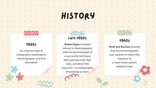
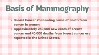
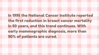
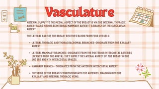
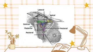
Breast
Breast
Anatomy
Anatomy
& Imaging
& Imaging
Techniques
Techniques
Soft Tissue Imaging
Mammography
Mammography
radiographic examination of the breast.
X-ray mammography has proved to be an accurate and simple method of
detecting breast cancer, but it is not simple to perform.
History
1920s
As a distinct type of
radiographic examination,
mammography was first
attempted.
late 1950s
Robert Egan renewed
interest in mammography
with his demonstration of
a successful technique
that used low kVp, high
mAs, and direct film
exposure—no radiographic
intensifying screens.
1960s
Wolf and Ruzicka showed
that xeromammography
was superior to direct film
exposure at
a much lower patient
radiation dose.
History
1992
the US government
mandated regulations
in the Mammography
Quality Standards Act
(MQSA).
1990
Xeromammography was
retired because single
screen-film mammography
provided better images at
even lower patient
radiation dose.
Breast Cancer 2nd leading cause of death from
cancer in women
Approximately 260,000 new cases of breast
cancer and 40,000 deaths from breast cancer are
reported in the United States.
Basis of Mammography
In 1995 the National Cancer Institute reported
the first reduction in breast cancer mortality
in 50 years, and this trend continues. With
early mammographic diagnosis, more than
90% of patients are cured.
Chapter 23, Box 23-1, Bushong 10th Ed., Page 375
50-69 y.o.: mortality reduction 16-35%
40-49 y.o.: mortality reduction 15-20%
–Lower incidence
–Rapidly growing tumors
–Dense breasts
Mortality Reduction
Screening
mammography
It is performed on
asymptomatic women with
the use of a two-view
protocol, usually medial
lateral oblique and cranial
caudad, to detect an
unsuspected cancer.
Screening mammography in
patients 50 years or older
reduces cancer mortality.
Types of
Types of
Mammography
Mammography
Sensitivity in women > 50 y.o.
98%fatty breast
84% dense breasts
Specificity
82-98%
Diagnostic Accuracy of
Screening Mammography
Diagnostic
mammography
It is performed on patients
with symptoms or elevated
risk factors. Two or three
views of each breast
may be required.
Types of
Types of
Mammography
Mammography
Surface Anatomy
Surface Anatomy
The breast is located on the anterior thoracic wall. It extends horizontally from
the lateral border of the sternum to the midaxillary line. Vertically, it spans
between the 2nd and 6th costal cartilages. It lies superficially to the pectoralis
major and serratus anterior muscles.
The breast can be considered to be composed of two regions:
Circular body –largest and most prominent part of the breast.
Axillary tail-smaller part, runs along the inferior lateral edge of the
pectoralis major towards the axillary fossa.
Surface Anatomy
Surface Anatomy
At the centr of the breast is the nipple, composed mostly of smooth muscle fibers.
Surrounding the nipple is a pigmented area of skin termed the AREOLAE. There are
numerous sebaceous glands within the areolae–the seen large during pregnancy,
secreting an oily substance that acts as a protective lubricant for the nipple.
Chapter 23, Fig. 23-1, Bushong 10th Ed., Page 376
Modified sweat glands. They consist of a series of
ducts and secretory lobules (15-20).
Each lobule consists of many alveoli drained by a
single lactiferous duct. These ducts converge at
the nipple like spokes of a wheel.
Mammary Gland
The connective tissue stromais a supporting structure which
surrounds the mammary glands. It has a fibrous and a fatty
component.
The fibrous stroma condenses to form suspensory ligaments
(of Cooper). These ligaments have two main functions:
Attach and secure the breast to the dermis and
underlying pectoral fascia.
Separate the secretory lobules of the breast.
Connective Tissue Stroma
The base of the breast lies on the pectoral fascia –a flat
sheet of connective tissue associated with the pectoralis
major muscle. It acts as an attachment point for the
suspensory ligaments.
There is a layer of loose connective tissue between the
breast and pectoral fascia–known as the retromammary
space. This is a potential space, often used in reconstructive
plastic surgery.
Pectoral Fascia
Vasculature
Vasculature
Arterial supply to the medial aspect of the breast is via the internal thoracic
artery (also known as internal mammary artery)–a branch of the subclavian
artery.
The lateral part of the breast receives blood from four vessels:
Lateral thoracic and thoracoacromial branches–originate from the axillary
artery.
Lateral mammary branches–originate from the posterior intercostal arteries
(derived from the aorta). They supply the lateral aspect of the breast in the
2nd 3rd and 4th intercostal spaces.
Mammary branch– originates from the anterior intercostal artery.
The veins of the breast correspond with the arteries, draining into the
axillary and internal thoracic veins.
Lymphatics
Lymphatics
The lymphatic drainage of the breast is of great clinical importance due to its
role in the metastasis of breast cancer cells.
There are three groups of lymph nodes that receive lymph from breast tissue–the
axillary nodes (75%), parasternal nodes (20%) and posterior intercostal nodes
(5%).
The skin of the breast also receives lymphatic drainage:
Skin–drains to the axillary, inferior deep cervical and infraclavicular nodes.
Nipple and areola–drains to the subareolar lymphatic plexus.
Nerve Supply
The breast is innervated by the anterior and lateral cutaneous branches of the
4th to 6th inter costal nerves. These nerves contain both sensory and
autonomic nerve fibres (the autonomic fibres regulate smooth muscle and
blood vessel tone).
It should be noted that these nerves do not control the production and secretion
of milk. This is regulated by the hormones prolactin and oxytocin, which are
secreted from the pituitary gland.
PHYSIOLOGY
BREASTS ARE EXOCRINE
GLANDS WHOSE
PRIMARY FUNCTION IS
LACTATION DURING
PREGNANCY
Physiology
Amastia—complete absence of one or both
breasts.
Athelia—complete absence of the nipple.
Amazia—absence of the breast tissue with
presence of the nipple.
Nipple inversion—nipple inverts inward.
Polymastia—accessory or supernumerary
breast.
Polythelia—accessory nipple; most common
breast anomaly
Congenital Breast Anomalies

Mammography: Breast Anatomy & Imaging Techniques.pdf

  • 1.
  • 2.
    Mammography Mammography radiographic examination ofthe breast. X-ray mammography has proved to be an accurate and simple method of detecting breast cancer, but it is not simple to perform.
  • 3.
    History 1920s As a distincttype of radiographic examination, mammography was first attempted. late 1950s Robert Egan renewed interest in mammography with his demonstration of a successful technique that used low kVp, high mAs, and direct film exposure—no radiographic intensifying screens. 1960s Wolf and Ruzicka showed that xeromammography was superior to direct film exposure at a much lower patient radiation dose.
  • 4.
    History 1992 the US government mandatedregulations in the Mammography Quality Standards Act (MQSA). 1990 Xeromammography was retired because single screen-film mammography provided better images at even lower patient radiation dose.
  • 5.
    Breast Cancer 2ndleading cause of death from cancer in women Approximately 260,000 new cases of breast cancer and 40,000 deaths from breast cancer are reported in the United States. Basis of Mammography
  • 6.
    In 1995 theNational Cancer Institute reported the first reduction in breast cancer mortality in 50 years, and this trend continues. With early mammographic diagnosis, more than 90% of patients are cured.
  • 7.
    Chapter 23, Box23-1, Bushong 10th Ed., Page 375
  • 8.
    50-69 y.o.: mortalityreduction 16-35% 40-49 y.o.: mortality reduction 15-20% –Lower incidence –Rapidly growing tumors –Dense breasts Mortality Reduction
  • 9.
    Screening mammography It is performedon asymptomatic women with the use of a two-view protocol, usually medial lateral oblique and cranial caudad, to detect an unsuspected cancer. Screening mammography in patients 50 years or older reduces cancer mortality. Types of Types of Mammography Mammography
  • 11.
    Sensitivity in women> 50 y.o. 98%fatty breast 84% dense breasts Specificity 82-98% Diagnostic Accuracy of Screening Mammography
  • 12.
    Diagnostic mammography It is performedon patients with symptoms or elevated risk factors. Two or three views of each breast may be required. Types of Types of Mammography Mammography
  • 13.
    Surface Anatomy Surface Anatomy Thebreast is located on the anterior thoracic wall. It extends horizontally from the lateral border of the sternum to the midaxillary line. Vertically, it spans between the 2nd and 6th costal cartilages. It lies superficially to the pectoralis major and serratus anterior muscles. The breast can be considered to be composed of two regions: Circular body –largest and most prominent part of the breast. Axillary tail-smaller part, runs along the inferior lateral edge of the pectoralis major towards the axillary fossa.
  • 14.
    Surface Anatomy Surface Anatomy Atthe centr of the breast is the nipple, composed mostly of smooth muscle fibers. Surrounding the nipple is a pigmented area of skin termed the AREOLAE. There are numerous sebaceous glands within the areolae–the seen large during pregnancy, secreting an oily substance that acts as a protective lubricant for the nipple.
  • 16.
    Chapter 23, Fig.23-1, Bushong 10th Ed., Page 376
  • 17.
    Modified sweat glands.They consist of a series of ducts and secretory lobules (15-20). Each lobule consists of many alveoli drained by a single lactiferous duct. These ducts converge at the nipple like spokes of a wheel. Mammary Gland
  • 18.
    The connective tissuestromais a supporting structure which surrounds the mammary glands. It has a fibrous and a fatty component. The fibrous stroma condenses to form suspensory ligaments (of Cooper). These ligaments have two main functions: Attach and secure the breast to the dermis and underlying pectoral fascia. Separate the secretory lobules of the breast. Connective Tissue Stroma
  • 19.
    The base ofthe breast lies on the pectoral fascia –a flat sheet of connective tissue associated with the pectoralis major muscle. It acts as an attachment point for the suspensory ligaments. There is a layer of loose connective tissue between the breast and pectoral fascia–known as the retromammary space. This is a potential space, often used in reconstructive plastic surgery. Pectoral Fascia
  • 21.
    Vasculature Vasculature Arterial supply tothe medial aspect of the breast is via the internal thoracic artery (also known as internal mammary artery)–a branch of the subclavian artery. The lateral part of the breast receives blood from four vessels: Lateral thoracic and thoracoacromial branches–originate from the axillary artery. Lateral mammary branches–originate from the posterior intercostal arteries (derived from the aorta). They supply the lateral aspect of the breast in the 2nd 3rd and 4th intercostal spaces. Mammary branch– originates from the anterior intercostal artery. The veins of the breast correspond with the arteries, draining into the axillary and internal thoracic veins.
  • 22.
    Lymphatics Lymphatics The lymphatic drainageof the breast is of great clinical importance due to its role in the metastasis of breast cancer cells. There are three groups of lymph nodes that receive lymph from breast tissue–the axillary nodes (75%), parasternal nodes (20%) and posterior intercostal nodes (5%). The skin of the breast also receives lymphatic drainage: Skin–drains to the axillary, inferior deep cervical and infraclavicular nodes. Nipple and areola–drains to the subareolar lymphatic plexus.
  • 24.
    Nerve Supply The breastis innervated by the anterior and lateral cutaneous branches of the 4th to 6th inter costal nerves. These nerves contain both sensory and autonomic nerve fibres (the autonomic fibres regulate smooth muscle and blood vessel tone). It should be noted that these nerves do not control the production and secretion of milk. This is regulated by the hormones prolactin and oxytocin, which are secreted from the pituitary gland.
  • 26.
    PHYSIOLOGY BREASTS ARE EXOCRINE GLANDSWHOSE PRIMARY FUNCTION IS LACTATION DURING PREGNANCY
  • 27.
    Physiology Amastia—complete absence ofone or both breasts. Athelia—complete absence of the nipple. Amazia—absence of the breast tissue with presence of the nipple. Nipple inversion—nipple inverts inward. Polymastia—accessory or supernumerary breast. Polythelia—accessory nipple; most common breast anomaly
  • 28.