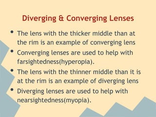
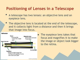
Thin Lenses
By: Emily Earls
T’Aundrai Smith
Objectives

•
•

Compare diverging and converging lenses
Relate concepts to eyeglasses and contact
lenses

•

Describe the positioning of lenses in
compound microscopes and telescopes
Introductory
At the end of this lesson, students should be
able to tell the difference between converging
and diverging lenses. Understand the purpose
in eyeglasses and contact lenses and what is
different about them. Also, know what the
lenses are for in compound microscopes and
telescopes.
Diverging & Converging Lenses

•
•

•
•

The lens with the thicker middle than at
the rim is an example of converging lens
Converging lenses are used to help with
farsightedness(hyperopia).

The lens with the thinner middle than it is
at the rim is an example of diverging lens
Diverging lenses are used to help with
nearsightedness(myopia).
Ray Diagrams

•
•

•

Ray diagrams for concave lenses inside and
outside the focal point give similar results; an
erect virtual image smaller than the object
Ray diagrams for convex lenses outside the
focal point give similar results; a real
inverted image
Ray diagrams inside the focal point gives a
virtual erect image that will be formed
•

Real inverted image

•

Virtual erect image
Positioning of Lenses in a Compound
Microscope

•
•

A compound microscope has two types of
lenses. It has objective lenses and an ocular
lens.
The objective lenses are located in the
rotating nosepiece, near the object being
magnified.

Objective Lens

•
Ocular Lens

The ocular lens is located in
the part you look through,
which magnifies the image.
Positioning of Lenses in a Telescope

•
•

A telescope has two lenses: an objective lens and an
eyepiece lens.
The objective lens is located at the end of the telescope,
and it collects light from a distance and then it brings
that image into focus.

● The eyepiece lens takes that
focus and magnifies it to make
the image or object look bigger
to the retina.
Eyeglasses vs. Contact Lenses

•

Eyeglasses are a frame that holds two pieces of glass

or plastic, which have been ground into lenses to
correct refractive errors

•

•

Contact lenses are worn directly on the cornea of the
eye
Eyeglasses and Contacts help with sight by adding or
subtracting focusing power to the eye’s cornea and lens
Bibliography
•
•
•
•

•

Craig , F. (2011, March 9). How Telescopes Work. Retrieved October 24,
2013, from http://science.howstuffworks.com/telescope1.htm

Microbus. (2007). The Microscope. Retrieved October 23, 2013, from
http://www.microscope-microscope.org/basic/microscope-parts.htm

Nave, C. R. (n.d.). Ray Diagrams for Lenses. Ray Diagrams for Lenses.
Retrieved October 28, 2013, from http://hyperphysics.phyastr.gsu.edu/hbase/geoopt/raydiag.html

University, O. S. (n.d.). Eye Glasses and Contact Lenses. Eye Glasses
and Contact Lenses. Retrieved October 23, 2013, from
http://medicalcenter.osu.edu/patientcare/healthcare_services/eye_care/gla
sses_contact_lenses/Pages/index.aspx

Thin lenses

  • 1.
    Thin Lenses By: EmilyEarls T’Aundrai Smith
  • 2.
    Objectives • • Compare diverging andconverging lenses Relate concepts to eyeglasses and contact lenses • Describe the positioning of lenses in compound microscopes and telescopes
  • 3.
    Introductory At the endof this lesson, students should be able to tell the difference between converging and diverging lenses. Understand the purpose in eyeglasses and contact lenses and what is different about them. Also, know what the lenses are for in compound microscopes and telescopes.
  • 4.
    Diverging & ConvergingLenses • • • • The lens with the thicker middle than at the rim is an example of converging lens Converging lenses are used to help with farsightedness(hyperopia). The lens with the thinner middle than it is at the rim is an example of diverging lens Diverging lenses are used to help with nearsightedness(myopia).
  • 6.
    Ray Diagrams • • • Ray diagramsfor concave lenses inside and outside the focal point give similar results; an erect virtual image smaller than the object Ray diagrams for convex lenses outside the focal point give similar results; a real inverted image Ray diagrams inside the focal point gives a virtual erect image that will be formed
  • 7.
  • 8.
    Positioning of Lensesin a Compound Microscope • • A compound microscope has two types of lenses. It has objective lenses and an ocular lens. The objective lenses are located in the rotating nosepiece, near the object being magnified. Objective Lens • Ocular Lens The ocular lens is located in the part you look through, which magnifies the image.
  • 9.
    Positioning of Lensesin a Telescope • • A telescope has two lenses: an objective lens and an eyepiece lens. The objective lens is located at the end of the telescope, and it collects light from a distance and then it brings that image into focus. ● The eyepiece lens takes that focus and magnifies it to make the image or object look bigger to the retina.
  • 10.
    Eyeglasses vs. ContactLenses • Eyeglasses are a frame that holds two pieces of glass or plastic, which have been ground into lenses to correct refractive errors • • Contact lenses are worn directly on the cornea of the eye Eyeglasses and Contacts help with sight by adding or subtracting focusing power to the eye’s cornea and lens
  • 11.
    Bibliography • • • • • Craig , F.(2011, March 9). How Telescopes Work. Retrieved October 24, 2013, from http://science.howstuffworks.com/telescope1.htm Microbus. (2007). The Microscope. Retrieved October 23, 2013, from http://www.microscope-microscope.org/basic/microscope-parts.htm Nave, C. R. (n.d.). Ray Diagrams for Lenses. Ray Diagrams for Lenses. Retrieved October 28, 2013, from http://hyperphysics.phyastr.gsu.edu/hbase/geoopt/raydiag.html University, O. S. (n.d.). Eye Glasses and Contact Lenses. Eye Glasses and Contact Lenses. Retrieved October 23, 2013, from http://medicalcenter.osu.edu/patientcare/healthcare_services/eye_care/gla sses_contact_lenses/Pages/index.aspx