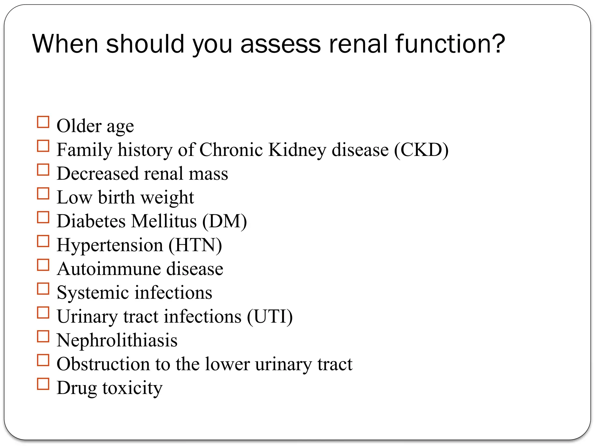
KIDNEY (RENAL) FUNCTIONS TESTS
RENAL CLEARENCE TESTS
When should you assess renal function?
 Older age
 Family history of Chronic Kidney disease (CKD)
 Decreased renal mass
 Low birth weight
 Diabetes Mellitus (DM)
 Hypertension (HTN)
 Autoimmune disease
 Systemic infections
 Urinary tract infections (UTI)
 Nephrolithiasis
 Obstruction to the lower urinary tract
 Drug toxicity
Biochemical Tests of Renal Function
 Measurement of GFR
 Clearance tests
 Plasma creatinine
 Urea, uric acid and β2-microglobulin
 Renal tubular function tests
 Osmolality measurements
 Specific proteinurea
 Glycosuria
 Aminoaciduria
 Urinalysis
 Appearance
 Specific gravity and osmolality
 pH
 osmolality
 Glucose
 Protein
 Urinary sediments
In acute and chronic renal failure, there is effectively a loss of function of
whole nephrons
 Filtration is essential to the formation of urine  tests of glomerular
function are almost always required in the investigation and
management of any patient with renal disease.
The most frequently used tests are those that assess either the GFR
or the integrity of the glomerular filtration barrier.
Biochemical Tests of renal function
GFR can be estimated by measuring the urinary excretion of a substance that is completely filtered from
the blood by the glomeruli and it is not secreted, reabsorbed or metabolized by the renal
tubules.
 Clearance is defined as the (hypothetical) quantity of blood or plasma completely cleared of a substance per
unit of time.
 Clearance of substances that are filtered exclusively or predominantly by the glomeruli but
neither reabsorbed nor secreted by other regions of the nephron can be used to measure GFR.
TheVolume of blood from which inulin is cleared or completely removed in one minute is known as the
inulin clearance and is equal to the GFR.
Measurement of inulin clearance requires the infusion of inulin into the blood and is not suitable for
routine clinical use
GFR =
(U V)
P
inulin
inulin

V is not urine volume, it is urine flow rate
Measurement of glomerular filtration rate
Glomerular filtration rate
1 to 2% of muscle creatine spontaneously converts to creatinine daily
and released into body fluids at a constant rate.
Endogenous creatinine produced is proportional to muscle mass, it is a
function of total muscle mass the production varies with age and sex
 Dietary fluctuations of creatinine intake cause only minor variation in
daily creatinine excretion of the same person.
 Creatinine released into body fluids at a constant rate and its plasma
levels maintained within narrow limits  Creatinine clearance may be
measured as an indicator of GFR.
Creatinine
An estimate of the GFR can be calculated from the creatinine content of a 24-hour urine collection, and
the plasma concentration within this period.
The volume of urine is measured, urine flow rate is calculated (ml/min) and the assay for creatinine is
performed on plasma and urine to obtain the concentration in mg per dl or per ml.
Creatinine clearance in adults is normally about of 120 ml/min,
The accurate measurement of creatinine clearance is difficult, especially in outpatients, since it is necessary
to obtain a complete and accurately timed sample of urine
Creatinine clearance and clinical utility
The most frequently used clearance test is based on the measurement of
creatinine.
 Small quantity of creatinine is reabsorbed by the tubules and other
quantities are actively secreted by the renal tubules  So creatinine
clearance is approximately 7% greater than inulin clearance.
The difference is not significant when GFR is normal but when the GFR
is low (less 10 ml/min), tubular secretion makes the major contribution
to creatinine excretion and the creatinine clearance significantly
overestimates the GFR.
Creatinine clearance and clinical utility
The 'clearance' of creatinine from plasma is directly related to the GFR if:
The urine volume is collected accurately
There are no ketones or heavy proteinuria present to interfere with the
creatinine determination.
It should be noted that the GFR decline with age (to a greater extent in males
than in females) and this must be taken into account when interpreting results.
Creatinine clearance and clinical utility
 Catabolism of proteins and nucleic acids results in formation of so
called nonprotein nitrogenous compounds.
 Urea , creatinine and uric acid
 In kidney dysfunction, the levels of these compounds are elevated in
plasma
Measurement of nonprotein nitrogen-containing compounds
Urea is the major nitrogen-containing metabolic product of protein catabolism in
humans,
 Its elimination in the urine represents the major route for nitrogen excretion.
 More than 90% of urea is excreted through the kidneys, with losses through the
GIT and skin
 Urea is filtered freely by the glomeruli
 Plasma urea concentration is often used as an index of renal glomerular function
 Urea production is increased by a high protein intake and it is decreased in
patients with a low protein intake or in patients with liver disease.
Plasma Urea
Many renal diseases with various glomerular, tubular, interstitial or vascular damage can cause an
increase in plasma urea concentration.
The reference interval for serum urea of healthy adults is 15-39 mg/dl.
 Plasma concentrations also tend to be slightly higher in males than females. High protein diet causes
significant increases in plasma urea concentrations and urinary excretion.
Measurement of plasma creatinine provides a more accurate assessment than urea because there are
many factors that affect urea level.
Nonrenal factors can affect the urea level (normal adults is level 15-39 mg/dl) like:
Mild dehydration,
high protein diet,
increased protein catabolism, muscle wasting as in starvation,
reabsorption of blood proteins after a GIT haemorrhage,
treatment with cortisol or its synthetic analogous
Plasma Urea
Renal handling of uric acid is complex and involves four sequential steps:
Glomerular filtration of virtually all the uric acid in capillary plasma entering the
glomerulus.
Reabsorption in the proximal convoluted tubule of about 98 to 100% of filtered uric
acid.
Subsequent secretion of uric acid into the lumen of the distal portion of the proximal
tubule.
Further reabsorption in the distal tubule.
 Hyperuricemia is defined by serum or plasma uric acid concentrations higher than 7.0 mg/dl
(0.42mmol/L) in men or greater than 6.0 mg/dl (0.36mmol/L) in women
Uric acid
 2-microglobulin is a small peptide (molecular weight 11.8 kDa),
β
It is present on the surface of most cells and in low concentrations in the plasma.
It is completely filtered by the glomeruli and is reabsorbed and catabolized by
proximal tubular cells.
The plasma concentration of 2-microglobulin is a good index of GFR in normal
β
people, being unaffected by diet or muscle mass.
It is increased in certain malignancies and inflammatory diseases.
Since it is normally reabsorbed and catabolized in the tubules, measurement of β2-
microglobulin excretion provides a sensitive method of assessing tubular integrity.
Plasma β2-microglobulin
Biochemical Tests of Renal Function
 Renal tubular function tests
Osmolality measurements
Specific proteinurea
Glycosuria
Aminoaciduria
Renal tubular function tests
• To ensure that important constituents such as water, sodium, glucose and a.a. are not lost
from the body, tubular reabsorption must be equally efficient
• Compared with the GFR as an assessment of glomerualr function, there are no easily
performed tests which measure tubular function in quantitative manner
• Investigation of tubular function:
1. Osmolality measurements in plasma and urine; normal urine: plasma osmolality ratio is
usually between 1.0-3.0
2. Specific proteinuria
3. Glycosuria
4. Aminoaciduria
The glomerular basement membrane does not usually allow passage of albumin and
large proteins.A small amount of albumin, usually less than 25 mg/24 hours, is found
in urine.
Urinary protein excretion in the normal adult should be less than 150
mg/day.
When larger amounts, in excess of 250 mg/24 hours, are detected, significant
damage to the glomerular membrane has occurred.
Quantitative urine protein measurements should always be made on complete 24-
hour urine collections.
Albumin excretion in the range 25-300 mg/24 hours is termed microalbuminuria
Proteinuria
 TYPES OF PROTEINURIA
 Glomerular proteinuria
 Tubular proteinuria
 Overflow proteinuria
Proteinuria
Glomerular proteinuria
 Glomerular proteinuria — Glomerular proteinuria is due to
increased filtration of macromolecules (such as albumin) across
the glomerular capillary wall.The proteinuria associated with
diabetic nephropathy and other glomerular diseases, as well as
more benign causes such as orthostatic or exercise-induced
proteinuria fall into this category. Most patients with benign
causes of isolated proteinuria excrete less than 1 to 2 g/day
Tubular proteinuria
 Low molecular weight proteins — such as ß2-microglobulin,
immunoglobulin light chains, retinol-binding protein, and
amino acids — have a molecular weight that is generally under
25,000 in comparison to the 69,000 molecular weight of
albumin.These smaller proteins can be filtered across the
glomerulus and are then almost completely reabsorbed in the
proximal tubule. Interference with proximal tubular
reabsorption, due to a variety of tubulointerstitial diseases or
even some primary glomerular diseases, can lead to increased
excretion of these smaller proteins
Overflow proteinuria
 Increased excretion of low molecular weight proteins can occur
with marked overproduction of a particular protein, leading to
increased glomerular filtration and excretion.This is almost
always due to immunoglobulin light chains in multiple myeloma,
but may also be due to lysozyme (in acute myelomonocytic
leukemia), myoglobin (in rhabdomyolysis), or hemoglobin (in
intravascular hemolysis
Biochemical Tests of Renal Function
 Urinalysis
 Appearance
 Specific gravity and osmolality
 pH
 Glucose
 Protein
 Urinary sediments
Biochemical testing of urine involves the use of commercially available disposable strips When the
strip is manually immersed in the urine specimen, the reagents react with a specific component of urine
in such a way that to form color
 Colour change produced is proportional to the concentration of the component being tested for.
To test a urine sample:
fresh urine is collected into a clean dry container
the sample is not centrifuged
 the disposable strip is briefly immersed in the urine specimen;
The colour of the test areas are compared with those provided on a colour chart
Urinalysis using disposable strips
polyurea
Increased osmotic load, e.g due to glucose
Increased water ingestion
Diabetes insipidus: - Failure of ADH production results in
marked polyuria (diabetes insipidus), which stimulates thirst
and greatly increases water intake
Nephrogenic diabetes insipidus: The kidneys’ lack of
response to ADH has similar effect ( failure of the tubules to
respond to Vassopressin (ADH
Nephrotic syndrome
 The nephrotic syndrome is caused by renal diseases that increase the
permeability across the glomerular filtration barrier. It is classically
characterized by four clinical features, but the first two are used
diagnostically because the last two may not be seen in all patients:
 Nephrotic range proteinuria — Urinary protein excretion greater than 50
mg/kg per day
 Hypoalbuminemia — Serum albumin concentration less than 3 g/dL (30 g/L)
 Edema
 Hyperlipidemia
SODIUM 135 to 145 mEq/L
POTASSIUM 3.5 to 5.5 mEq/L
CHLORIDES 100 to 110 mEq/L
BICARBONATE 24 to 26 mEq/L
CALCIUM 8.6 to 10 mgs/dl
MAGNESIUM 1.6 to 2.4 mgs/dl
PHOSPHORUS 3.0 to 5.0 mgs/dl
URIC ACID 2.5 to 6.0 mgs/dl
pH 7.4
CREATININE 0.8 to 1.4 mgs/dl
Normal values of Internal Chemical
Environmentcontrolled by the Kidneys
:
15 to 20 mgs/dl
BUN (Blood Urea Nitrogen)

KIDNEY (RENAL) FUNCTIONS TESTS-PPT =anna.pptx

  • 1.
    KIDNEY (RENAL) FUNCTIONSTESTS RENAL CLEARENCE TESTS
  • 2.
    When should youassess renal function?  Older age  Family history of Chronic Kidney disease (CKD)  Decreased renal mass  Low birth weight  Diabetes Mellitus (DM)  Hypertension (HTN)  Autoimmune disease  Systemic infections  Urinary tract infections (UTI)  Nephrolithiasis  Obstruction to the lower urinary tract  Drug toxicity
  • 3.
    Biochemical Tests ofRenal Function  Measurement of GFR  Clearance tests  Plasma creatinine  Urea, uric acid and β2-microglobulin  Renal tubular function tests  Osmolality measurements  Specific proteinurea  Glycosuria  Aminoaciduria  Urinalysis  Appearance  Specific gravity and osmolality  pH  osmolality  Glucose  Protein  Urinary sediments
  • 4.
    In acute andchronic renal failure, there is effectively a loss of function of whole nephrons  Filtration is essential to the formation of urine  tests of glomerular function are almost always required in the investigation and management of any patient with renal disease. The most frequently used tests are those that assess either the GFR or the integrity of the glomerular filtration barrier. Biochemical Tests of renal function
  • 5.
    GFR can beestimated by measuring the urinary excretion of a substance that is completely filtered from the blood by the glomeruli and it is not secreted, reabsorbed or metabolized by the renal tubules.  Clearance is defined as the (hypothetical) quantity of blood or plasma completely cleared of a substance per unit of time.  Clearance of substances that are filtered exclusively or predominantly by the glomeruli but neither reabsorbed nor secreted by other regions of the nephron can be used to measure GFR. TheVolume of blood from which inulin is cleared or completely removed in one minute is known as the inulin clearance and is equal to the GFR. Measurement of inulin clearance requires the infusion of inulin into the blood and is not suitable for routine clinical use GFR = (U V) P inulin inulin  V is not urine volume, it is urine flow rate Measurement of glomerular filtration rate
  • 6.
  • 7.
    1 to 2%of muscle creatine spontaneously converts to creatinine daily and released into body fluids at a constant rate. Endogenous creatinine produced is proportional to muscle mass, it is a function of total muscle mass the production varies with age and sex  Dietary fluctuations of creatinine intake cause only minor variation in daily creatinine excretion of the same person.  Creatinine released into body fluids at a constant rate and its plasma levels maintained within narrow limits  Creatinine clearance may be measured as an indicator of GFR. Creatinine
  • 8.
    An estimate ofthe GFR can be calculated from the creatinine content of a 24-hour urine collection, and the plasma concentration within this period. The volume of urine is measured, urine flow rate is calculated (ml/min) and the assay for creatinine is performed on plasma and urine to obtain the concentration in mg per dl or per ml. Creatinine clearance in adults is normally about of 120 ml/min, The accurate measurement of creatinine clearance is difficult, especially in outpatients, since it is necessary to obtain a complete and accurately timed sample of urine Creatinine clearance and clinical utility
  • 9.
    The most frequentlyused clearance test is based on the measurement of creatinine.  Small quantity of creatinine is reabsorbed by the tubules and other quantities are actively secreted by the renal tubules  So creatinine clearance is approximately 7% greater than inulin clearance. The difference is not significant when GFR is normal but when the GFR is low (less 10 ml/min), tubular secretion makes the major contribution to creatinine excretion and the creatinine clearance significantly overestimates the GFR. Creatinine clearance and clinical utility
  • 10.
    The 'clearance' ofcreatinine from plasma is directly related to the GFR if: The urine volume is collected accurately There are no ketones or heavy proteinuria present to interfere with the creatinine determination. It should be noted that the GFR decline with age (to a greater extent in males than in females) and this must be taken into account when interpreting results. Creatinine clearance and clinical utility
  • 11.
     Catabolism ofproteins and nucleic acids results in formation of so called nonprotein nitrogenous compounds.  Urea , creatinine and uric acid  In kidney dysfunction, the levels of these compounds are elevated in plasma Measurement of nonprotein nitrogen-containing compounds
  • 12.
    Urea is themajor nitrogen-containing metabolic product of protein catabolism in humans,  Its elimination in the urine represents the major route for nitrogen excretion.  More than 90% of urea is excreted through the kidneys, with losses through the GIT and skin  Urea is filtered freely by the glomeruli  Plasma urea concentration is often used as an index of renal glomerular function  Urea production is increased by a high protein intake and it is decreased in patients with a low protein intake or in patients with liver disease. Plasma Urea
  • 13.
    Many renal diseaseswith various glomerular, tubular, interstitial or vascular damage can cause an increase in plasma urea concentration. The reference interval for serum urea of healthy adults is 15-39 mg/dl.  Plasma concentrations also tend to be slightly higher in males than females. High protein diet causes significant increases in plasma urea concentrations and urinary excretion. Measurement of plasma creatinine provides a more accurate assessment than urea because there are many factors that affect urea level. Nonrenal factors can affect the urea level (normal adults is level 15-39 mg/dl) like: Mild dehydration, high protein diet, increased protein catabolism, muscle wasting as in starvation, reabsorption of blood proteins after a GIT haemorrhage, treatment with cortisol or its synthetic analogous Plasma Urea
  • 14.
    Renal handling ofuric acid is complex and involves four sequential steps: Glomerular filtration of virtually all the uric acid in capillary plasma entering the glomerulus. Reabsorption in the proximal convoluted tubule of about 98 to 100% of filtered uric acid. Subsequent secretion of uric acid into the lumen of the distal portion of the proximal tubule. Further reabsorption in the distal tubule.  Hyperuricemia is defined by serum or plasma uric acid concentrations higher than 7.0 mg/dl (0.42mmol/L) in men or greater than 6.0 mg/dl (0.36mmol/L) in women Uric acid
  • 15.
     2-microglobulin isa small peptide (molecular weight 11.8 kDa), β It is present on the surface of most cells and in low concentrations in the plasma. It is completely filtered by the glomeruli and is reabsorbed and catabolized by proximal tubular cells. The plasma concentration of 2-microglobulin is a good index of GFR in normal β people, being unaffected by diet or muscle mass. It is increased in certain malignancies and inflammatory diseases. Since it is normally reabsorbed and catabolized in the tubules, measurement of β2- microglobulin excretion provides a sensitive method of assessing tubular integrity. Plasma β2-microglobulin
  • 16.
    Biochemical Tests ofRenal Function  Renal tubular function tests Osmolality measurements Specific proteinurea Glycosuria Aminoaciduria
  • 17.
    Renal tubular functiontests • To ensure that important constituents such as water, sodium, glucose and a.a. are not lost from the body, tubular reabsorption must be equally efficient • Compared with the GFR as an assessment of glomerualr function, there are no easily performed tests which measure tubular function in quantitative manner • Investigation of tubular function: 1. Osmolality measurements in plasma and urine; normal urine: plasma osmolality ratio is usually between 1.0-3.0 2. Specific proteinuria 3. Glycosuria 4. Aminoaciduria
  • 18.
    The glomerular basementmembrane does not usually allow passage of albumin and large proteins.A small amount of albumin, usually less than 25 mg/24 hours, is found in urine. Urinary protein excretion in the normal adult should be less than 150 mg/day. When larger amounts, in excess of 250 mg/24 hours, are detected, significant damage to the glomerular membrane has occurred. Quantitative urine protein measurements should always be made on complete 24- hour urine collections. Albumin excretion in the range 25-300 mg/24 hours is termed microalbuminuria Proteinuria
  • 19.
     TYPES OFPROTEINURIA  Glomerular proteinuria  Tubular proteinuria  Overflow proteinuria Proteinuria
  • 20.
    Glomerular proteinuria  Glomerularproteinuria — Glomerular proteinuria is due to increased filtration of macromolecules (such as albumin) across the glomerular capillary wall.The proteinuria associated with diabetic nephropathy and other glomerular diseases, as well as more benign causes such as orthostatic or exercise-induced proteinuria fall into this category. Most patients with benign causes of isolated proteinuria excrete less than 1 to 2 g/day
  • 21.
    Tubular proteinuria  Lowmolecular weight proteins — such as ß2-microglobulin, immunoglobulin light chains, retinol-binding protein, and amino acids — have a molecular weight that is generally under 25,000 in comparison to the 69,000 molecular weight of albumin.These smaller proteins can be filtered across the glomerulus and are then almost completely reabsorbed in the proximal tubule. Interference with proximal tubular reabsorption, due to a variety of tubulointerstitial diseases or even some primary glomerular diseases, can lead to increased excretion of these smaller proteins
  • 22.
    Overflow proteinuria  Increasedexcretion of low molecular weight proteins can occur with marked overproduction of a particular protein, leading to increased glomerular filtration and excretion.This is almost always due to immunoglobulin light chains in multiple myeloma, but may also be due to lysozyme (in acute myelomonocytic leukemia), myoglobin (in rhabdomyolysis), or hemoglobin (in intravascular hemolysis
  • 23.
    Biochemical Tests ofRenal Function  Urinalysis  Appearance  Specific gravity and osmolality  pH  Glucose  Protein  Urinary sediments
  • 24.
    Biochemical testing ofurine involves the use of commercially available disposable strips When the strip is manually immersed in the urine specimen, the reagents react with a specific component of urine in such a way that to form color  Colour change produced is proportional to the concentration of the component being tested for. To test a urine sample: fresh urine is collected into a clean dry container the sample is not centrifuged  the disposable strip is briefly immersed in the urine specimen; The colour of the test areas are compared with those provided on a colour chart Urinalysis using disposable strips
  • 25.
    polyurea Increased osmotic load,e.g due to glucose Increased water ingestion Diabetes insipidus: - Failure of ADH production results in marked polyuria (diabetes insipidus), which stimulates thirst and greatly increases water intake Nephrogenic diabetes insipidus: The kidneys’ lack of response to ADH has similar effect ( failure of the tubules to respond to Vassopressin (ADH
  • 26.
    Nephrotic syndrome  Thenephrotic syndrome is caused by renal diseases that increase the permeability across the glomerular filtration barrier. It is classically characterized by four clinical features, but the first two are used diagnostically because the last two may not be seen in all patients:  Nephrotic range proteinuria — Urinary protein excretion greater than 50 mg/kg per day  Hypoalbuminemia — Serum albumin concentration less than 3 g/dL (30 g/L)  Edema  Hyperlipidemia
  • 27.
    SODIUM 135 to145 mEq/L POTASSIUM 3.5 to 5.5 mEq/L CHLORIDES 100 to 110 mEq/L BICARBONATE 24 to 26 mEq/L CALCIUM 8.6 to 10 mgs/dl MAGNESIUM 1.6 to 2.4 mgs/dl PHOSPHORUS 3.0 to 5.0 mgs/dl URIC ACID 2.5 to 6.0 mgs/dl pH 7.4 CREATININE 0.8 to 1.4 mgs/dl Normal values of Internal Chemical Environmentcontrolled by the Kidneys : 15 to 20 mgs/dl BUN (Blood Urea Nitrogen)

Editor's Notes

  • #3 Nephrolithiasis: The process of forming a kidney stone, a stone in the kidney