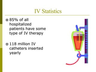
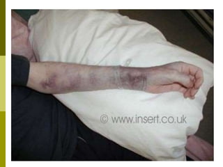
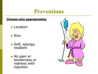
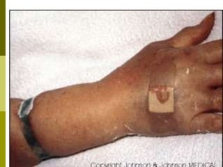
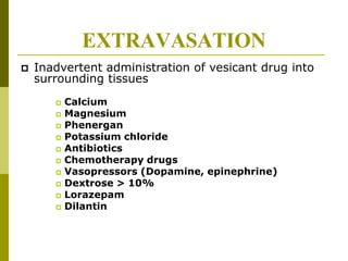
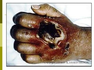
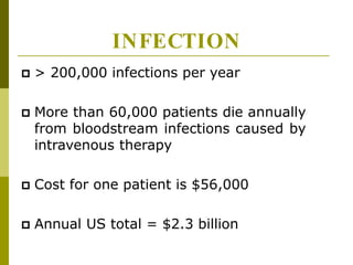
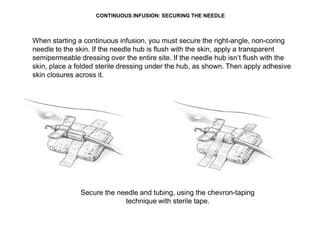
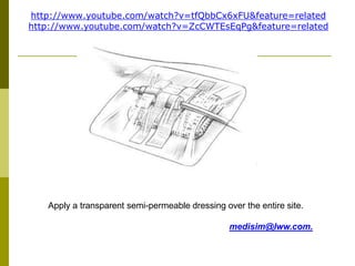
INTRAVENOU
S THERAPY
IV Statistics
 85% of all
hospitalized
patients have some
type of IV therapy
 118 million IV
catheters inserted
yearly
COMPLICATIONS
PHLEBITIS
 Inflammation of the vein
wall—precursor to sepsis
 What causes phlebitis?
 IV left in too long
 Cannula too large
 Vein in poor condition
 Acidic solution or high
osmolality
 Infusion rate too fast
Preventions
Choose vein appropriately
 Location
 Size
 Soft, spongy,
resilient
 No pain or
tenderness or
redness with
injection
INFILTRATION
 Leaking of nonvesicant fluid into
tissues surrounding the vein
 Check IV site every two hours
 Complications
 Nerve compression requiring
fasciotomy
EXTRAVASATION
 Inadvertent administration of vesicant drug into
surrounding tissues
 Calcium
 Magnesium
 Phenergan
 Potassium chloride
 Antibiotics
 Chemotherapy drugs
 Vasopressors (Dopamine, epinephrine)
 Dextrose > 10%
 Lorazepam
 Dilantin
INFECTION
 Cellulitis: An acute, spreading, bacterial
infection below the surface of the skin
characterized by redness (erythema),
warmth, swelling, and pain. Usually
localized.
 Sepsis: clinical symptoms of systemic
illness, such as fever, chills, malaise,
hypotension, and mental status changes.
Sepsis can be life threatening.
INFECTION
 > 200,000 infections per year
 More than 60,000 patients die annually
from bloodstream infections caused by
intravenous therapy
 Cost for one patient is $56,000
 Annual US total = $2.3 billion
Causes
 Poor insertion site
 Squad starts
 Unsterile start
 IV left in too long—
change q 96 hours!
 Hub contamination
Cellulitis
Prevention
 Hand washing
 Sterile technique
 Catheter size
 Insertion site
 Site inspection every two hours
 Encourage patient to report any
discomfort
Patient’s Worst Nightmare!!!!
Other sites to avoid include:
• veins below a previous I.V. infiltration
• veins below a phlebitic area
• sclerosed or thrombosed veins
• areas of skin inflammation, disease, bruising,
or breakdown
• an arm affected by a radical mastectomy,
edema, blood clot, or infection
• an arm with an arteriovenous shunt or fistula.
Muscle Man IV!
STARTING AN IV




Talk with patient
Gather equipment
Set up fluid and tubing on pump
Check patient order and ID band &
allergies
Wash your hands!!
Select a vein
Select a catheter size



1. Apply tourniquet 5-6 inches above insertion site
2. Never leave tourniquet on longer than one minute
3. Then Remove tourniquet and prepare equipment
STARTING AN IV (CONT.)



 Open equipment and connect flush to J-
loop
Loosen caps of IV and J-loop but leave in
place for sterility. (They should just slide off when
you pick up the device).
Cleanse skin with chlorhexidine gluconate
solution in back & forth motion X 30
seconds
Allow to dry for 30 seconds
1. Put on Gloves!!!


Immobilize vein
Position needle 10-15 degree angle over
site
Insert cannula bevel up
Watch for blood backflow
Advance cannula
Only try twice before calling another RN
to help




STARTING AN IV (CONT.)
Advance cannula while holding stylet
stationary
Release tourniquet!!
22. Withdraw stylet while putting pressure on
vein above injection site
Stabilize the hub of the canula
23. Insert tubing or prn adaptor
Apply pressure above
insertion site to slow
bleeding
Stabilize the hub of the canula
while inserting the tubing
Saline flush is already attached and
tubing flushed and ready
It may get messy sometimes, but with experience
this will be minimized
• Flush with saline to clear tubing and insure IV has not infiltrated.
3. Stabilize tubing with tape to prevent IV from pulling out while
applying the sterile dressing.
1. Apply clear sterile dressing. Cover site and hub, not tubing
Leave the end of the hub
of the canula outside the
dressing so that tubing
can be changed without
removing the dressing.
27. Date, time and initial site and tubing
STARTING AN IV (CONT.)
3. Document!
What is wrong
with this picture?
Dartmouth
Power Port
CONTINUOUS INFUSION: SECURING THE NEEDLE
When starting a continuous infusion, you must secure the right-angle, non-coring
needle to the skin. If the needle hub is flush with the skin, apply a transparent
semipermeable dressing over the entire site. If the needle hub isn’t flush with the
skin, place a folded sterile dressing under the hub, as shown. Then apply adhesive
skin closures across it.
Secure the needle and tubing, using the chevron-taping
technique with sterile tape.
Apply a transparent semi-permeable dressing over the entire site.
medisim@lww.com.
http://www.youtube.com/watch?v=tfQbbCx6xFU&feature=related
http://www.youtube.com/watch?v=ZcCWTEsEqPg&feature=related

IV Therapy.pptx

  • 1.
  • 2.
    IV Statistics  85%of all hospitalized patients have some type of IV therapy  118 million IV catheters inserted yearly
  • 3.
  • 4.
    PHLEBITIS  Inflammation ofthe vein wall—precursor to sepsis  What causes phlebitis?  IV left in too long  Cannula too large  Vein in poor condition  Acidic solution or high osmolality  Infusion rate too fast
  • 7.
    Preventions Choose vein appropriately Location  Size  Soft, spongy, resilient  No pain or tenderness or redness with injection
  • 8.
    INFILTRATION  Leaking ofnonvesicant fluid into tissues surrounding the vein  Check IV site every two hours  Complications  Nerve compression requiring fasciotomy
  • 10.
    EXTRAVASATION  Inadvertent administrationof vesicant drug into surrounding tissues  Calcium  Magnesium  Phenergan  Potassium chloride  Antibiotics  Chemotherapy drugs  Vasopressors (Dopamine, epinephrine)  Dextrose > 10%  Lorazepam  Dilantin
  • 12.
    INFECTION  Cellulitis: Anacute, spreading, bacterial infection below the surface of the skin characterized by redness (erythema), warmth, swelling, and pain. Usually localized.  Sepsis: clinical symptoms of systemic illness, such as fever, chills, malaise, hypotension, and mental status changes. Sepsis can be life threatening.
  • 13.
    INFECTION  > 200,000infections per year  More than 60,000 patients die annually from bloodstream infections caused by intravenous therapy  Cost for one patient is $56,000  Annual US total = $2.3 billion
  • 14.
    Causes  Poor insertionsite  Squad starts  Unsterile start  IV left in too long— change q 96 hours!  Hub contamination
  • 16.
  • 17.
    Prevention  Hand washing Sterile technique  Catheter size  Insertion site  Site inspection every two hours  Encourage patient to report any discomfort
  • 18.
  • 22.
    Other sites toavoid include: • veins below a previous I.V. infiltration • veins below a phlebitic area • sclerosed or thrombosed veins • areas of skin inflammation, disease, bruising, or breakdown • an arm affected by a radical mastectomy, edema, blood clot, or infection • an arm with an arteriovenous shunt or fistula.
  • 23.
  • 24.
    STARTING AN IV     Talkwith patient Gather equipment Set up fluid and tubing on pump Check patient order and ID band & allergies Wash your hands!! Select a vein Select a catheter size   
  • 25.
    1. Apply tourniquet5-6 inches above insertion site 2. Never leave tourniquet on longer than one minute 3. Then Remove tourniquet and prepare equipment
  • 26.
    STARTING AN IV(CONT.)     Open equipment and connect flush to J- loop Loosen caps of IV and J-loop but leave in place for sterility. (They should just slide off when you pick up the device). Cleanse skin with chlorhexidine gluconate solution in back & forth motion X 30 seconds Allow to dry for 30 seconds
  • 27.
    1. Put onGloves!!!
  • 28.
      Immobilize vein Position needle10-15 degree angle over site Insert cannula bevel up Watch for blood backflow Advance cannula Only try twice before calling another RN to help     STARTING AN IV (CONT.)
  • 33.
    Advance cannula whileholding stylet stationary Release tourniquet!!
  • 34.
    22. Withdraw styletwhile putting pressure on vein above injection site Stabilize the hub of the canula
  • 35.
    23. Insert tubingor prn adaptor Apply pressure above insertion site to slow bleeding Stabilize the hub of the canula while inserting the tubing Saline flush is already attached and tubing flushed and ready
  • 36.
    It may getmessy sometimes, but with experience this will be minimized
  • 37.
    • Flush withsaline to clear tubing and insure IV has not infiltrated. 3. Stabilize tubing with tape to prevent IV from pulling out while applying the sterile dressing.
  • 38.
    1. Apply clearsterile dressing. Cover site and hub, not tubing Leave the end of the hub of the canula outside the dressing so that tubing can be changed without removing the dressing.
  • 39.
    27. Date, timeand initial site and tubing
  • 40.
    STARTING AN IV(CONT.) 3. Document!
  • 41.
    What is wrong withthis picture?
  • 42.
  • 46.
  • 50.
    CONTINUOUS INFUSION: SECURINGTHE NEEDLE When starting a continuous infusion, you must secure the right-angle, non-coring needle to the skin. If the needle hub is flush with the skin, apply a transparent semipermeable dressing over the entire site. If the needle hub isn’t flush with the skin, place a folded sterile dressing under the hub, as shown. Then apply adhesive skin closures across it. Secure the needle and tubing, using the chevron-taping technique with sterile tape.
  • 51.
    Apply a transparentsemi-permeable dressing over the entire site. medisim@lww.com. http://www.youtube.com/watch?v=tfQbbCx6xFU&feature=related http://www.youtube.com/watch?v=ZcCWTEsEqPg&feature=related