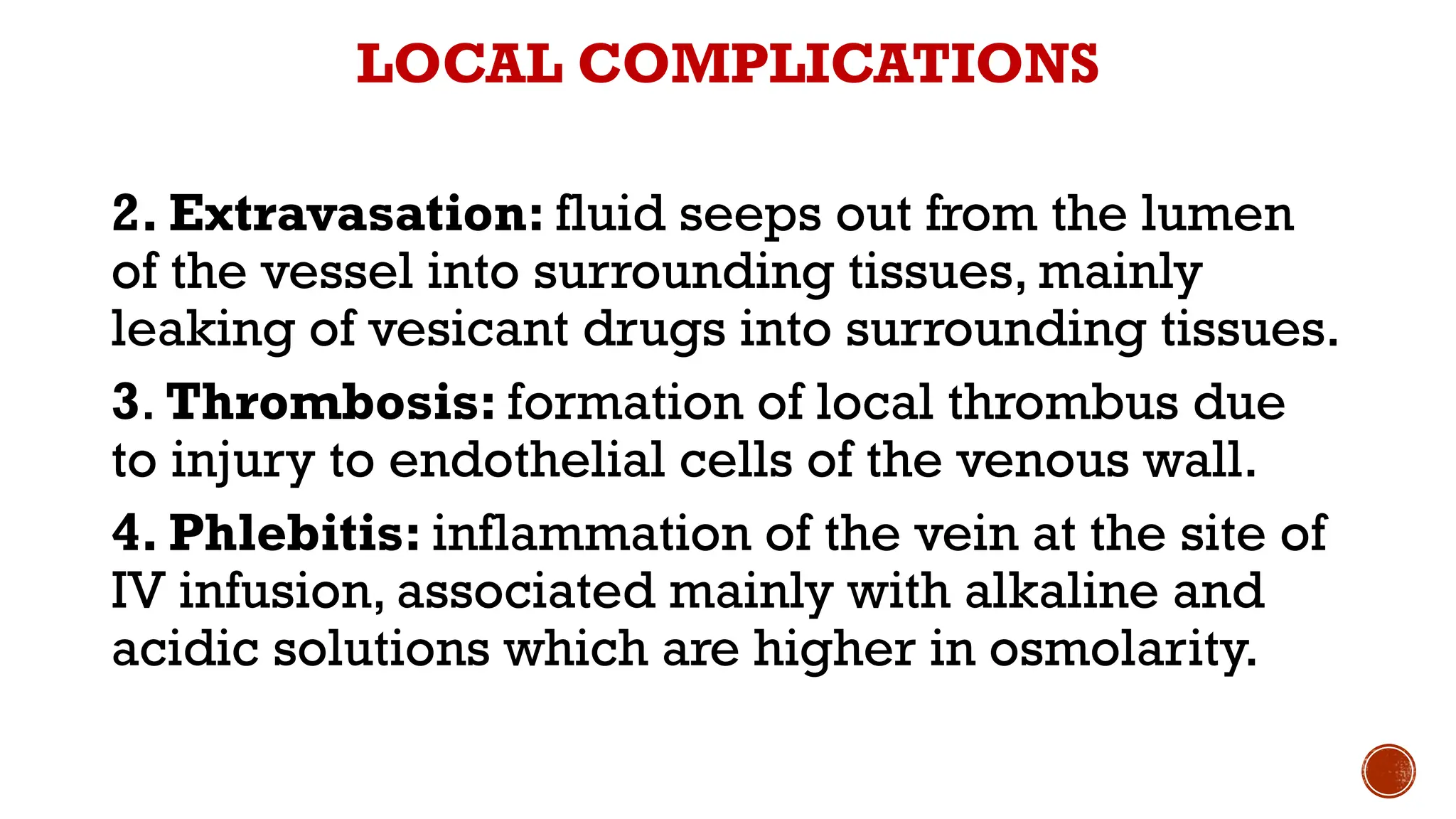
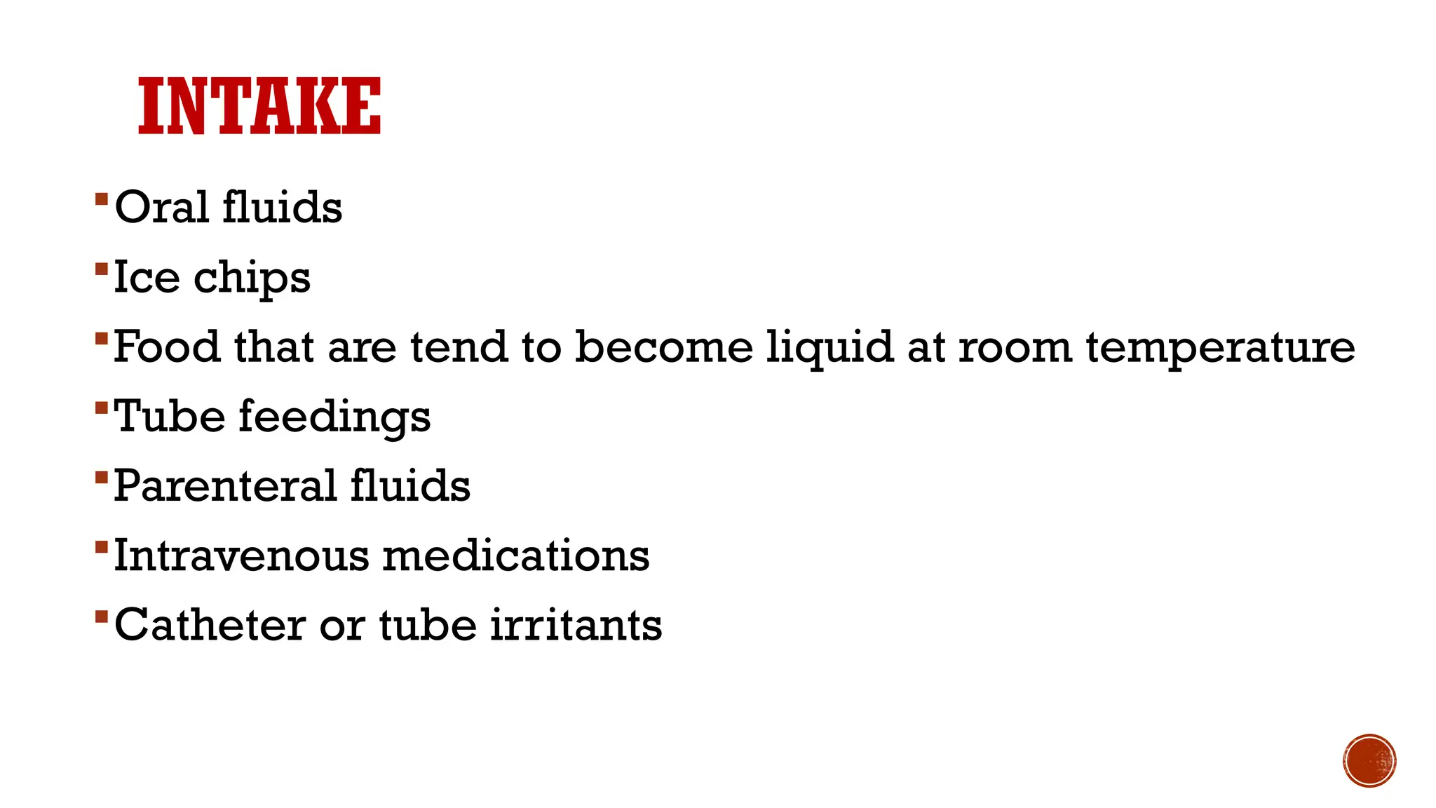
INTRAVENOUS
THERAPY
Babli Shama
M.Sc. MSN
TUTOR
NINS, GUWAHATI
INTRAVENOUS THERAPY
Replacement of fluids and electrolytes intravenously is one
of the most commonly used treatment modalities for fluid
and electrolyte imbalances.
IV therapy is a way to give fluid, medicine, nutrition, or
blood directly into the bloodstream through the vein.
FACTORS TO CONSIDER IN SELECTION OF
SITE FOR INTRAVENOUS CANNULATION
Non-dominant hand.
Most distal part of vein initially.
Extension and splint if cannulation is to be done near
the joints.
Veins that feel Soft, full, large, and naturally splinted
bones are preferred.
Avoids veins on the extremities. an injured or past
surgical intervention.
FACTORS TO CONSIDER IN SELECTION OF
SITE FOR INTRAVENOUS CANNULATION
VASCULAR ACCESS DEVICES
Peripheral short catheter: used for administration of IV
therapy that is expected to last only for a week.
TPN and chemotherapy drugs can’t administered through
them.
VASCULAR ACCESS DEVICES
Midline peripheral catheters: These catheters can be used
for IV therapies that may extend from 2 weeks to 6 weeks.
Inserted just above or below the antecubital fossa into the
cephalic or proximal basilic veins.
VASCULAR ACCESS DEVICES
Central venous devices: the possible sites for insertion of
CVL are the internal jugular, femoral, and subclavian.
A variety of IV fluids, blood products, TPN, and medications
can be administered through the central venous devices.
COMMONLY USED VEINS FOR
INTRAVENOUS CANNULATION
• The commonly used
venipuncture sites are
metacarpal, basilic and
cephalic veins
• The antecubital basilic and
median veins are usually used
for blood draws and bolus
injection of medications.
TYPES OF IV FLUIDS
IV fluids are classified on the bases of the concentration
of solute in the solvent.
1. Crystalloids
a) Isotonic solutions
b) Hypotonic solutions
c) Hypertonic solutions
2. Colloids
CRYSTALLOIDS
Crystalloids contain small molecules that can
cross the semipermeable membrane and enter
in the intercellular space.
They are used to increase fluid volume both in
interstitial and intervascular space.
Crystalloids are distinguished based on their
tonicity into isotonic, hypotonic, and hypertonic
solutions.
1. ISOTONIC SOLUTIONS:
They have same concentration of solutes as blood
plasma.
These solutions expand the vascular volume
initially.They are used to treat hypovolemia, burns,
fluid loss in diarrhoea and mild metabolic acidosis.
Examples
RL (lactated Ringer’s ) balanced electrolyte
solution.
0.9% NaCl (normal saline)
It can be used temporarily to expand the ECF volume,
but not given as a routine maintenance solution as it
conation Na+ and Cl+ in excess.
D5 (Dextrose 5% in water):
Dextrose 5% is avoided in patients who have risk of
developing cerebral edema, because after
metabolization of dextrose free water molecules leads
to the expansion of ICF and ECF volume.
2. HYPOTONIC SOLUTIONS
They have less concentration of solutes then blood plasma.
Hypotonic solutions are used to treat patients with
dehydration or elevated sodium levels.
As the concentration of solutes is low in these fluids, it attracts
water into cell and helps in treating cellular dehydration.
Promote elimination by kidneys.
Not administered to the patients with risk of cerebral edema
and 3rd
space fluid shift.
EXAMPLES:
0.33% NaCl (0ne third normal saline)
0.45%NaCl (half normal saline)
3. HYPERTONIC SOLUTIONS
They have higher concentrations of solutes than blood
plasma.
Hypertonic solutions expand the vascular volume by
pulling out fluids from the intracellular and interstitial
compartments.
Also used for peripheral parenteral nutrition and to
replace nutrients and electrolytes.
Avoided for patients with kidney and heart failure.
EXAMPLES:
D5NS - 5% dextrose in normal saline
D5 1/2 NS – 5% dextrose in 0.45% NaCl
D5 RL – 5% dextrose in lactated ringer’s
COLLOIDS
Also known as plasma expenders or volume
expenders.
Colloids contain large particles that do not cross the
semipermeable membrane and remain in the IV
space.
They draw fluids from extravascular spaces due to
their high oncotic pressure, hence increasing the
intravascular volume.
Examples:
Dextran & albumin.
CALCULATION OF INTRAVENOUS FLUID PLAN
COMPLICATIONS OF IV FLUID
THERAPY
LOCAL COMPLICATIONS
1. Infiltration: when IV cannula is dislodged from
veins and fluids are infused into surrounding
tissues.
LOCAL COMPLICATIONS
2. Extravasation: fluid seeps out from the lumen
of the vessel into surrounding tissues, mainly
leaking of vesicant drugs into surrounding tissues.
3. Thrombosis: formation of local thrombus due
to injury to endothelial cells of the venous wall.
4. Phlebitis: inflammation of the vein at the site of
IV infusion, associated mainly with alkaline and
acidic solutions which are higher in osmolarity.
LOCAL COMPLICATIONS
5.Thrombophlebitis: when thrombosis is
accompanied by inflammation.
6.Occlusion: mainly caused by clots due to
inappropriate flushing or very slow flow
rate.
7. Dislodgement: unintentional slipping out
of catheter from situ.
SYSTEMIC COMPLICATIONS
1. Air Embolism: large amount of air enters into
patients veins, it is a serious complication that may
lead to respiratory distress, weak pulse, increased
CVP, decreased BP, and loss of consciousness with
dislodgement into various parts of body
2. Hematoma: leakage of blood into surrounding soft
tissues. Minor hematomas May resolve itself within 2
weeks and warm compressions are effective in
resolving them.
3. Systemic infection: local infection or not using
proper aseptic techniques may lead to local and
systemic infection.
SYSTEMIC COMPLICATIONS
4. Speed shock & circulatory overload: patient may feel
anxious, respiratory distress, increased BP and CVP and
neck engorgement, circulatory overload and speed shock
may lead to congestive heart failure or even the death of
patient.
5. Allergic reactions: when patient is allergic to catheter
or the medications which are inserted via it.
6.Vasovagal reaction: patient experience vasospasm due
to pain, vein may suddenly collapse during venipuncture
and patient may experience dizziness, diaphoresis, pain,
nausea, hypotension or even faint. Nurse have to check BP
and report immediately while performing necessary
MEASURING FLUID INTAKE AND
OUTPUT
Measurement and recording of all fluid intake and
output during a 24–hour period provides important
data about the client's fluid and electrolyte balance.
 The unit of measurement of intake and output is mL
(milliliter).To measure fluid intake, nurses convert
household measures such as glass, cup, or soup
bowl to metric units. Gauge fluid balance and give
valuable information about your patient's condition.
MEASUREMENT OF VOLUME
1 tablespoon (tbsp) = 15 millilitres(ml)
3 teaspoons(tsp) = 15 millilitres(ml)
1 cup(C) = 240 millilitres(ml)
8 ounces(oz) = 240 millilitres(ml)
1 teaspoon(tsp) = 5 millilitres(ml)
1 cup(C) = 8 ounces(oz)
16 ounces(oz) = 1 pound(lb)
1 ounce (oz) = 30milliliters(ml)
INTAKE
Oral fluids
Ice chips
Food that are tend to become liquid at room temperature
Tube feedings
Parenteral fluids
Intravenous medications
Catheter or tube irritants
OUTPUT
Urine
Vomitus and liquid faeces
Tube drainage
Wound
Drainage
Draining fistulas
DO’S AND DON'TS
• Those patients have undergone surgery or if he has a
medical condition or take medication that can affect
fluid intake or loss. Measure and record all intake and
output.
• At least every 8 hours, record the type and amount of all
fluids he's received and describe the route as oral,
parenteral, rectal, or by enteric tube.
DO’S AND DON'TS
• Ice chips are recorded as fluid at approximately
half their volume.
• Record the type and amount of all fluids the patient
has lost and the route. Describe them as urine,
liquid stool, vomitus, tube drainage, and any fluid
aspirated from a body cavity.
DO’S AND DON'TS
• If irrigating a nasogastric or another tube or the
bladder, measure the amount instilled and subtract it
from total output.
• An accurate measurement, keep toilet paper out of
your patient's urine. Measure drainage in a calibrated
container. Evaluate trends over 24 to 48 hours.
• Don't assess output by amount only. Consider color,
color changes, and odor too. Don't use the same
graduated container for more than one patient
IV therapy, Type of IV fluids, Fluid calculation

IV therapy, Type of IV fluids, Fluid calculation

  • 1.
  • 2.
    INTRAVENOUS THERAPY Replacement offluids and electrolytes intravenously is one of the most commonly used treatment modalities for fluid and electrolyte imbalances. IV therapy is a way to give fluid, medicine, nutrition, or blood directly into the bloodstream through the vein.
  • 3.
    FACTORS TO CONSIDERIN SELECTION OF SITE FOR INTRAVENOUS CANNULATION Non-dominant hand. Most distal part of vein initially. Extension and splint if cannulation is to be done near the joints. Veins that feel Soft, full, large, and naturally splinted bones are preferred. Avoids veins on the extremities. an injured or past surgical intervention.
  • 4.
    FACTORS TO CONSIDERIN SELECTION OF SITE FOR INTRAVENOUS CANNULATION
  • 5.
    VASCULAR ACCESS DEVICES Peripheralshort catheter: used for administration of IV therapy that is expected to last only for a week. TPN and chemotherapy drugs can’t administered through them.
  • 6.
    VASCULAR ACCESS DEVICES Midlineperipheral catheters: These catheters can be used for IV therapies that may extend from 2 weeks to 6 weeks. Inserted just above or below the antecubital fossa into the cephalic or proximal basilic veins.
  • 7.
    VASCULAR ACCESS DEVICES Centralvenous devices: the possible sites for insertion of CVL are the internal jugular, femoral, and subclavian. A variety of IV fluids, blood products, TPN, and medications can be administered through the central venous devices.
  • 8.
    COMMONLY USED VEINSFOR INTRAVENOUS CANNULATION • The commonly used venipuncture sites are metacarpal, basilic and cephalic veins • The antecubital basilic and median veins are usually used for blood draws and bolus injection of medications.
  • 12.
    TYPES OF IVFLUIDS IV fluids are classified on the bases of the concentration of solute in the solvent. 1. Crystalloids a) Isotonic solutions b) Hypotonic solutions c) Hypertonic solutions 2. Colloids
  • 13.
    CRYSTALLOIDS Crystalloids contain smallmolecules that can cross the semipermeable membrane and enter in the intercellular space. They are used to increase fluid volume both in interstitial and intervascular space. Crystalloids are distinguished based on their tonicity into isotonic, hypotonic, and hypertonic solutions.
  • 14.
    1. ISOTONIC SOLUTIONS: Theyhave same concentration of solutes as blood plasma. These solutions expand the vascular volume initially.They are used to treat hypovolemia, burns, fluid loss in diarrhoea and mild metabolic acidosis. Examples RL (lactated Ringer’s ) balanced electrolyte solution.
  • 15.
    0.9% NaCl (normalsaline) It can be used temporarily to expand the ECF volume, but not given as a routine maintenance solution as it conation Na+ and Cl+ in excess. D5 (Dextrose 5% in water): Dextrose 5% is avoided in patients who have risk of developing cerebral edema, because after metabolization of dextrose free water molecules leads to the expansion of ICF and ECF volume.
  • 16.
    2. HYPOTONIC SOLUTIONS Theyhave less concentration of solutes then blood plasma. Hypotonic solutions are used to treat patients with dehydration or elevated sodium levels. As the concentration of solutes is low in these fluids, it attracts water into cell and helps in treating cellular dehydration. Promote elimination by kidneys. Not administered to the patients with risk of cerebral edema and 3rd space fluid shift. EXAMPLES: 0.33% NaCl (0ne third normal saline) 0.45%NaCl (half normal saline)
  • 17.
    3. HYPERTONIC SOLUTIONS Theyhave higher concentrations of solutes than blood plasma. Hypertonic solutions expand the vascular volume by pulling out fluids from the intracellular and interstitial compartments. Also used for peripheral parenteral nutrition and to replace nutrients and electrolytes. Avoided for patients with kidney and heart failure.
  • 18.
    EXAMPLES: D5NS - 5%dextrose in normal saline D5 1/2 NS – 5% dextrose in 0.45% NaCl D5 RL – 5% dextrose in lactated ringer’s
  • 19.
    COLLOIDS Also known asplasma expenders or volume expenders. Colloids contain large particles that do not cross the semipermeable membrane and remain in the IV space. They draw fluids from extravascular spaces due to their high oncotic pressure, hence increasing the intravascular volume. Examples: Dextran & albumin.
  • 20.
  • 21.
    COMPLICATIONS OF IVFLUID THERAPY LOCAL COMPLICATIONS 1. Infiltration: when IV cannula is dislodged from veins and fluids are infused into surrounding tissues.
  • 22.
    LOCAL COMPLICATIONS 2. Extravasation:fluid seeps out from the lumen of the vessel into surrounding tissues, mainly leaking of vesicant drugs into surrounding tissues. 3. Thrombosis: formation of local thrombus due to injury to endothelial cells of the venous wall. 4. Phlebitis: inflammation of the vein at the site of IV infusion, associated mainly with alkaline and acidic solutions which are higher in osmolarity.
  • 23.
    LOCAL COMPLICATIONS 5.Thrombophlebitis: whenthrombosis is accompanied by inflammation. 6.Occlusion: mainly caused by clots due to inappropriate flushing or very slow flow rate. 7. Dislodgement: unintentional slipping out of catheter from situ.
  • 24.
    SYSTEMIC COMPLICATIONS 1. AirEmbolism: large amount of air enters into patients veins, it is a serious complication that may lead to respiratory distress, weak pulse, increased CVP, decreased BP, and loss of consciousness with dislodgement into various parts of body 2. Hematoma: leakage of blood into surrounding soft tissues. Minor hematomas May resolve itself within 2 weeks and warm compressions are effective in resolving them. 3. Systemic infection: local infection or not using proper aseptic techniques may lead to local and systemic infection.
  • 25.
    SYSTEMIC COMPLICATIONS 4. Speedshock & circulatory overload: patient may feel anxious, respiratory distress, increased BP and CVP and neck engorgement, circulatory overload and speed shock may lead to congestive heart failure or even the death of patient. 5. Allergic reactions: when patient is allergic to catheter or the medications which are inserted via it. 6.Vasovagal reaction: patient experience vasospasm due to pain, vein may suddenly collapse during venipuncture and patient may experience dizziness, diaphoresis, pain, nausea, hypotension or even faint. Nurse have to check BP and report immediately while performing necessary
  • 26.
    MEASURING FLUID INTAKEAND OUTPUT Measurement and recording of all fluid intake and output during a 24–hour period provides important data about the client's fluid and electrolyte balance.  The unit of measurement of intake and output is mL (milliliter).To measure fluid intake, nurses convert household measures such as glass, cup, or soup bowl to metric units. Gauge fluid balance and give valuable information about your patient's condition.
  • 27.
    MEASUREMENT OF VOLUME 1tablespoon (tbsp) = 15 millilitres(ml) 3 teaspoons(tsp) = 15 millilitres(ml) 1 cup(C) = 240 millilitres(ml) 8 ounces(oz) = 240 millilitres(ml) 1 teaspoon(tsp) = 5 millilitres(ml) 1 cup(C) = 8 ounces(oz) 16 ounces(oz) = 1 pound(lb) 1 ounce (oz) = 30milliliters(ml)
  • 28.
    INTAKE Oral fluids Ice chips Foodthat are tend to become liquid at room temperature Tube feedings Parenteral fluids Intravenous medications Catheter or tube irritants
  • 29.
    OUTPUT Urine Vomitus and liquidfaeces Tube drainage Wound Drainage Draining fistulas
  • 30.
    DO’S AND DON'TS •Those patients have undergone surgery or if he has a medical condition or take medication that can affect fluid intake or loss. Measure and record all intake and output. • At least every 8 hours, record the type and amount of all fluids he's received and describe the route as oral, parenteral, rectal, or by enteric tube.
  • 31.
    DO’S AND DON'TS •Ice chips are recorded as fluid at approximately half their volume. • Record the type and amount of all fluids the patient has lost and the route. Describe them as urine, liquid stool, vomitus, tube drainage, and any fluid aspirated from a body cavity.
  • 32.
    DO’S AND DON'TS •If irrigating a nasogastric or another tube or the bladder, measure the amount instilled and subtract it from total output. • An accurate measurement, keep toilet paper out of your patient's urine. Measure drainage in a calibrated container. Evaluate trends over 24 to 48 hours. • Don't assess output by amount only. Consider color, color changes, and odor too. Don't use the same graduated container for more than one patient