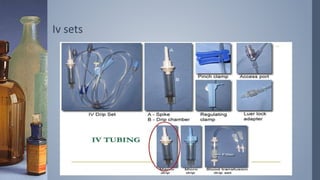
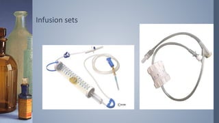
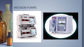
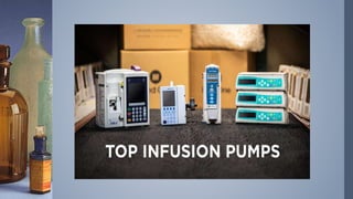
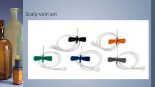
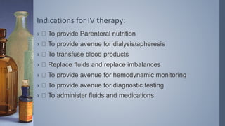
2. Historical background:
In the 1830s, a lethal strain of cholera wracked much of
Europe. This type of cholera was called “Russian cholera,”
or “blue cholera,” for the dusky cyanotic complexion of its.
Mr. Thomas Latta, implemente d this iv therapy.
3. Intravenous therapy :
› Intravenous therapy or IV therapy is the infusion of liquid
substances directly into a vein. The word intravenous
simply means "within a vein”.
› Iv therapy constitutes the administration of liquid
substances directly into a vein and general
4. Infusion therapy :
› Infusion therapy is defined as the parenteral infusion of
fluids, electrolytes, blood components, nutrients, or
medications to prevent or treat deficiencies.
› Infusion therapy may be dispensed by I.V.,
subcutaneous, intraosseous or intrathecal routes of
administration.
5. Blood component therapy:
› Common blood components that may be transfused
include red blood cells, fresh-frozen plasma, platelets,
and clotting factors .
6. PARENTERAL NUTRITION
› Parenteral nutrient solution composition is determined
individually to meet each patient’s nutritional needs.
› Parenteral solutions include electrolytes, dextrose, amino
acids, vitamins, and various trace elements.
› These solutions are referred to as total parenteral
nutrition (TPN).
› here are also solutions referred to as total nutrient
admixture (TNA) solutions that provide a nutrient mix that
includes electrolytes, dextrose, amino acids, vitamins,
trace elements, and fats.
7. › The concentration of nutrients in TPN solutions, IVFEs,
and TNA solutions, is determined by I.V. delivery route.
› The solutions are less concentrated when they are
delivered by a peripheral I.V. route and are more
concentrated when delivered by a central I.V. route.
8. Fluid compartments
EXTRACELLULAR FLUID (ECF) Approximately 80% of
extracellular fluid is interstitial, which occupies the microscopic
spaces between the cells. 20% is plasma, which is the liquid
portion of the blood.
INTRACELLULAR FLUID ( ICF) This is known as cytosol
and is fluid.
Substances move around from areas of high concentration to
low concentration and a concentration gradient will exist
between the two.
They move passively.
Movement is facilitated through process of osmosis or
diffusion.
10. 1. Colloid
› • Fluids with large molecules which donot pass the cell
membranes, when infused remain mainly in the
intravascular compartment.
› • It expands intravascular volume and draw fluid from
extravascular spaces. Eg. Albumin, hemacel, dextran.
11. 2. Crystalloid
› • It contains small molecules flowing easily through cell
membranes, allowing for transfer from blood stream into cells
and body tissues.
› • It increases the blood volume in both interstitial and
intravascular spaces.
› It is further divided into:
› a. Isotonic: eg. 0.9% Sodium Chloride, Ringer Lactate,5%
dextrose in water.
› b.Hypotonic: eg. 0.45% sodium chloride,0.33% Sodium
Chloride,o.2% sodium chloride,2.5% dextrose in water.
› c.Hypertonic: eg. 3% Sodium Chloride,5% Sodium Chloride.
23. IMPLANTABLE PORTS:
1. A port (Port-a-Cath or MediPort) is a central venous line
that does not have an external connector; instead, it has
a small reservoir that is covered with silicone rubber and
is implanted under the skin.
24. VERICOSE VEINS /SCLERO THERAPY:
› Sclerotherapy is a procedure used to treat blood vessels
vascular malformations and also those of the lymphatic
system.
› A medicine is injected into the vessels, which makes
them shrink.
› In adults, sclerotherapy is often used to treat spider
veins, smaller varicose veins and hemorrhoids.
25.
26. Indications for IV therapy:
› To provide Parenteral nutrition
› To provide avenue for dialysis/apheresis
› To transfuse blood products
› Replace fluids and replace imbalances
› To provide avenue for hemodynamic monitoring
› To provide avenue for diagnostic testing
› To administer fluids and medications
27. Principles of IV therapy:
› 1. maintenance :
› • Fluids are given for compensating ongoing sensible
losses.
› • It includes urine output, fecal matter, respiration,
perspiration.
› • Its requirement is higher in children than adults
because of higher metabolic rate, more body surface
area, higher respiratory rate
28. › ¼ NS,1/4 NS+ D5, ½ NS, 1/2NS+D5 are the most
commonly used fluid for maintenance.
› • Normal range for children is (70-150)ml/hr.
29. 2. replacement
› It includes fluid to meet ongoing losses due to medical
treatment.Eg. Patient with chest tube drain, umcontrolled
vomotong, continue diarrhea,CSF shunt, etc. Ringer
lactate, Normal saline are preferred.
30. Procedure :
1. Confirm Patient’s Identification
2. Review Physician’s Order
3. Explanation about the procedure
4. Perform Hand Hygiene
5. Gather Equipments a. Cardex b. Tray containing gloves,
syringe with normal saline, marker medicine. c. IV drip fluid
d. Buroset
6. Inspect administration set
7. Place IV level on the IVF bottle
8. Hang solution container on the pole.
31. 9. Open IV set aseptically.
10. Fill the drip chamber half.
11. Expel air bubble if any.
12. Connect tubing to catheter and initiate infusion.
13. Infuse as per the rate and amount.
14. Observe for complications like signs of infection, phlebitis,
infiltration, kin color changes, fluid and electrolyte overload, etc.
15. Never allow the bottle to get empty completely.
16. Quickly remove the spike from the old IV solution without
touching the tip and insert spike into the new intravenous
bottle.
17. Document accurately.
18. IV tubing should be changed every 48-72 hours and
cannula every 72 hours.
32. SIDE EFFECTS OF IV THERAPY
› infection
› Infilteration
› Phlebitis and thrombophlebitis
› Extravasation
› hypersensitivity
33. Infilteration
› infiltration occurs when I.V. fluid or medications leak into
the surrounding tissue. Infiltration can be caused by
improper placement or dislodgment of the catheter.
Patient movement can cause the catheter to slip out or
through the blood vessel lumen.
34. Phlebitis
› Phlebitis is inflammation of a vein. It is usually associated
with acidic or alkaline solutions or solutions that have a
high osmolarity. Phlebitis can also occur as a result of
vein trauma during insertion, use of an inappropriate I.V.
catheter size for the vein, or prolonged use of the same
I.V. site.
35. Extravasation :
› Extravasation is the leaking of vesicant drugs into
surrounding tissue. Extravasation can cause severe local
tissue damage, possibly leading to delayed healing,
infection, tissue necrosis, disfigurement, loss of function,
and even amputation.