
Download as PDF, PPTX

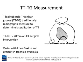
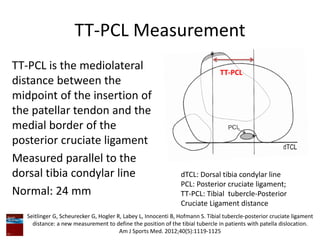

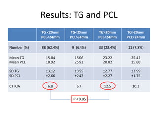
This study evaluated 141 patients with patellar pathology to determine if the tibial tubercle-trochlear groove (TT-TG) distance measured on MRI corresponds to measurements on CT scans. They also examined the relationship between the TT-TG distance and the tibial tubercle-posterior cruciate ligament (TT-PCL) distance. They found poor agreement between TT-TG measurements on MRI versus CT. The TT-PCL distance measures true tibial tubercle lateralization while the TT-TG reflects lateralization and knee joint rotation. Patients can be grouped based on their TT-TG and TT-PCL distances which may help explain outcomes after tibial tubercle surgery.


