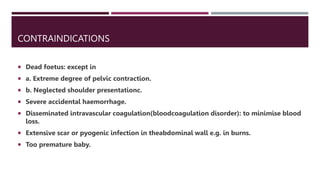
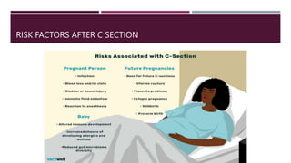
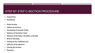
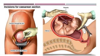
CESAREAN SECTION
RAMESH KUMAR DEEPAK SUMAN
GROUP 408A
INTRODUCTION
 Cesarean section is a fetal delivery through an open abdominal incision (laparotomy) and an
incision in the uterus (hysterotomy). The first cesarean documented occurred in 1020 AD, and
since then, the procedure has evolved tremendously. It is now the most common surgery
performed in the United States, with over 1 million women delivered by cesarean every year. The
cesarean delivery rate rose from 5% in 1970 to 31.9% in 2016. Though there are continuing
efforts to reduce the rate of cesarean sections, experts do not anticipate a significant drop for at
least a decade or two. While it confers risks of both immediate and long-term complications, for
some women, cesarean delivery can be the safest or even the only way to deliver a healthy
newborn.
DEFINITION
 It is an operative procedure whereby the fetuses after the end of 28th
week are delivered through an incision on the abdominal and uterine
walls.
MATERNAL INDICATIONS FOR CESAREAN
 Prior cesarean delivery
 Maternal request
 Pelvic deformity or cephalopelvic disproportion
 Previous perineal trauma
 Prior pelvic or anal/rectal reconstructive surgery
 Herpes simplex or HIV infection
 Cardiac or pulmonary disease
 Cerebral aneurysm or arteriovenous malformation
 Pathology requiring concurrent intraabdominal surgery
 Perimortem cesarean
UTERINE/ANATOMIC INDICATIONS FOR CESAREAN
 Abnormal placentation (such as placenta previa, placenta accreta)
 Placental abruption
 Prior classical hysterotomy
 Prior full-thickness myomectomy
 History of uterine incision dehiscence
 Invasive cervical cancer
 Prior trachelectomy
 Genital tract obstructive mass
 Permanent cerclage
FETAL INDICATIONS FOR CESAREAN
 Nonreassuring fetal status (such as abnormal umbilical cord Doppler study) or abnormal fetal
heart tracing
 Umbilical cord prolapse
 Failed operative vaginal delivery
 Malpresentation
 Macrosomia
 Congenital anomaly
 Thrombocytopenia
 Prior neonatal birth trauma
THE PRIMARY PERSONNEL FOR A CESAREAN SECTION
CONSISTS OF:
 The surgeon
 The surgeon’s assistant
 The anesthesiologist or anesthetist
 A scrub nurse or technician
 A circulating nurse
 Someone to care for the neonate
TYPES OF C SECTION
 Classical caesarean section
 This is rarely performed. It involves a vertical incision made through the visceral
peritoneum and the contractile part of the uterus above the bladder
 Indications

 Gestational age less than 32 weeks before the lower segement are formed
 Placental praevia
 Fetus in a transverse lie
 Shoulder presentation
 Advantages of Classical caesarean section
 It doesn't take much time to perform It can be employed when general
anaesthesia is not available
 Disadvantages of Classical caesarean section
 Rupture of a uterine scar in subsequent pregnancy
 Haemorrhage is greater
 Small bowel adhesion to the anterior suture line
 Delayed wound healing
 Lower segment caesarean section
 This is possible by means of transverse incision through the lower uterine
segment.
 Advantages
 Less danger of infection or haemorrhage
 Less incidence of uterine rupture in subsequent pregnancies
 It is the caesarean section mostly employed by obstetricians
METHODS OF C SECTION
 Elective caesarean section
 This type is used when the caesarean section is performed at a scheduled
time, such with a known fetus pelvic disproportion. The patient is
usually admitted to the hospital the day prior to surgery. This allows for
laboratory investigations and provides an opportunity to rule out
presence of infection
 Emergency caesarean section
 Here, there is no indication of caesarean section prior to the surgery. It is
usually done when a woman must have laboured with failure and there
is an urgent need to save the life of both the mother and the child or
either
CONTRAINDICATIONS
 Dead foetus: except in
 a. Extreme degree of pelvic contraction.
 b. Neglected shoulder presentationc.
 Severe accidental haemorrhage.
 Disseminated intravascular coagulation(bloodcoagulation disorder): to minimise blood
loss.
 Extensive scar or pyogenic infection in theabdominal wall e.g. in burns.
 Too premature baby.
RISK FACTORS AFTER C SECTION
STEP BY STEP C-SECTION PROCEDURE
 Preparation
 Anesthesia
 Initial incision
 Follow-up incisions
 Suctioning of amniotic fluids
 Delivery of the baby's head
 Delivery of the baby's shoulders and body
 Birth of the baby
 Cutting of the umbilical cord
 Delivery of the placenta
 Closing the incision
 Recovery
DELIVERY OF THE PLACENTA
 The next steps are the delivery of the placenta, followed by the suturing of the uterus
and all the layers that were cut during the surgery. Once the placenta has been
removed, it will be examined by your doctor.7 Closing up everything that's been cut
through to get to the baby is usually the longest part of the cesarean section, which
in total typically takes about 30 to 60 minutes to complete.
 During this time you can usually have your baby with you to breastfeed or hold.
However, don’t feel pressure to begin breastfeeding immediately, you can start any
time in the first hours after your baby is born—a small delay won’t cause any harm.
Simply enjoying your baby however works best for you is fine. It may also be possible
for your support person to hold the baby close to your face if you are unable to hold
your baby.
SUTURING
 • Double row suture
 • first row - continuous
 mucous - muscular
 sutures
 • second row - continuous
 muscular-muscular
 sutures
 • peritonization - restoring
 the integrity of the plica
 vesico-uterina
CONCLUDING PART
 Peritoneal toileting is done and the blood clots
 are removed
 • The tubes and ovaries are examined
 • After being satisfied that the uterus is well
 contracted, the abdomen is closed in layers
 • The vagina is cleansed of blood clots
 • The blood loss is commonly between 500 and
 • 1000 ml
UTERINE RUPTURE, PERIOPERATIVE AND PERINATAL MORBIDITY AFTER
SINGLE-LAYER AND DOUBLE-LAYER CLOSURE AT CESAREAN DELIVERY.
 Single-layer uterine closure is associated with decreased infectious morbidity in the
index surgery, but not uterine rupture or other adverse outcomes in the subsequent
gestation.
FIRST-BIRTH CESAREAN AND PLACENTAL ABRUPTION OR PREVIA AT
SECOND BIRTH
 Among our study cohort, abruptio placentae complicated 11.5 per 1000
and placenta previa 5.2 per 1000 singleton deliveries at second births. In
logistic regression analyses adjusted for maternal age, women with
first-birth cesareans had significantly increased risk of abruptio
placentae (OR 1.3, 95% CI 1.1, 1.5), and placenta previa (OR 1.4, 95% CI
1.1, 1.6) at second births, compared with women with prior vaginal
deliveries.
POSTOPERATIVE CARE.
 Observation for the first 6–8 hours is important. Periodic check up of pulse, BP,
 amount of vaginal bleeding and behavior of the uterus is done and recorded
 • Fluid: Sodium chloride (0.9%) or Ringer’s lactate drip is continued until at least 2
 – 2.5 liters of the solution are infused. Blood transfusion is required if the blood loss
 is more than average during the operation (average blood loss in cesarean section is
 approximately 0.5 to 1 liter)
 • Oxytocics: Injection oxytocin 5 units IM or IV (slow) or methergin 0.2 mg IM is
 given and may be repeated
 • Prophylactic antibiotic (cephalosporins, metronidazole) for all cesarean delivery
 is given for 2–3 days. Therapeutic antibiotic is given when indicated
 Analgesics in the form of pethidine hydrochloride 75-100 mg is administered and
 may have to be repeated
 • Ambulation:The patient can sit on the bed or even get out of bed to evacuate the
 bladder, providednthe general condition permits. She is encouraged to move her
 legs and ankles and to breathe deeply to minimize leg vein thrombosis and
 pulmonary embolism
 • Baby is put to the breast for feeding after 3–4 hours when mother is stable and
 relieved of pain
 Day 1: • Oral feeding in the form of plain or electrolyte water or
 raw tea may be given. Active bowel sounds are observed by the
 end of the day.
 • Day 2: • Light solid diet of the patient’s choice is given. • Bowel
 care: 3–4 teaspoons of lactulose is given at bed time, if the
 bowels do not move spontaneously.
 • Day 5 or Day 6: The abdominal skin stitches are to be removed
 on the D-5 (in transverse) or D-6 (in longitudinal).
 • Discharge: The patient is discharged on the day following
 removal of the stitches, if otherwise fit. Usual advices like those
 following vaginal delivery are given. Depending on postoperative
 recovery and availability of care at home, patient may be
 discharged as early as third to as late as seventh postoperative day.
THANKS FOR YOUR ATTENTION

Cesarean section

  • 1.
    CESAREAN SECTION RAMESH KUMARDEEPAK SUMAN GROUP 408A
  • 2.
    INTRODUCTION  Cesarean sectionis a fetal delivery through an open abdominal incision (laparotomy) and an incision in the uterus (hysterotomy). The first cesarean documented occurred in 1020 AD, and since then, the procedure has evolved tremendously. It is now the most common surgery performed in the United States, with over 1 million women delivered by cesarean every year. The cesarean delivery rate rose from 5% in 1970 to 31.9% in 2016. Though there are continuing efforts to reduce the rate of cesarean sections, experts do not anticipate a significant drop for at least a decade or two. While it confers risks of both immediate and long-term complications, for some women, cesarean delivery can be the safest or even the only way to deliver a healthy newborn.
  • 3.
    DEFINITION  It isan operative procedure whereby the fetuses after the end of 28th week are delivered through an incision on the abdominal and uterine walls.
  • 4.
    MATERNAL INDICATIONS FORCESAREAN  Prior cesarean delivery  Maternal request  Pelvic deformity or cephalopelvic disproportion  Previous perineal trauma  Prior pelvic or anal/rectal reconstructive surgery  Herpes simplex or HIV infection  Cardiac or pulmonary disease  Cerebral aneurysm or arteriovenous malformation  Pathology requiring concurrent intraabdominal surgery  Perimortem cesarean
  • 5.
    UTERINE/ANATOMIC INDICATIONS FORCESAREAN  Abnormal placentation (such as placenta previa, placenta accreta)  Placental abruption  Prior classical hysterotomy  Prior full-thickness myomectomy  History of uterine incision dehiscence  Invasive cervical cancer  Prior trachelectomy  Genital tract obstructive mass  Permanent cerclage
  • 6.
    FETAL INDICATIONS FORCESAREAN  Nonreassuring fetal status (such as abnormal umbilical cord Doppler study) or abnormal fetal heart tracing  Umbilical cord prolapse  Failed operative vaginal delivery  Malpresentation  Macrosomia  Congenital anomaly  Thrombocytopenia  Prior neonatal birth trauma
  • 7.
    THE PRIMARY PERSONNELFOR A CESAREAN SECTION CONSISTS OF:  The surgeon  The surgeon’s assistant  The anesthesiologist or anesthetist  A scrub nurse or technician  A circulating nurse  Someone to care for the neonate
  • 8.
    TYPES OF CSECTION  Classical caesarean section  This is rarely performed. It involves a vertical incision made through the visceral peritoneum and the contractile part of the uterus above the bladder  Indications   Gestational age less than 32 weeks before the lower segement are formed  Placental praevia  Fetus in a transverse lie  Shoulder presentation
  • 9.
     Advantages ofClassical caesarean section  It doesn't take much time to perform It can be employed when general anaesthesia is not available  Disadvantages of Classical caesarean section  Rupture of a uterine scar in subsequent pregnancy  Haemorrhage is greater  Small bowel adhesion to the anterior suture line  Delayed wound healing
  • 10.
     Lower segmentcaesarean section  This is possible by means of transverse incision through the lower uterine segment.  Advantages  Less danger of infection or haemorrhage  Less incidence of uterine rupture in subsequent pregnancies  It is the caesarean section mostly employed by obstetricians
  • 11.
    METHODS OF CSECTION  Elective caesarean section  This type is used when the caesarean section is performed at a scheduled time, such with a known fetus pelvic disproportion. The patient is usually admitted to the hospital the day prior to surgery. This allows for laboratory investigations and provides an opportunity to rule out presence of infection  Emergency caesarean section  Here, there is no indication of caesarean section prior to the surgery. It is usually done when a woman must have laboured with failure and there is an urgent need to save the life of both the mother and the child or either
  • 12.
    CONTRAINDICATIONS  Dead foetus:except in  a. Extreme degree of pelvic contraction.  b. Neglected shoulder presentationc.  Severe accidental haemorrhage.  Disseminated intravascular coagulation(bloodcoagulation disorder): to minimise blood loss.  Extensive scar or pyogenic infection in theabdominal wall e.g. in burns.  Too premature baby.
  • 13.
  • 15.
    STEP BY STEPC-SECTION PROCEDURE  Preparation  Anesthesia  Initial incision  Follow-up incisions  Suctioning of amniotic fluids  Delivery of the baby's head  Delivery of the baby's shoulders and body  Birth of the baby  Cutting of the umbilical cord  Delivery of the placenta  Closing the incision  Recovery
  • 17.
    DELIVERY OF THEPLACENTA  The next steps are the delivery of the placenta, followed by the suturing of the uterus and all the layers that were cut during the surgery. Once the placenta has been removed, it will be examined by your doctor.7 Closing up everything that's been cut through to get to the baby is usually the longest part of the cesarean section, which in total typically takes about 30 to 60 minutes to complete.  During this time you can usually have your baby with you to breastfeed or hold. However, don’t feel pressure to begin breastfeeding immediately, you can start any time in the first hours after your baby is born—a small delay won’t cause any harm. Simply enjoying your baby however works best for you is fine. It may also be possible for your support person to hold the baby close to your face if you are unable to hold your baby.
  • 18.
    SUTURING  • Doublerow suture  • first row - continuous  mucous - muscular  sutures  • second row - continuous  muscular-muscular  sutures  • peritonization - restoring  the integrity of the plica  vesico-uterina
  • 19.
    CONCLUDING PART  Peritonealtoileting is done and the blood clots  are removed  • The tubes and ovaries are examined  • After being satisfied that the uterus is well  contracted, the abdomen is closed in layers  • The vagina is cleansed of blood clots  • The blood loss is commonly between 500 and  • 1000 ml
  • 20.
    UTERINE RUPTURE, PERIOPERATIVEAND PERINATAL MORBIDITY AFTER SINGLE-LAYER AND DOUBLE-LAYER CLOSURE AT CESAREAN DELIVERY.  Single-layer uterine closure is associated with decreased infectious morbidity in the index surgery, but not uterine rupture or other adverse outcomes in the subsequent gestation.
  • 21.
    FIRST-BIRTH CESAREAN ANDPLACENTAL ABRUPTION OR PREVIA AT SECOND BIRTH  Among our study cohort, abruptio placentae complicated 11.5 per 1000 and placenta previa 5.2 per 1000 singleton deliveries at second births. In logistic regression analyses adjusted for maternal age, women with first-birth cesareans had significantly increased risk of abruptio placentae (OR 1.3, 95% CI 1.1, 1.5), and placenta previa (OR 1.4, 95% CI 1.1, 1.6) at second births, compared with women with prior vaginal deliveries.
  • 23.
    POSTOPERATIVE CARE.  Observationfor the first 6–8 hours is important. Periodic check up of pulse, BP,  amount of vaginal bleeding and behavior of the uterus is done and recorded  • Fluid: Sodium chloride (0.9%) or Ringer’s lactate drip is continued until at least 2  – 2.5 liters of the solution are infused. Blood transfusion is required if the blood loss  is more than average during the operation (average blood loss in cesarean section is  approximately 0.5 to 1 liter)  • Oxytocics: Injection oxytocin 5 units IM or IV (slow) or methergin 0.2 mg IM is  given and may be repeated  • Prophylactic antibiotic (cephalosporins, metronidazole) for all cesarean delivery  is given for 2–3 days. Therapeutic antibiotic is given when indicated
  • 24.
     Analgesics inthe form of pethidine hydrochloride 75-100 mg is administered and  may have to be repeated  • Ambulation:The patient can sit on the bed or even get out of bed to evacuate the  bladder, providednthe general condition permits. She is encouraged to move her  legs and ankles and to breathe deeply to minimize leg vein thrombosis and  pulmonary embolism  • Baby is put to the breast for feeding after 3–4 hours when mother is stable and  relieved of pain
  • 25.
     Day 1:• Oral feeding in the form of plain or electrolyte water or  raw tea may be given. Active bowel sounds are observed by the  end of the day.  • Day 2: • Light solid diet of the patient’s choice is given. • Bowel  care: 3–4 teaspoons of lactulose is given at bed time, if the  bowels do not move spontaneously.  • Day 5 or Day 6: The abdominal skin stitches are to be removed  on the D-5 (in transverse) or D-6 (in longitudinal).  • Discharge: The patient is discharged on the day following  removal of the stitches, if otherwise fit. Usual advices like those  following vaginal delivery are given. Depending on postoperative  recovery and availability of care at home, patient may be  discharged as early as third to as late as seventh postoperative day.
  • 26.
    THANKS FOR YOURATTENTION