Download to read offline
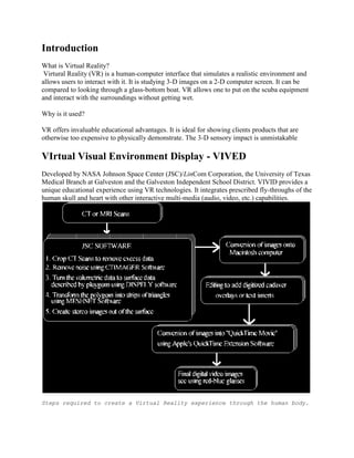


Virtual reality (VR) allows users to interact with simulated 3D environments through specialized computer hardware and software. This document describes the process of creating VR simulations of the human skull and heart using medical imaging data. CT and MRI scans were converted into 3D models and rendered into stereo image sequences. These sequences could be viewed through VR headsets or monitors to provide an interactive educational experience, allowing students to fly through virtual tours of the human body. While high resolution VR is possible, limitations remain in hardware and software. Researchers continue working to improve VR technology for medical education applications.


![Virtual surgery [new].ppt](https://cdn.slidesharecdn.com/ss_thumbnails/finalppt-111113045537-phpapp01-thumbnail.jpg?width=640&height=640&fit=bounds)


