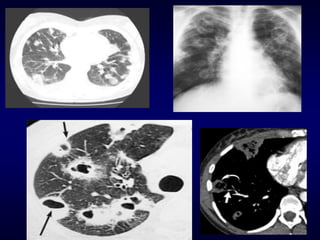
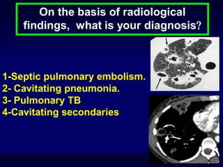
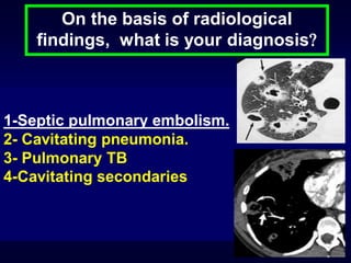
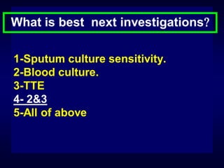
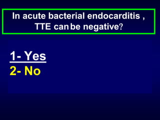
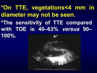
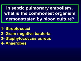
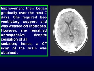
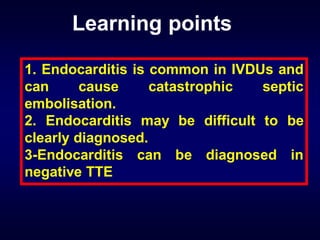
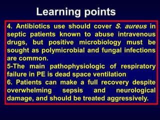
Interactive Case Presentation

Gamal Rabie Agmy ,MD ,FCCP
Professor of Chest Diseases, Assiut University
A 36-year-old female presented
with a 3-day history of increasing
shortness of breath, cough with
yellowish sputum, haemoptysis,
pleuretic chest pain, nausea,
vomiting and rigors.
The patient was an intravenous
drug user (IVDU) who smoked 20
cigarettes per day and denied
drinking alcohol. Her only past
medical history was of mild
asthma, for which salbutamol
was taken as needed.
The patient appeared distressed
and unwell. Her temperature was
39.3°C, and she was tachycardic (120
beats per minute), hypotensive (BP
85/55 mHg) and hypoxic
(O2
saturation 82% on air with a
respiratory rate of 32 breaths per
minute).
*There were needle marks in both groins and
forearms.
*Heart sounds were normal with no murmurs.
*Chest examination demonstrated scattered
crepitations all over the chest

*Abdominal and neurological examinations
were unremarkable
Initial investigations were as follows:
white
blood
cells
13.2×10⁹·L-1,
neutrophils 9.7×10⁹·L-1, haemoglobin 11.3
g·dL-1, platelets 71×109·L-1, INR of 1.5,
creatinine 2.3mg/DL, urea 84 mg/DL,
alkaline phosphatase 195 IU·L-1, alanine
aminotransferase 63 IU·L-1, and Creactive protein 285 mg·L-1.
Initial
Arterial
blood
gases
on
air showed : pH 7.60, partial pressure of O2 50
mmHg, partial pressure of CO2 28 mmHg and
bicarbonate 24 mLeq/L.
Depending on clinical picture and
investigations; what is your diagnosis
1-Pulmonary TB
2-Nosocomial pneumonia.
3- severe Pneumonia in immunocompromised
host
4-Simple Pneumonia in immunocompromised
host
Depending on clinical picture and
investigations; what is your diagnosis
1-Pulmonary TB
2-Nosocomial pneumonia.
3- severe Pneumonia in immunocompromised
host
4-Simple Pneumonia in immunocompromised
host
Can you Interpret the arterial blood gases
(PH 7.60, Pa O2 50 mmHg, Pa CO2 28 mmHg and
bicarbonate 24 mLeq/L )

1- Acute type 1 respiratory failure
2- Acute on top of chronic type 1 respiratory
failure
3- Chronic type 1 respiratory failure
4- Type 11 respiratory failure
Can you Interpret the arterial blood gases
(PH 7.60, Pa O2 50 mmHg, Pa CO2 28 mmHg and
bicarbonate 24 mLeq/L )

1- Acute type 1 respiratory failure
2- Acute on top of chronic type 1 respiratory
failure
3- Chronic type 1 respiratory failure

4- Type 11 respiratory failure
The patient was initially treated for severe
pneumonia and a chest radiography
followed by computed (CT) scan of the chest
were obtained .
On the basis of radiological
findings, what is your diagnosis

1-Septic pulmonary embolism.
2- Cavitating pneumonia.
3- Pulmonary TB
4-Cavitating secondaries
On the basis of radiological
findings, what is your diagnosis

1-Septic pulmonary embolism.
2- Cavitating pneumonia.
3- Pulmonary TB
4-Cavitating secondaries
What is best next investigations
1-Sputum culture sensitivity.
2-Blood culture.
3-TTE
4- 2&3
5-All of above
What is best next investigations
1-Sputum culture sensitivity.
2-Blood culture.
3-TTE
4- 2&3
5-All of above
In acute bacterial endocarditis ,
TTE can be negative

1- Yes
2- No
In acute bacterial endocarditis ,
TTE can be negative

1- Yes
2- No
*On TTE, vegetations<4 mm in
diameter may not be seen.
*The sensitivity of TTE compared
with TOE is 40–63% versus 90–
100%.
In septic pulmonary embolism ,
what is the commonest organism
demonstrated by blood culture
1- Streptococci
2- Gram negative bacteria
3- Staphylococcus aureus
4- Anaerobes
In septic pulmonary embolism ,
what is the commonest organism
demonstrated by blood culture
1- Streptococci
2- Gram negative bacteria
3- Staphylococcus aureus
4- Anaerobes
S. aureus is the main agent,
followed by various streptococci,
aerobic
Gram-negative
rods,
anaerobic cocci and bacilli
On the basis of the CT findings, the
diagnosis of infected pulmonary emboli was
considered
and
right-sided
infective
endocarditis (IE) was suspected. This was
supported by positive blood culture result.
However, the transthoracic echocardiogram
(TTE) showed no vegetations.
Despite treatment with
appropriate highdose
intravenous antibiotics, the
patient deteriorated
progressively,
becoming
confused and agitated. She
remained
febrile,
hypotensive and hypoxic.
Approximately, 72 hours
post
admission,
she
developed a rash
The patient's level of consciousness continued to
deteriorate such that the airway could no longer be
protected. Subsequently, she was transferred to the
intensive care unit (ICU) where she was sedated and
intubated. Inotropic support was required.
For the next 10 days, despite appropriate
antibiotic and supportive therapy, the
patient failed to improve. She developed
spontaneous
pneumothoraces
and
several other complications, including
anaemia, profound hypoalbuminaemia
(albumin 9 g·L-1), massive oedema of all
limbs and severe lower limb ulceration.
Improvement then began
gradually over the next 7
days. She required less
ventilatory support and
was weaned off inotropes.
However, she remained
unresponsive
despite
cessation of all
sedation; hence, a CT
scan of the brain was
obtained .
Suggest possible mechanisms that could
explain systemic embolisation in right-sided IE.

1. Concurrent involvement of both left and right
ventricles.
2. Paradoxical embolism.
3. Acquired pulmonary arteriovenous malformation.
4. Metastatic as part of generalised septicaemia.
5-All of above
Suggest possible mechanisms that could
explain systemic embolisation in right-sided IE.

1. Concurrent involvement of both left and right
ventricles.
2. Paradoxical embolism.
3. Acquired pulmonary arteriovenous malformation.
4. Metastatic as part of generalised septicaemia.
5-All of above
Over the course of several weeks,
the patient gradually regained
consciousness, intelligent speech,
and motor function on the left side.
After 8 weeks in hospital, she was
transferred to a rehabilitation
facility. On discharge from this
facility,
she
was
able
to
communicate intelligently, mobilise
without assistance and was fully
independent.
The main pathophysiologic mechanism
of RF in pulmonary embolism is :

•
•
•
•

1-Shunt
2-Dead space ventilation
3-Hypoventilation
4-Diffusion defect
The main pathophysiologic mechanism
of RF in pulmonary embolism is :

•
•
•
•

1-Shunt
2-Dead space ventilation
3-Hypoventilation
4-Diffusion defect
Optimal V/Q matching
Dead Space
Shunt
Learning points
1. Endocarditis is common in IVDUs and
can
cause
catastrophic
septic
embolisation.
2. Endocarditis may be difficult to be
clearly diagnosed.
3-Endocarditis can be diagnosed in
negative TTE
Learning points
4. Antibiotics use should cover S. aureus in
septic patients known to abuse intravenous
drugs, but positive microbiology must be
sought as polymicrobial and fungal infections
are common.
5-The main pathophysiologic of respiratory
failure in PE is dead space ventilation
6. Patients can make a full recovery despite
overwhelming
sepsis
and
neurological
damage, and should be treated aggressively.
Interactive case presentation

Interactive case presentation

  • 2.
    Interactive Case Presentation GamalRabie Agmy ,MD ,FCCP Professor of Chest Diseases, Assiut University
  • 3.
    A 36-year-old femalepresented with a 3-day history of increasing shortness of breath, cough with yellowish sputum, haemoptysis, pleuretic chest pain, nausea, vomiting and rigors.
  • 4.
    The patient wasan intravenous drug user (IVDU) who smoked 20 cigarettes per day and denied drinking alcohol. Her only past medical history was of mild asthma, for which salbutamol was taken as needed.
  • 5.
    The patient appeareddistressed and unwell. Her temperature was 39.3°C, and she was tachycardic (120 beats per minute), hypotensive (BP 85/55 mHg) and hypoxic (O2 saturation 82% on air with a respiratory rate of 32 breaths per minute).
  • 6.
    *There were needlemarks in both groins and forearms. *Heart sounds were normal with no murmurs. *Chest examination demonstrated scattered crepitations all over the chest *Abdominal and neurological examinations were unremarkable
  • 7.
    Initial investigations wereas follows: white blood cells 13.2×10⁹·L-1, neutrophils 9.7×10⁹·L-1, haemoglobin 11.3 g·dL-1, platelets 71×109·L-1, INR of 1.5, creatinine 2.3mg/DL, urea 84 mg/DL, alkaline phosphatase 195 IU·L-1, alanine aminotransferase 63 IU·L-1, and Creactive protein 285 mg·L-1.
  • 8.
    Initial Arterial blood gases on air showed :pH 7.60, partial pressure of O2 50 mmHg, partial pressure of CO2 28 mmHg and bicarbonate 24 mLeq/L.
  • 9.
    Depending on clinicalpicture and investigations; what is your diagnosis 1-Pulmonary TB 2-Nosocomial pneumonia. 3- severe Pneumonia in immunocompromised host 4-Simple Pneumonia in immunocompromised host
  • 10.
    Depending on clinicalpicture and investigations; what is your diagnosis 1-Pulmonary TB 2-Nosocomial pneumonia. 3- severe Pneumonia in immunocompromised host 4-Simple Pneumonia in immunocompromised host
  • 11.
    Can you Interpretthe arterial blood gases (PH 7.60, Pa O2 50 mmHg, Pa CO2 28 mmHg and bicarbonate 24 mLeq/L ) 1- Acute type 1 respiratory failure 2- Acute on top of chronic type 1 respiratory failure 3- Chronic type 1 respiratory failure 4- Type 11 respiratory failure
  • 12.
    Can you Interpretthe arterial blood gases (PH 7.60, Pa O2 50 mmHg, Pa CO2 28 mmHg and bicarbonate 24 mLeq/L ) 1- Acute type 1 respiratory failure 2- Acute on top of chronic type 1 respiratory failure 3- Chronic type 1 respiratory failure 4- Type 11 respiratory failure
  • 13.
    The patient wasinitially treated for severe pneumonia and a chest radiography followed by computed (CT) scan of the chest were obtained .
  • 15.
    On the basisof radiological findings, what is your diagnosis 1-Septic pulmonary embolism. 2- Cavitating pneumonia. 3- Pulmonary TB 4-Cavitating secondaries
  • 16.
    On the basisof radiological findings, what is your diagnosis 1-Septic pulmonary embolism. 2- Cavitating pneumonia. 3- Pulmonary TB 4-Cavitating secondaries
  • 17.
    What is bestnext investigations 1-Sputum culture sensitivity. 2-Blood culture. 3-TTE 4- 2&3 5-All of above
  • 18.
    What is bestnext investigations 1-Sputum culture sensitivity. 2-Blood culture. 3-TTE 4- 2&3 5-All of above
  • 19.
    In acute bacterialendocarditis , TTE can be negative 1- Yes 2- No
  • 20.
    In acute bacterialendocarditis , TTE can be negative 1- Yes 2- No
  • 21.
    *On TTE, vegetations<4mm in diameter may not be seen. *The sensitivity of TTE compared with TOE is 40–63% versus 90– 100%.
  • 22.
    In septic pulmonaryembolism , what is the commonest organism demonstrated by blood culture 1- Streptococci 2- Gram negative bacteria 3- Staphylococcus aureus 4- Anaerobes
  • 23.
    In septic pulmonaryembolism , what is the commonest organism demonstrated by blood culture 1- Streptococci 2- Gram negative bacteria 3- Staphylococcus aureus 4- Anaerobes
  • 24.
    S. aureus isthe main agent, followed by various streptococci, aerobic Gram-negative rods, anaerobic cocci and bacilli
  • 25.
    On the basisof the CT findings, the diagnosis of infected pulmonary emboli was considered and right-sided infective endocarditis (IE) was suspected. This was supported by positive blood culture result. However, the transthoracic echocardiogram (TTE) showed no vegetations.
  • 26.
    Despite treatment with appropriatehighdose intravenous antibiotics, the patient deteriorated progressively, becoming confused and agitated. She remained febrile, hypotensive and hypoxic. Approximately, 72 hours post admission, she developed a rash
  • 27.
    The patient's levelof consciousness continued to deteriorate such that the airway could no longer be protected. Subsequently, she was transferred to the intensive care unit (ICU) where she was sedated and intubated. Inotropic support was required.
  • 28.
    For the next10 days, despite appropriate antibiotic and supportive therapy, the patient failed to improve. She developed spontaneous pneumothoraces and several other complications, including anaemia, profound hypoalbuminaemia (albumin 9 g·L-1), massive oedema of all limbs and severe lower limb ulceration.
  • 29.
    Improvement then began graduallyover the next 7 days. She required less ventilatory support and was weaned off inotropes. However, she remained unresponsive despite cessation of all sedation; hence, a CT scan of the brain was obtained .
  • 30.
    Suggest possible mechanismsthat could explain systemic embolisation in right-sided IE. 1. Concurrent involvement of both left and right ventricles. 2. Paradoxical embolism. 3. Acquired pulmonary arteriovenous malformation. 4. Metastatic as part of generalised septicaemia. 5-All of above
  • 31.
    Suggest possible mechanismsthat could explain systemic embolisation in right-sided IE. 1. Concurrent involvement of both left and right ventricles. 2. Paradoxical embolism. 3. Acquired pulmonary arteriovenous malformation. 4. Metastatic as part of generalised septicaemia. 5-All of above
  • 32.
    Over the courseof several weeks, the patient gradually regained consciousness, intelligent speech, and motor function on the left side.
  • 33.
    After 8 weeksin hospital, she was transferred to a rehabilitation facility. On discharge from this facility, she was able to communicate intelligently, mobilise without assistance and was fully independent.
  • 34.
    The main pathophysiologicmechanism of RF in pulmonary embolism is : • • • • 1-Shunt 2-Dead space ventilation 3-Hypoventilation 4-Diffusion defect
  • 35.
    The main pathophysiologicmechanism of RF in pulmonary embolism is : • • • • 1-Shunt 2-Dead space ventilation 3-Hypoventilation 4-Diffusion defect
  • 36.
  • 37.
  • 38.
  • 39.
    Learning points 1. Endocarditisis common in IVDUs and can cause catastrophic septic embolisation. 2. Endocarditis may be difficult to be clearly diagnosed. 3-Endocarditis can be diagnosed in negative TTE
  • 40.
    Learning points 4. Antibioticsuse should cover S. aureus in septic patients known to abuse intravenous drugs, but positive microbiology must be sought as polymicrobial and fungal infections are common. 5-The main pathophysiologic of respiratory failure in PE is dead space ventilation 6. Patients can make a full recovery despite overwhelming sepsis and neurological damage, and should be treated aggressively.