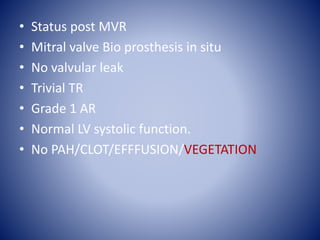
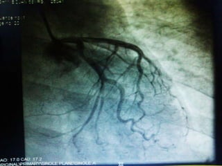
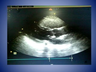
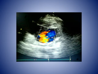
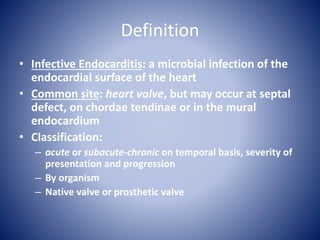
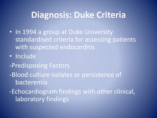
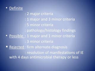
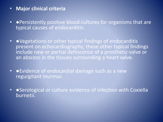
2. Patient Chief Complaints
• Mrs Lakshmi Bojan 58 years old female came
with chief complaints of
• H/O Fever on & off since 3 months
• Myalgia+
• Severe Headache since 33 months
• Generalized Weakness since 2 weeks
• Dry cough since 1 week
3. Fever
• low grade
• Intermittent
• Not associated with chills and Rigors
• 8 months
• Patient has shown to almost all hospitals in
South India.
4. – No h/o seizure
– No h/o bladder, bowel disturbance
– No h/o regurgitation of feeds
– No h/o burning micturition, increased frequency.
– No h/o abdominal pain, vomiting, diarrhea.
– No h/o cough with expectoration, breathlessness
– No h/o skin ulcers, rash, jaundice
5. Past history
– Not a known diabetic, hypertensive, epileptic,
COPD, IHD.
– No previous similar episodes.
6. Personal History
• Not a smoker and Not an alcoholic
Family history
• No Family history of Cardiac Ailment
8. General Examination
• Conscious
• Oriented
• febrile +
• Clubbing+ Grade 1
• Pallor +
• No cyanosis
• Not icteric
• No pedal edema
• No petechiae
• No Splinter
hemorrhages
• No Oslers node
• No Janeway lesion
• No Retinal hemorrhage
9. • CVS:
– S1, S2 heard,
– A pan systolic murmur heard in the Left
Parasternal Area,
– S3 Gallop +.
• RS:
– Normal vesicular breath sounds heard, No Added
Sounds
• P/A
– Soft, Right Hypochondriac tenderness +, Bowel
sounds +, no organomegaly
• CNS
WNL
10. So patient was admitted in ward on 26th jan and
started evaluating as PUO.
INVESTIGATIONS
WBC- 17.8
Hb - 9.2 g/dl
Platelet count- 3,70,000
CRP -37.7
Blood cultures and Urine Cultures were
negative.
11. • On 27th Jan Patient became breathless and
started desaturating so she was shifted to
MICU and was intubated and managed as
Acute Coronary syndrome as Trop T was 116.2
and CPK MB was 9.96. Pro BNP was 7868.
Patient was started on antiplatelets and
anticoagulants. Then patient was extubated
on 28th Jan.
Chest Xray showed Flash Pulmonary Edema
and patient started having cough with
productive sputum with blood tinge and used
to become breathless on and off
12.
13. • On 29th Jan Screening Echo was done which
showed IHD , RWMA +, Myxomatous MV,
dIlated LA, moderae MR, Grade 1 AR,
moderate TR, Severe PAH (PASP 62mmHg), EF
50%, no clots /vegetations.
• On 31st Jan, Lasix infusion was started and
diuresis done , patient was put on NIV.
14. • Work up for PUO started
• Brucella IgM and IgG – Negative
• Procalcitonin -18.030
• MPS – Negative.
• Sputum for AFB- Negative.
• Upper GI endoscopy done showed pangastritis
• Sigmoidoscopy done showed anal fissures
and hemmorhoids.
• ANA blot- Negative
• Leptospira IgM and IgG – Negative .
15. • USG Abdomen showed mild hepatomegaly,
mild bilateral pleural effusion , dilated IVC and
Hepatic veins. Patient improved
symptomatically with diuresis.
• Trans Esophageal Echocardiography done on
1ST Feb showed Mitral valve Vegetation
measuring 1.2 x 0.7 cm attached to AML, Flail
AML, non coapting leaflets with severe MR,
Aortic vlve – tri leaflets , No AR, pulmonary
valve normal, no PR, Tricuspid valve- mild TR,
Left Atrium mildly dilated , no LA appendage
clot.
16.
17.
18. • Then patient was advised to get CAG done
shifted to ward on same day.
• Patient was started on Inj VANCOMYCIN on 4th
Feb under ICU Supervision. Patient tolerated
antibiotics well then patient was advised to
continue Inj Vancomycin and Inj Ceftriaxone
on OPD basis. Central Line Insertion was done
on 6th Feb, Patient was discharged with central
line in situ on 6th Feb and was explained about
the need of MVR once the infection settles .
19. • Patient came back on 10th feb and undergone
CAG on 13th feb which showed Normal
Coronaries.
• Patient underwent MVR with 27mm BiOCAR
VALVE ON 17th Feb.
• Post op Echo done on 10th march showed
20. • Status post MVR
• Mitral valve Bio prosthesis in situ
• No valvular leak
• Trivial TR
• Grade 1 AR
• Normal LV systolic function.
• No PAH/CLOT/EFFFUSION/VEGETATION
21.
22. Follow up
Echo done on 29th july
• Status post MVR
• Mitral valve Bio prosthesis in situ
• No valvular and paravalvular leak
• Mild TR
• Moderate PAH
• Grade 1 AR
• No RWMA
• Normal LV systolic function.
• No PAH/CLOT/EFFFUSION/VEGETATION
26. Definition
• Infective Endocarditis: a microbial infection of the
endocardial surface of the heart
• Common site: heart valve, but may occur at septal
defect, on chordae tendinae or in the mural
endocardium
• Classification:
– acute or subacute-chronic on temporal basis, severity of
presentation and progression
– By organism
– Native valve or prosthetic valve
28. Diagnosis: Duke Criteria
• In 1994 a group at Duke University
standardised criteria for assessing patients
with suspected endocarditis
• Include
-Predisposing Factors
-Blood culture isolates or persistence of
bacteremia
-Echocardiogram findings with other clinical,
laboratory findings
29. • Definite
: 2 major criteria
: 1 major and 3 minor criteria
: 5 minor criteria
: pathology/histology findings
• Possible : 1 major and 1 minor criteria
: 3 minor criteria
Rejected: firm alternate diagnosis
: resolution of manifestations of IE
with 4 days antimicrobial therapy or less
30. • Major clinical criteria
• ●Persistently positive blood cultures for organisms that are
typical causes of endocarditis.
• ●Vegetations or other typical findings of endocarditis
present on echocardiography; these other typical findings
include new or partial dehiscence of a prosthetic valve or
an abscess in the tissues surrounding a heart valve.
• ●Evidence of endocardial damage such as a new
regurgitant murmur.
• ●Serological or culture evidence of infection with Coxiella
burnetii.
31. • Minor clinical criteria — Minor clinical criteria include the following
• ●Fever
• ●The presence of a predisposing valvular condition or intravenous drug abuse.
• ●"Vascular phenomenon" such as emboli to organs or the brain, hemorrhages in
the mucous membranes around the eyes. Cerebral microhemorrhages detected by
magnetic resonance imaging (MRI) are NOT considered vascular phenomena by
the modified Duke criteria, even though they are more common in patients with IE
than in age matched controls.
• ●"Immunologic phenomenon" such as glomerulonephritis, or lesions such as
Roth's spots (in the retina of the eyes) or "Osler's nodes (nodules on the fingers or
toes)
• ●Positive blood cultures that do not meet the strict definitions of a major
criterion.
• Other minor criteria (including hematuria and splenomegaly) have been proposed
by the St. Thomas group, but important considerations and validations of these
and other criteria have not been undertaken.
35. Echocardiography
• Trans Thoracic Echocardiograpy (TTE)
– rapid, non-invasive – excellent specificity (98%) but poor
sensitivity
– obesity, chronic obstructive pulmonary disease and chest
wall deformities
• Transesophageal Echo (TOE)
– more invasive, sensitivity up to 95%, useful for prosthetic
valves and to evaluate myocardial invasion
– Negative predictive valve of 92%
– TOE more cost effective in those with S. aureus catheter-
associated bacteremia and bacteremia/fever and recent
IVDA
36. Culture Negative Endocarditis
• Blood culture-negative IE is defined as endocarditis without
etiology following inoculation of three independent blood
samples in a standard blood culture system with negative
cultures after seven days of incubation and subculturing.
• 5-7% of patients with endocarditis will have sterile blood
cultures
• 1 Year study from France
– 44 of 88 cases of CNE, negative cultures were associated
with prior administration of antibiotics
• Withhold empirical therapy until cultures drawn
37. • ●Cultures are negative in patients with IE for three
major reasons:
• •Previous administration of antimicrobial agents
• •Inadequate microbiological techniques
• •Infection with highly fastidious bacteria or
nonbacterial pathogens (eg, fungi)
38. • ●The incidence of culture-negative IE is higher in
developing countries.
• ●HACEK organisms can be easily isolated with current
blood culture systems
• ●The local prevalence of infection with pathogens such
as C. burnetii and Bartonella spp, the most common
agents of culture-negative endocarditis, varies widely
in different geographic locations and epidemiologic
settings.
• ●Serology and PCR help on blood samples or removed
valves help to identify fastidious pathogens.
39. Therapy
• Streptococci/Enterococci
– Determine MIC of Penicillin
– Penicillin +/- aminoglycoside
– Ceftriaxone alone
– Vancomycin +/- aminoglycoside
HACEK Group
– Cefotaxime/ceftriaxone