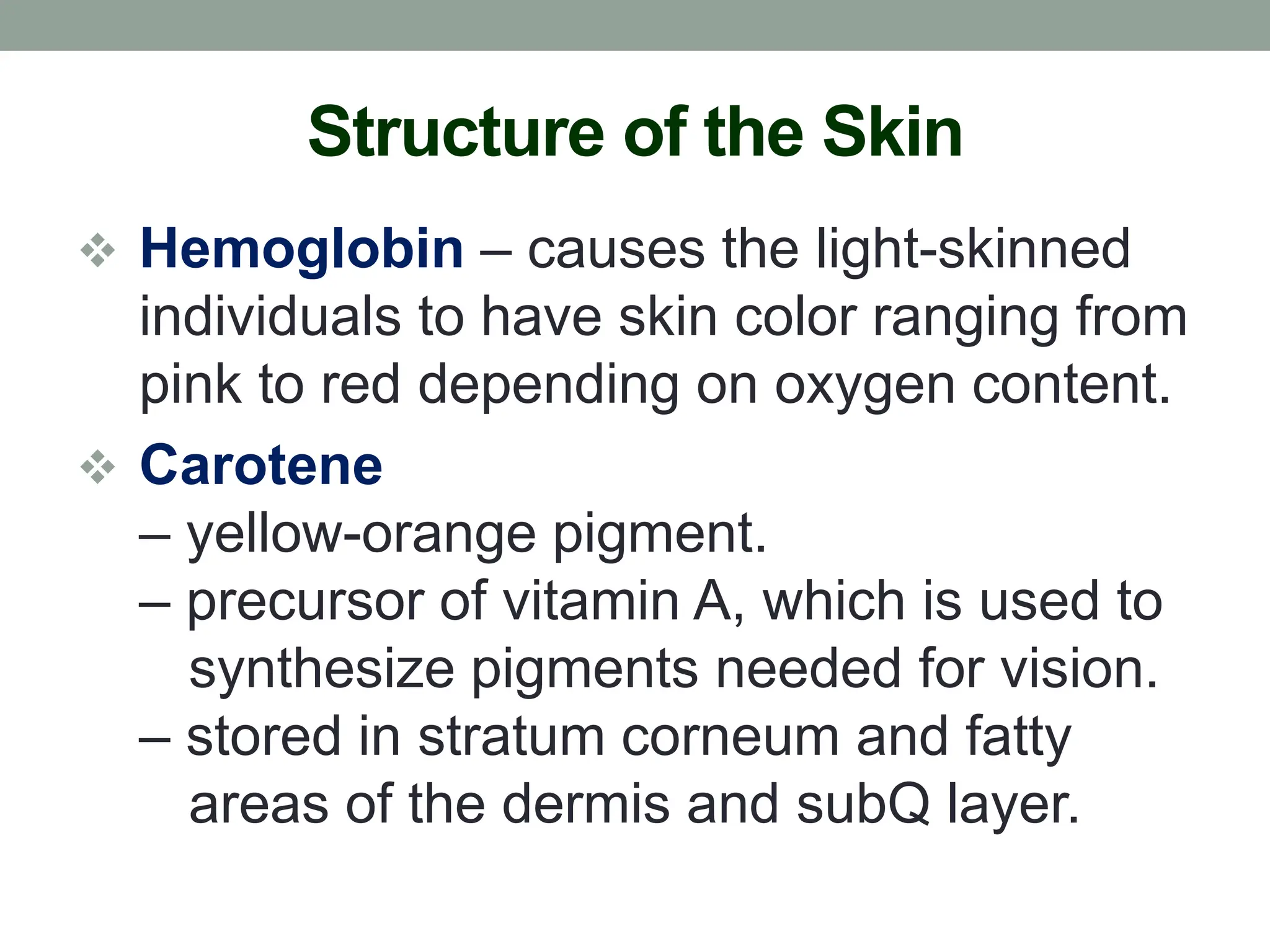
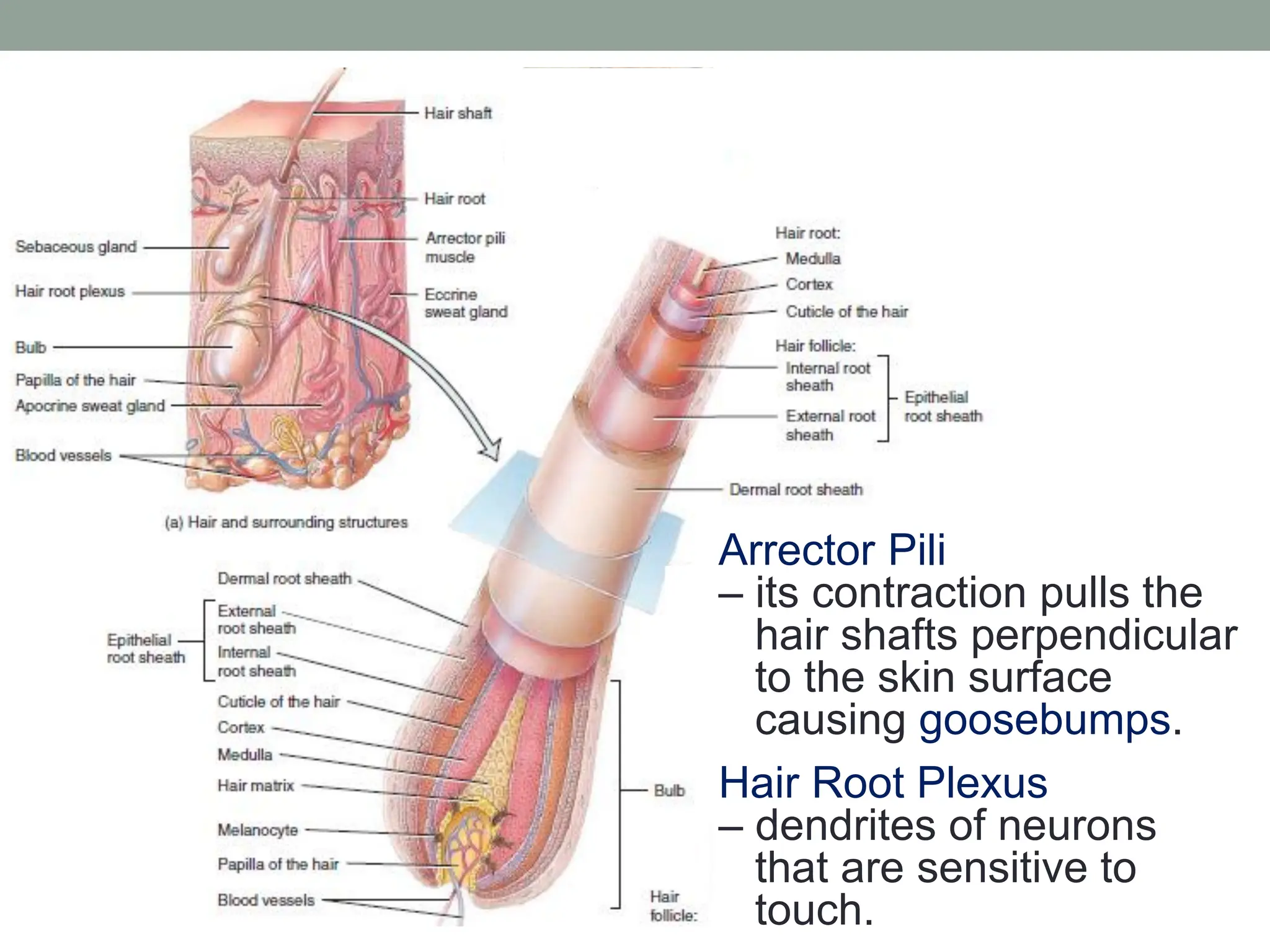
INTEGUMENTARY
SYSTEM
Reference:
Tortora, G.J. & Derrickson, B. (2012). Principles of anatomy and Physiology
(13th ed.). John Wiley & Sons, Inc.
Prepared by:
Maria Lourdes P. Sagun
Functions of the Integumentary
System
• Regulates body temperature.
• Stores blood.
• Protects body from external environment.
• Detects cutaneous sensations.
• Excretes and absorbs substances.
• Synthesizes vitamin D.
Structure of the Skin
Skin – cutaneous membrane. It covers the
external surface of the body and is
the largest organ of the body.
Epidermis – consists of epithelial tissue.
Dermis – composed of connective tissue.
Hypodermis – consists of areolar and
adipose tissues. It also contains blood
vessels and nerve endings (pacinian
corpuscles) that are sensitive to pressure.
Sectional View of Skin and Subcutaneous Layer
Structure of the Skin
Principal Types of Cells in the Epidermis
Keratinocytes
– produce keratin, which protect the skin
and underlying tissues from abrasions,
heat, microbes, and chemicals.
– produce lamellar granules, which
release a water-repellent sealant that
decreases water entry and loss and
inhibits the entry of foreign materials.
Structure of the Skin
Melanocytes – produce melanin, which
contributes to skin color and absorbs
ultraviolet light.
Langerhans Cells – epidermal dendritic
cells. They help other cells of the immune
system recognize microbe and destroy it.
Merkel Cells & Merkel (tactile) disc –
detect touch sensations.
Cells in the Epidermis
Structure of the Skin
Layers of the Epidermis
Stratum Basale/Stratum Germinativum
– some cells in this layer are stem cells,
which produce new keratinocytes.
– the keratin intermediate filaments
(tonofilaments) form the keratin that
protects the deeper layers from injury.
 Skin Graft – transfer of a patch of
healthy skin to cover a wound.
Structure of the Skin
Stratum Spinosum
– keratinocytes have the same organelles
as cells of the stratum basale and some
retain their ability to divide.
– at each spinelike projection, keratin
intermediate filaments insert into
desmosomes, which tightly join the cells
to one another. This provides strength
and flexibility to the skin.
Structure of the Skin
Stratum Granulosum
– keratinocytes are undergoing apoptosis.
– a distinctive feature is the presence of
darkly staining granules of protein,
keratohyalin and lamellar granules,
which fuse with the plasma membrane
and release a lipid-rich secretion.
– marks the transition between the active
strata and the dead cells.
Structure of the Skin
Stratum Lucidum – dead keratinocytes
that contain large amounts of keratin and
thickened plasma membranes, which
provides additional level of toughness.
Stratum Corneum – multiple layers of
dead cells protect deeper layers from
injury and microbial invasion.
Callus – abnormal thickening the stratum
corneum.
Structure of the Skin
Keratinization – newly formed cells in the
stratum basale are slowly pushed to the
surface. As cells move from an epidermal
layer to the next, they accumulate keratin.
Dandruff – excessive amount of keratinized
cells shed from the scalp.
 Psoriasis – keratinocytes divide and move
more quickly than normal from stratum
basale to stratum corneum (7 to 10 days).
Structure of the Skin
Division of Dermis (Tissue Structure):
Papillary Region – consists of thin
collagen and fine elastic fibers. Its surface
area is increased by dermal papillae.
Capillary Loops – blood vessels.
Meissner Corpuscles – tactile receptors.
Free Nerve Endings – initiate signals that
give rise to sensations of warmth,
coolness, pain, tickling and itching.
Structure of the Skin
Reticular Region – regular orientation of
the large collagen fibers helps the skin
resist stretching. Blood vessels, nerves,
hair follicles, oil glands and sudoriferous
glands occupy the spaces between fibers.
 Stretch Marks – internal scarring due to
the internal damage of the collagenous,
vascular structure of the dermis when
the skin is stretched too much.
Structure of the Skin
Epidermal Ridges – straight lines or pattern
of loops and whorls, as on the tips of the
digits. They increase the grip of the hand
or foot by increasing friction. They as well
increase the number of tactile receptors.
Fingerprints/Footprint – formed from the
sweat and ridges.
 Tension Lines – indicate the predominant
direction of underlying collagen fibers.
Structure of the Skin
Structural Basis of Skin Color
 Melanin – causes the skin’s color to vary
from pale yellow to reddish-brown to black
 Pheomelanin – yellow to red
 Eumelanin – brown to black
Freckles – patches of accumulated melanin
Age (Liver) Spots – flat blemishes
Nevus (Mole) – localized overgrowth of
melanocytes
Structure of the Skin
Melanocytes synthesize melanin from the
amino acid tyrosine in the presence of an
enzyme tyrosinase. Synthesis occurs in an
organelle melanosome.
 Albinism – inherited inability to produce
melanin. Melanocytes are unable to
synthesize tyrosinase.
 Vitiligo – an immune system malfunction
in which antibodies attack melanocytes.
Structure of the Skin
 Hemoglobin – causes the light-skinned
individuals to have skin color ranging from
pink to red depending on oxygen content.
 Carotene
– yellow-orange pigment.
– precursor of vitamin A, which is used to
synthesize pigments needed for vision.
– stored in stratum corneum and fatty
areas of the dermis and subQ layer.
Structure of the Skin
 Cyanotic – mucous membrane, nail beds
and skin appear bluish due to inadequate
amount of oxygen.
 Jaundice – yellowish appearance to skin
and eyes due to buildup of bilirubin.
 Erythema – redness of the skin caused by
engorgement of capillaries in the dermis
due injury, exposure to heat, infection,
inflammation or allergic reaction.
Structure of the Skin
 Pallor – paleness of the skin because of
shock and anemia.
Tattoing – permanent coloration of the skin
in which a foreign pigment is deposited
with a needle into the dermis.
Body Piercing – insertion of jewelry through
an artificial opening. Complications are
infections, allergic reactions, and
anatomical damage.
Accesory Structures of the Skin
 Hairs – genetic and hormonal influences
determines the thickness and pattern of
hair distribution.
Layers of Cells:
 Medulla
 Cortex
 Cuticle
Accesory Structures of the Skin
Hair Follicle – surrounds the root hair, which
is made up of epithelial root sheath.
Dermal Root Sheath – dense dermis that
surrounds the hair follicle.
Bulb – base of hair follicle and dermal root
sheath. It housed the papilla of the hair
(contains areolar connective tissue and
blood vessels), and the hair matrix
(germinal layer of cells).
Arrector Pili
– its contraction pulls the
hair shafts perpendicular
to the skin surface
causing goosebumps.
Hair Root Plexus
– dendrites of neurons
that are sensitive to
touch.
Accesory Structures of the Skin
Stages of Hair Growth Cycle:
 Growth – cells of the hair matrix divide.
Scalp hair is in this stage for 2 to 6 years.
 Regression – cells of the hair matrix stop
dividing, hair follicle atrophies (shrinks), or
the hair stops growing. It is in 2 to 3 weeks.
 Resting – old hair root falls out or pushed
out of the hair follicle, and a new hair
begins to grow in its place in ~3 months.
Accesory Structures of the Skin
Alopecia – partial or complete lack of hair,
may result from genetic factors, aging,
endocrine disorders, chemotherapy or
skin disease.
 Hair Removal
 Depilatory – substance which dissolves
the protein in the hair shaft.
 Electrolysis – electric current is used to
destroy the hair matrix.
Accesory Structures of the Skin
Types of Hairs:
 Lanugo – very fine, nonpigmented, downy
hairs produce by the hair follicles in the
fifth month of development.
 Terminal Hairs – long, coarse, heavily
pigmented hairs that replaced the lanugo
of the eyebrows, eyelashes and scalp.
 Vellus Hairs – short, fine, pale hairs that
replace the lanugo on rest of the body.
Accesory Structures of the Skin
Hair Color
 Dark-Colored Hair – contain mostly
eumelanin (brown to black).
 Blond and Red Hair – contain pheomelanin
(yellow to red).
 Gray Hair – result of a progressive decline
in melanin production.
 White Hair – result from lack of melanin,
accumulation of air bubbles in the shaft.
Accesory Structures of the Skin
 Hirsutism – excessive body hair or body
hair in areas that usually are not hairy. It
may be caused by a tumor of the adrenal
gland, testes, or ovaries produces an
excessive amount of androgens.
 Androgenic Alopecia – androgens inhibit
hair growth.
Minoxidil – first drug approved for
enhancing scalp hair growth.
Accesory Structures of the Skin
 Sebaceous Glands – secrete sebum.
 coats the surface of hairs
 prevents excessive evaporation of water
 keeps the skin soft and pliable
 inhibits the growth of some bacteria
 Acne – inflammation of oil glands. It
occurs in sebaceous follicles that have
been colonized by bacteria.
 Cystic Acne – the infection may destroy
and displace epidermal cells.
Accesory Structures of the Skin
 Sudoriferous Glands
 Eccrine Sweat Gland
– help regulate body temperature.
• insensible perspiration
• sensible perspiration
– eliminate wastes such as urea, uric
acid and ammonia.
– release sweat in response to an
emotional stress (emotional sweating)
Accesory Structures of the Skin
 Apocrine Sweat Gland
– apocrine sweat appears milky or
yellowish in color. It contains the same
components as eccrine sweat plus lipids
and proteins.
– secrete sweat during sexual activities
and are inactive during thermoregulatory
sweating.
– do not begin to function until puberty
Accesory Structures of the Skin
 Ceruminous Glands
– produce waxy lubricating secretion.
Cerumen – yellowish material from the
secretion of ceruminous and oil glands,
providing barrier to the entry of foreign
bodies and insects, waterproofs canal,
and prevents bacteria and fungi.
 Impacted Cerumen – abnormally large
amount of cerumen in the external
auditory canal.
Accesory Structures of the Skin
 Nails
 Nail Body – visible portion of the nail.
 Free Edge – extend past the distal end.
 Nail Root – buried in a fold of the skin.
 Lunula – crescent-shaped area of the
proximal end of the nail body.
 Hyponychium – nail bed.
 Eponychium – cuticle.
 Nail Matrix – produce new nail cells.
Accesory Structures of the Skin
Functions of Nails
 protect the distal end of the digits
 provide support and counterpressure to the
palmar surface of the fingers to enhance
touch perception and manipulation
 allow us to grasp and manipulate small
objects, and can be used to scratch and
groom the body

Integumentary System (Human Anatomy and Physiology)

  • 1.
    INTEGUMENTARY SYSTEM Reference: Tortora, G.J. &Derrickson, B. (2012). Principles of anatomy and Physiology (13th ed.). John Wiley & Sons, Inc. Prepared by: Maria Lourdes P. Sagun
  • 2.
    Functions of theIntegumentary System • Regulates body temperature. • Stores blood. • Protects body from external environment. • Detects cutaneous sensations. • Excretes and absorbs substances. • Synthesizes vitamin D.
  • 3.
    Structure of theSkin Skin – cutaneous membrane. It covers the external surface of the body and is the largest organ of the body. Epidermis – consists of epithelial tissue. Dermis – composed of connective tissue. Hypodermis – consists of areolar and adipose tissues. It also contains blood vessels and nerve endings (pacinian corpuscles) that are sensitive to pressure.
  • 4.
    Sectional View ofSkin and Subcutaneous Layer
  • 5.
    Structure of theSkin Principal Types of Cells in the Epidermis Keratinocytes – produce keratin, which protect the skin and underlying tissues from abrasions, heat, microbes, and chemicals. – produce lamellar granules, which release a water-repellent sealant that decreases water entry and loss and inhibits the entry of foreign materials.
  • 6.
    Structure of theSkin Melanocytes – produce melanin, which contributes to skin color and absorbs ultraviolet light. Langerhans Cells – epidermal dendritic cells. They help other cells of the immune system recognize microbe and destroy it. Merkel Cells & Merkel (tactile) disc – detect touch sensations.
  • 7.
    Cells in theEpidermis
  • 8.
    Structure of theSkin Layers of the Epidermis Stratum Basale/Stratum Germinativum – some cells in this layer are stem cells, which produce new keratinocytes. – the keratin intermediate filaments (tonofilaments) form the keratin that protects the deeper layers from injury.  Skin Graft – transfer of a patch of healthy skin to cover a wound.
  • 9.
    Structure of theSkin Stratum Spinosum – keratinocytes have the same organelles as cells of the stratum basale and some retain their ability to divide. – at each spinelike projection, keratin intermediate filaments insert into desmosomes, which tightly join the cells to one another. This provides strength and flexibility to the skin.
  • 10.
    Structure of theSkin Stratum Granulosum – keratinocytes are undergoing apoptosis. – a distinctive feature is the presence of darkly staining granules of protein, keratohyalin and lamellar granules, which fuse with the plasma membrane and release a lipid-rich secretion. – marks the transition between the active strata and the dead cells.
  • 11.
    Structure of theSkin Stratum Lucidum – dead keratinocytes that contain large amounts of keratin and thickened plasma membranes, which provides additional level of toughness. Stratum Corneum – multiple layers of dead cells protect deeper layers from injury and microbial invasion. Callus – abnormal thickening the stratum corneum.
  • 13.
    Structure of theSkin Keratinization – newly formed cells in the stratum basale are slowly pushed to the surface. As cells move from an epidermal layer to the next, they accumulate keratin. Dandruff – excessive amount of keratinized cells shed from the scalp.  Psoriasis – keratinocytes divide and move more quickly than normal from stratum basale to stratum corneum (7 to 10 days).
  • 14.
    Structure of theSkin Division of Dermis (Tissue Structure): Papillary Region – consists of thin collagen and fine elastic fibers. Its surface area is increased by dermal papillae. Capillary Loops – blood vessels. Meissner Corpuscles – tactile receptors. Free Nerve Endings – initiate signals that give rise to sensations of warmth, coolness, pain, tickling and itching.
  • 15.
    Structure of theSkin Reticular Region – regular orientation of the large collagen fibers helps the skin resist stretching. Blood vessels, nerves, hair follicles, oil glands and sudoriferous glands occupy the spaces between fibers.  Stretch Marks – internal scarring due to the internal damage of the collagenous, vascular structure of the dermis when the skin is stretched too much.
  • 16.
    Structure of theSkin Epidermal Ridges – straight lines or pattern of loops and whorls, as on the tips of the digits. They increase the grip of the hand or foot by increasing friction. They as well increase the number of tactile receptors. Fingerprints/Footprint – formed from the sweat and ridges.  Tension Lines – indicate the predominant direction of underlying collagen fibers.
  • 17.
    Structure of theSkin Structural Basis of Skin Color  Melanin – causes the skin’s color to vary from pale yellow to reddish-brown to black  Pheomelanin – yellow to red  Eumelanin – brown to black Freckles – patches of accumulated melanin Age (Liver) Spots – flat blemishes Nevus (Mole) – localized overgrowth of melanocytes
  • 18.
    Structure of theSkin Melanocytes synthesize melanin from the amino acid tyrosine in the presence of an enzyme tyrosinase. Synthesis occurs in an organelle melanosome.  Albinism – inherited inability to produce melanin. Melanocytes are unable to synthesize tyrosinase.  Vitiligo – an immune system malfunction in which antibodies attack melanocytes.
  • 19.
    Structure of theSkin  Hemoglobin – causes the light-skinned individuals to have skin color ranging from pink to red depending on oxygen content.  Carotene – yellow-orange pigment. – precursor of vitamin A, which is used to synthesize pigments needed for vision. – stored in stratum corneum and fatty areas of the dermis and subQ layer.
  • 20.
    Structure of theSkin  Cyanotic – mucous membrane, nail beds and skin appear bluish due to inadequate amount of oxygen.  Jaundice – yellowish appearance to skin and eyes due to buildup of bilirubin.  Erythema – redness of the skin caused by engorgement of capillaries in the dermis due injury, exposure to heat, infection, inflammation or allergic reaction.
  • 21.
    Structure of theSkin  Pallor – paleness of the skin because of shock and anemia. Tattoing – permanent coloration of the skin in which a foreign pigment is deposited with a needle into the dermis. Body Piercing – insertion of jewelry through an artificial opening. Complications are infections, allergic reactions, and anatomical damage.
  • 22.
    Accesory Structures ofthe Skin  Hairs – genetic and hormonal influences determines the thickness and pattern of hair distribution. Layers of Cells:  Medulla  Cortex  Cuticle
  • 23.
    Accesory Structures ofthe Skin Hair Follicle – surrounds the root hair, which is made up of epithelial root sheath. Dermal Root Sheath – dense dermis that surrounds the hair follicle. Bulb – base of hair follicle and dermal root sheath. It housed the papilla of the hair (contains areolar connective tissue and blood vessels), and the hair matrix (germinal layer of cells).
  • 24.
    Arrector Pili – itscontraction pulls the hair shafts perpendicular to the skin surface causing goosebumps. Hair Root Plexus – dendrites of neurons that are sensitive to touch.
  • 25.
    Accesory Structures ofthe Skin Stages of Hair Growth Cycle:  Growth – cells of the hair matrix divide. Scalp hair is in this stage for 2 to 6 years.  Regression – cells of the hair matrix stop dividing, hair follicle atrophies (shrinks), or the hair stops growing. It is in 2 to 3 weeks.  Resting – old hair root falls out or pushed out of the hair follicle, and a new hair begins to grow in its place in ~3 months.
  • 26.
    Accesory Structures ofthe Skin Alopecia – partial or complete lack of hair, may result from genetic factors, aging, endocrine disorders, chemotherapy or skin disease.  Hair Removal  Depilatory – substance which dissolves the protein in the hair shaft.  Electrolysis – electric current is used to destroy the hair matrix.
  • 27.
    Accesory Structures ofthe Skin Types of Hairs:  Lanugo – very fine, nonpigmented, downy hairs produce by the hair follicles in the fifth month of development.  Terminal Hairs – long, coarse, heavily pigmented hairs that replaced the lanugo of the eyebrows, eyelashes and scalp.  Vellus Hairs – short, fine, pale hairs that replace the lanugo on rest of the body.
  • 28.
    Accesory Structures ofthe Skin Hair Color  Dark-Colored Hair – contain mostly eumelanin (brown to black).  Blond and Red Hair – contain pheomelanin (yellow to red).  Gray Hair – result of a progressive decline in melanin production.  White Hair – result from lack of melanin, accumulation of air bubbles in the shaft.
  • 29.
    Accesory Structures ofthe Skin  Hirsutism – excessive body hair or body hair in areas that usually are not hairy. It may be caused by a tumor of the adrenal gland, testes, or ovaries produces an excessive amount of androgens.  Androgenic Alopecia – androgens inhibit hair growth. Minoxidil – first drug approved for enhancing scalp hair growth.
  • 30.
    Accesory Structures ofthe Skin  Sebaceous Glands – secrete sebum.  coats the surface of hairs  prevents excessive evaporation of water  keeps the skin soft and pliable  inhibits the growth of some bacteria  Acne – inflammation of oil glands. It occurs in sebaceous follicles that have been colonized by bacteria.  Cystic Acne – the infection may destroy and displace epidermal cells.
  • 31.
    Accesory Structures ofthe Skin  Sudoriferous Glands  Eccrine Sweat Gland – help regulate body temperature. • insensible perspiration • sensible perspiration – eliminate wastes such as urea, uric acid and ammonia. – release sweat in response to an emotional stress (emotional sweating)
  • 32.
    Accesory Structures ofthe Skin  Apocrine Sweat Gland – apocrine sweat appears milky or yellowish in color. It contains the same components as eccrine sweat plus lipids and proteins. – secrete sweat during sexual activities and are inactive during thermoregulatory sweating. – do not begin to function until puberty
  • 33.
    Accesory Structures ofthe Skin  Ceruminous Glands – produce waxy lubricating secretion. Cerumen – yellowish material from the secretion of ceruminous and oil glands, providing barrier to the entry of foreign bodies and insects, waterproofs canal, and prevents bacteria and fungi.  Impacted Cerumen – abnormally large amount of cerumen in the external auditory canal.
  • 34.
    Accesory Structures ofthe Skin  Nails  Nail Body – visible portion of the nail.  Free Edge – extend past the distal end.  Nail Root – buried in a fold of the skin.  Lunula – crescent-shaped area of the proximal end of the nail body.  Hyponychium – nail bed.  Eponychium – cuticle.  Nail Matrix – produce new nail cells.
  • 35.
  • 36.
    Accesory Structures ofthe Skin Functions of Nails  protect the distal end of the digits  provide support and counterpressure to the palmar surface of the fingers to enhance touch perception and manipulation  allow us to grasp and manipulate small objects, and can be used to scratch and groom the body