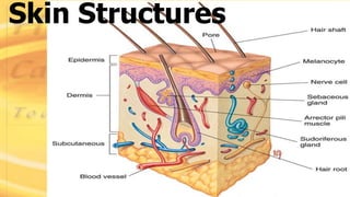
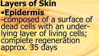
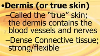
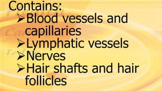
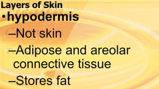
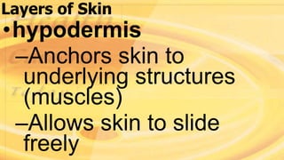
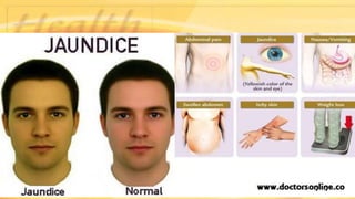
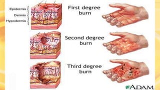
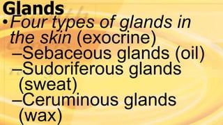
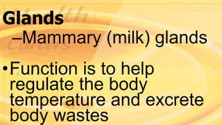
INTEGUMENTARY
SYSTEM
Structure and Function
•Integumentary system is
composed of the skin and
accessory structures (hair,
hair follicles, nails, glands
& nerves)
Structure and Function
•Functions of the
integumentary system
–Protects the other body
systems from injury and
infection:
Structure and Function
•Mechanical damage
•Chemical damage
•Thermal damage
•Ultraviolet damage
•Defense against
microorganisms
•Helps the body maintain
homeostasis by regulating
temperature, retaining
body fluids, and
eliminating wastes.
•Insulates and cushions
deeper organs
Skin Structures
Skin
•The largest organ of the
body
•1.5 – 2 sq meters
•4 kilograms, 7-15% of
total body weight
Skin
•Varies in thickness from
1/50 inch (0.5 mm) in the
eyelids to 1/4 inch (6.3
mm) in the soles of the
feet
•Changes in the skin often
indicate the presence of
other body system
disorders including
anemia, respiratory
disorders, liver disorders,
cancer, and shock
•Each inch of skin contains
15 ft of blood vessels
•The body sloughs off
about 500 million cells a
day; 1 ½ pounds per year
•The top layer is full of
keratin to prevent water
loss
•The rich capillary network
and sweat glands help
regulate heat loss from
surface
•It is a mini-excretory
system- loses urea, water
and salts
•Cutaneous sensory
receptors provide info
about the environment
•Manufactures proteins
important to immunity and
synthesizes Vitamin A, D &
K. (vit D synthesis very
important)
Layers of Skin
•Epidermis
-Outermost layer of the
skin. that is composed of
a surface of dead cells
with an underlying layer
Layers of Skin
•Epidermis
-composed of a surface of
dead cells with an under-
lying layer of living cells;
complete regeneration
approx. 35 days
•Made up of stratified
squamous cells that are
capable of keratinizing
(hard/tough)
•Melanocytes (pigment
cells); increase in sun
exposure will cause
these cells to produce
more melanin
•Dermis (or true skin)
–Called the “true” skin;
the dermis contains the
blood vessels and nerves
–Dense Connective tissue;
strong/flexible
Contains:
Blood vessels and
capillaries
Lymphatic vessels
Nerves
Hair shafts and hair
follicles
Contains:
Sensory receptors
Sudorferous glands-
sweat
Sebaceous glands- oil
Contains:
**The epidermis and
dermis are firmly
connected but can be
separated by friction
Layers of Skin
•hypodermis
–Not skin
–Adipose and areolar
connective tissue
–Stores fat
Layers of Skin
•hypodermis
–Anchors skin to
underlying structures
(muscles)
–Allows skin to slide
freely
Layers of Skin
•hypodermis
–Shock absorber,
insulator
–Thickens with weight
gain
Types of Skin
•Thick skin- only on palms
& soles
--Thick epidermis distinct
stratum lucidum & thick
stratum corneus; Lacks hair
follicles & sebaceous
glands
•Stretch marks- extreme
stretching that produces a
silvery white scar
•Blister- separation of
epidermal and dermal
layers by fluid filled pocket
•Flexure lines- skin
markings; dermal folds at
or near a joint; deep
creases
Skin Color
•Pigments:
–Keratin
•Yellow to orange
•Accumulates in stratum
corneum and fatty
tissue of hypodermis
Copyright 2003 by Mosby, Inc. All rights
reserved.
- Melanin
Only pigment in the skin
Yellow to reddish-brown
to black
Synthesis depends on
enzyme in melanocytes
- Melanin
Racial differences vary
on amount of melanin
made
Local accumulation in
freckles and moles
- Melanin
Exposure to sunlight
increases manufacturing
of melanin to prolonged
exposure; tan
Copyright 2003 by Mosby, Inc. All rights
reserved.
Abnormal
color/homeostatic
imbalances
•Erythema (redness)
–Congestion of blood in
vessels
–Burns
–Embarrassment
(blushing)
–Fever
–Hypertension
–Inflammation
–Allergy
Copyright 2003 by Mosby, Inc. All rights
reserved.
•Albinism
•Absence of pigment
color
•Vitiligo
•Loss of pigment in
certain areas, usually
acquired
Copyright 2003 by Mosby, Inc. All rights
reserved.
•Cyanosis (blue)
•Insufficient oxygen
•Jaundice (yellow)
•Liver disorder, biliary
disorder, presence of bile
in blood, diseases invol-
ving destruction of RBCs
Copyright 2003 by Mosby, Inc. All rights
reserved.
Copyright 2003 by Mosby, Inc. All rights
reserved.
Skin Injuries
•Blisters: injury damages
chemical bond of skin
layers at dermal/
epidermal junction
Skin Injuries
•Callous: abnormally thick
stratum corneum
•Burns:1st, 2nd, 3rd & 4th
degree depending on depth
of burn (UV light, corrosive
chemicals, electricity)
Copyright 2003 by Mosby, Inc. All rights
reserved.
Copyright 2003 by Mosby, Inc. All rights
reserved.
Accessory Structures
• Hair/Hair
follicle
• Glands
• Nails
• Nerves
Hair and Hair Follicles
•Skin has hair in all areas
except the soles of the
feet and palms of the
hands
Hair and Hair Follicles
•Composed of keratin and
proteins; also dead
epidermal cells
Hair and Hair Follicles
•Hair serves to block
foreign particles from
entering the body through
structures such as the
nose and eyes
Hair and Hair Follicles
•The visible portion is
called the shaft
•The hair follicle is the root
with its covering
–Anagen: growing follicle
–Telogen: resting follicle
Hair
•Growth:
–Lanugo: fetal hair and
lost at birth
–Puberty: coarse hair;
grows ½” per month
•Arrector Pili= hair
muscles
Nails
•Lunula is white due to
–thickened stratum
basale
•Cuticle (eponychium)
•Nail root- buried under
skin layers
Copyright 2003 by Mosby, Inc. All rights
reserved.
Copyright 2003 by Mosby, Inc. All rights
reserved.
Glands
•Four types of glands in
the skin (exocrine)
–Sebaceous glands (oil)
–Sudoriferous glands
(sweat)
–Ceruminous glands
(wax)
Glands
–Mammary (milk) glands
•Function is to help
regulate the body
temperature and excrete
body wastes
Sebaceous (Oil) glands
•Secretory in dermis- open
into hair shafts
•Acne- bacterial
inflammation of glands
–Secretions stimulated by
hormones at puberty
Sebaceous (Oil) glands
–Associated with
blackheads
•Sebum- combination of
cholesterol, proteins, fats,
and salts
Sebaceous (Oil) glands
–Keeps hair/skin soft and
pliable
–Inhibits growth of
bacteria and fungi
Sudoriferous glands
•Apocrine (sweat) glands-
secrete at hair follicle and
active at puberty
–In dermis with ducts that
open into hair follicles
–Axilla –Perianal
–Areole -Periumbilical
Sudoriferous glands
–Eyelids
–External ear canal
–External ear canal
Sudoriferous glands
–Also consists of ducts
and secretory coils, but
these glands are larger
than eccrine glands and
open onto hair follicles.
Eccrine (sweat glands)
Most areas of the body
In dermis with ducts to
surface
Regulates body temp w/
perspiration
(500 mL p/day)
•Modified sweat glands
•Located in auditory canal
•Cerumen- contains
secretions of oil and wax
glands; barrier for entrance
of foreign bodies
•Impacted cerumen may
reduce hearing

Integumentary system