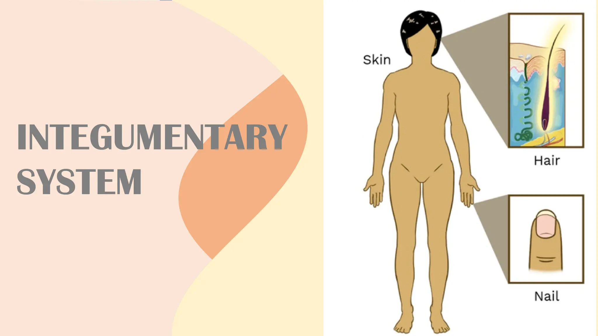
INTEGUMENTARY SYSTEM
- Consistsof the skin and accessory
structures, such as hair, glands, and
nails.
- Integument means covering, and the
integumentary system is one of the
more familiar systems of the body to
everyone because it covers the outside
of the body and is easily observed.
3.
INTEGUMENTARY SYSTEM
- Appearancein the integumentary system can
indicate physiological imbalances in the body.
- Some disorders such as acne or warts, affect
just the integumentary system.
- Other disorders affect different parts of the
body but are reflected in the integumentary
system, providing useful signs for diagnosis.
SKIN
- The skinrests on the subcutaneous tissue,
which is also a layer of connective tissue.
- The subcutaneous tissue is not part of the skin,
but it does connect the skin to underlying
muscle or bone.
- The skin is made up of two major tissue layers:
(1) epidermis and (2) dermis.
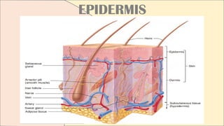
EPIDERMIS
- Meaning uponthe dermis
- Is the most superficial layer of the skin. It is a
layer of epithelial tissue that rests on the
dermis.
- Is a stratified squamous epithelium; in its
deepest layers, new cells are produced by
mitosis.
- The many cells of the epidermis prevent water
loss and resist abrasion.
13.
EPIDERMIS
- The outermostcells protect the cells
underneath, and the deeper, replicating cells
replace cells lost from the surface.
KERATINIZATION
- A process where the cells change shape and
chemical composition.
- The term keratinization reflects the fact that the
cells become filled with the protein keratin,
which makes them more rigid and durable.
1
2
3
4
5
STRATUM BASALE
Consists ofcuboidal or
columnar cells that undergo
mitotic division about every
19 days. One daughter cell
becomes a new stratum
basale cell and can divide
again. The other daughter
cell is pushed toward the
surface, a journey that
takes about 40-56 days.
Five strata
from
deepest to
the most
superficial
16.
1
2
3
4
5
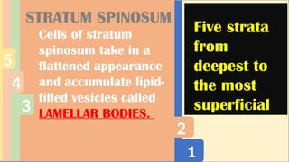
STRATUM SPINOSUM
Cells ofstratum
spinosum take in a
flattened appearance
and accumulate lipid-
filled vesicles called
LAMELLAR BODIES.
Five strata
from
deepest to
the most
superficial
1
2
3
4
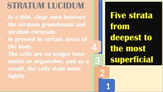
STRATUM LUCIDUM
- Isa thin, clear zone between
the stratum granulosum and
stratum corneum.
- Is present in certain areas of
the body.
- The cells are no longer have
nuclei or organelles, and as a
result, the cells stain more
lightly.
Five strata
from
deepest to
the most
superficial
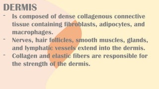
DERMIS
- Is composedof dense collagenous connective
tissue containing fibroblasts, adipocytes, and
macrophages.
- Nerves, hair follicles, smooth muscles, glands,
and lymphatic vessels extend into the dermis.
- Collagen and elastic fibers are responsible for
the strength of the dermis.
22.
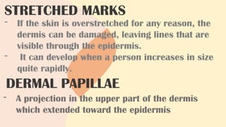
STRETCHED MARKS
- Ifthe skin is overstretched for any reason, the
dermis can be damaged, leaving lines that are
visible through the epidermis.
- It can develop when a person increases in size
quite rapidly.
DERMAL PAPILLAE
- A projection in the upper part of the dermis
which extended toward the epidermis
23.
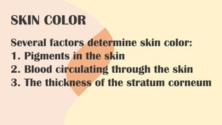
SKIN COLOR
Several factorsdetermine skin color:
1. Pigments in the skin
2. Blood circulating through the skin
3. The thickness of the stratum corneum
24.
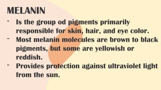
MELANIN
- Is thegroup od pigments primarily
responsible for skin, hair, and eye color.
- Most melanin molecules are brown to black
pigments, but some are yellowish or
reddish.
- Provides protection against ultraviolet light
from the sun.
25.
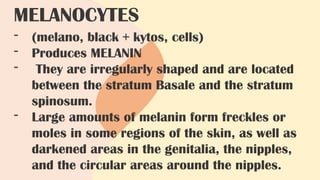
MELANOCYTES
- (melano, black+ kytos, cells)
- Produces MELANIN
- They are irregularly shaped and are located
between the stratum Basale and the stratum
spinosum.
- Large amounts of melanin form freckles or
moles in some regions of the skin, as well as
darkened areas in the genitalia, the nipples,
and the circular areas around the nipples.
26.
- For example,ALBINISM is a recessive genetic
trait that causes a deficiency or an absence of
melanin, resulting in fair skin, white hair, and
unpigmented irises in the eyes.
- Exposure to ultraviolet light, in sunlight,
stimulates melanocytes to increase melanin
production. The result is a suntan. The
production of melanin in response to ultraviolet
light is a protective measure, reducing DNA
damage in the cell.
27.
- Estrogen andmelanocyte-stimulating hormone
can cause an increase in melanin production
during pregnancy in the mother.
- Blood flowing through the skin imparts a
reddish hue, and when blood flow increases, the
red color intensifies.
- A decrease in the blood O, content produces
bluish discoloration of the skin, called
CYANOSIS
- Birthmarks are congenital disorders of the
blood vessels in the dermis
28.
CAROTENE
- Is ayellow pigment found in plants such as
squash and carrots.
- Humans normally ingest carotene and use it as
a source of Vitamin A.
- It is a lipid-soluble, and when consumed, it
accumulates in the lipids of the stratum
corneum and the adipocytes of the dermis and
subcutaneous tissue.
29.
- The subcutaneoustissue is not part of the
skin but instead attached the skin to
underlying bone and muscle and supplies it
with blood vessels and nerves.
- Is a loose connective tissue, including
adipose tissue that contains about half the
body’s stored lipids.
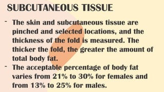
SUBCUTANEOUS TISSUE
30.
- The skinand subcutaneous tissue are
pinched and selected locations, and the
thickness of the fold is measured. The
thicker the fold, the greater the amount of
total body fat.
- The acceptable percentage of body fat
varies from 21% to 30% for females and
from 13% to 25% for males.
SUBCUTANEOUS TISSUE
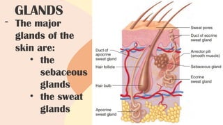
31.
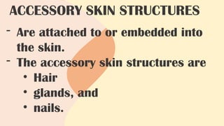
- Are attachedto or embedded into
the skin.
- The accessory skin structures are
• Hair
• glands, and
• nails.
ACCESSORY SKIN STRUCTURES
H A IR
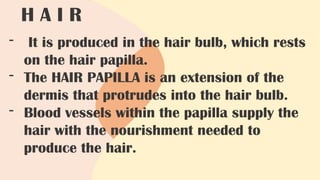
- It is produced in the hair bulb, which rests
on the hair papilla.
- The HAIR PAPILLA is an extension of the
dermis that protrudes into the hair bulb.
- Blood vessels within the papilla supply the
hair with the nourishment needed to
produce the hair.
34.
H A IR
- The duration of each stage depends on the
individual hair.
- Eyelashes grow for about 30 days and rest
for 105 days
- Scalp hairs grow for 3 yrs and rest for 1-2
years.
35.
H A IR COLOR
- Is determined by varying amounts and types
of melanin.
- The production and distribution of melanin
by melanocytes occurs in the hair bulb by
the same method as in the skin.
- With age, the amount of melanin in hair can
decrease, causing the hair color to become
faded, or the hair can contain no melanin
and be white.
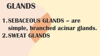
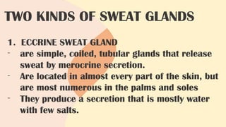
TWO KINDS OFSWEAT GLANDS
1. ECCRINE SWEAT GLAND
- are simple, coiled, tubular glands that release
sweat by merocrine secretion.
- Are located in almost every part of the skin, but
are most numerous in the palms and soles
- They produce a secretion that is mostly water
with few salts.
39.
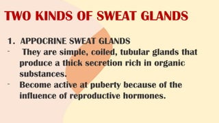
TWO KINDS OFSWEAT GLANDS
1. APPOCRINE SWEAT GLANDS
- They are simple, coiled, tubular glands that
produce a thick secretion rich in organic
substances.
- Become active at puberty because of the
influence of reproductive hormones.
40.
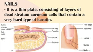
NAILS
- It isa thin plate, consisting of layers of
dead stratum corneum cells that contain a
very hard type of keratin.
41.
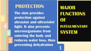
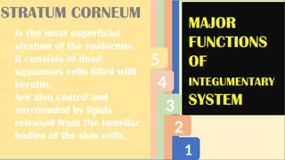
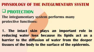
PHYSIOLOGY OF THEINTEGUMENTARY SYSTEM
PROTECTION
The integumentary system performs many
protective functions:
1. The intact skin plays an important role in
reducing water loss because its lipids act as a
barrier to the diffusion of water from the deeper
tissues of the body to the surface of the epidermis.
42.
PHYSIOLOGY OF THEINTEGUMENTARY SYSTEM
2. The skin acts as a barrier that prevents
microorganisms and other foreign
substances from entering the body.
Secretions from skin glands also produce
an environment unsuitable for some
microorganisms.
43.
PHYSIOLOGY OF THEINTEGUMENTARY SYSTEM
3. The stratified squamous epithelium of the
skin protects underlying structures against
abrasion.
4. Melanin absorbs ultraviolet light and
protects underlying structures from its
damaging effects.
44.
PHYSIOLOGY OF THEINTEGUMENTARY SYSTEM
5. Hair protects in several ways: The hair on
the head acts as a heat insulator, eyebrows
keep sweat out of the eyes, eyelashes protect
the eyes from foreign objects, and hair in the
nose and ears prevents the entry of dust and
other materials.
6. The nails protect the ends of the fingers
and toes from damage.
45.
PHYSIOLOGY OF THEINTEGUMENTARY SYSTEM
SENSATION
- Many sensory receptors are associated with
the skin. Receptors in the epidermis and
dermis can detect pain, heat, cold, and
pressure.
- Although hair does not have a nerve supply,
sensory receptors around the hair follicle
can detect the movement of a hair.
46.
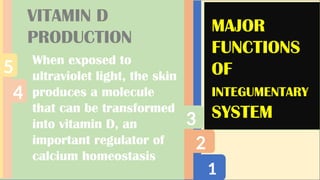
PHYSIOLOGY OF THEINTEGUMENTARY SYSTEM
VITAMIN D PRODUCTION
- When the skin is exposed to ultraviolet light,
a precursor molecule of vitamin D is
formed. The precursor is carried by the
blood to the liver, where it is modified, and
then to the kidneys, where the precursor is
modified further to form active vitamin D.
47.
PHYSIOLOGY OF THEINTEGUMENTARY SYSTEM
VITAMIN D PRODUCTION
- Adequate levels of vitamin D are
necessary because active vitamin D
stimulates the small intestine to absorb
calcium and phosphate, the substances
necessary for normal bone growth and
normal muscle function.
48.
PHYSIOLOGY OF THEINTEGUMENTARY SYSTEM
TEMPERATURE REGULATION
- Body temperature is normally maintained at
about 37°C (98.6°F).
- Regulation of body temperature is important
because the rate of chemical reactions
within the body can be increased or
decreased by changes in body temperature.
49.
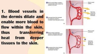
1. Blood vesselsin
the dermis dilate and
enable more blood to
flow within the skin,
thus transferring
heat from deeper
tissues to the skin.
50.
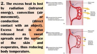
2. The excessheat is lost
by radiation (infrared
energy), convection (air
movement), or
conduction (direct
contact with an object).
Excess heat is also
released as sweat
spreads over the surface
of the skin and
evaporates, thus reducing
body temperature.
51.
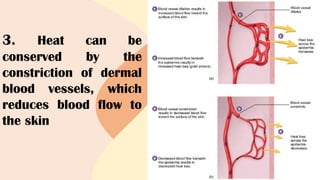
3. Heat canbe
conserved by the
constriction of dermal
blood vessels, which
reduces blood flow to
the skin
52.
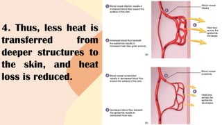
4. Thus, lessheat is
transferred from
deeper structures to
the skin, and heat
loss is reduced.
53.
PHYSIOLOGY OF THEINTEGUMENTARY SYSTEM
EXCRETION
- The integumentary system plays a minor role in
excretion, the removal of waste products from
the body. In addition to water and salts, sweat
contains small amounts of waste products, such
as urea, uric acid, and ammonia.
- Even though the body can lose large amounts of
sweat, the sweat glands do not play a significant
role in the excretion of waste products.
54.
INTEGUMENTARY SYSTEM ASA
DIAGNOSTIC AID
- The integumentary system is useful
in diagnosis because it is observed
easily and often reflects events
occurring in other parts of the body.
55.
Here are afew examples of skin illnesses
indicated by skin
1. CYANOSIS – a bluish color to the
skin caused by decreased blood O2
content, is an indication of impaired
circulatory or respiratory function.
56.
Here are afew examples of skin illnesses
indicated by skin
2. JAUNDICE
- A yellowish skin color.
- Can occur when the liver is
damaged by a disease, such as viral
hepatitis
57.
Here are afew examples of skin illnesses
indicated by skin
3. RASHES AND LESIONS
- Can be a symptom of problems
elsewhere in the body.
- The development of a rash can also
indicate an allergic reaction to
foods or to drugs, such as penicillin
58.
- In vitaminA deficiency, the skin produces excess
keratin and assumes a characteristic sandpaper
texture
- Subcutaneous - tissue in iron-deficiency anemia,
the nails lose their normal contour and become
flat or concave (spoon-shaped)
- Hair concentrates many substances that can be
detected by laboratory analysis, and a patient's
hair can be compared with a "normal" hair for
certain diagnoses.
59.
BURNS
- A burnis an injury to a tissue caused by
heat, cold, friction, chemicals, electricity, or
radiation. Burns are classified according to
their depth.
- In partial-thickness burns, part of the
stratum Basale remains viable, and
regeneration of the epidermis occurs from
within the burn area, as well as from the
edges of the burn.
60.
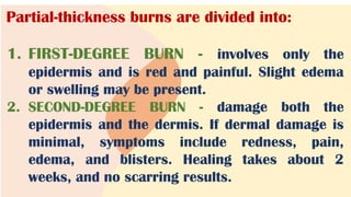
Partial-thickness burns aredivided into:
1. FIRST-DEGREE BURN - involves only the
epidermis and is red and painful. Slight edema
or swelling may be present.
2. SECOND-DEGREE BURN - damage both the
epidermis and the dermis. If dermal damage is
minimal, symptoms include redness, pain,
edema, and blisters. Healing takes about 2
weeks, and no scarring results.
61.
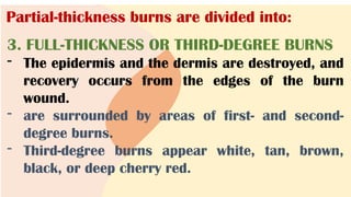
Partial-thickness burns aredivided into:
3. FULL-THICKNESS OR THIRD-DEGREE BURNS
- The epidermis and the dermis are destroyed, and
recovery occurs from the edges of the burn
wound.
- are surrounded by areas of first- and second-
degree burns.
- Third-degree burns appear white, tan, brown,
black, or deep cherry red.
62.
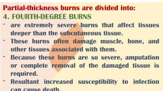
Partial-thickness burns aredivided into:
4. FOURTH-DEGREE BURNS
- are extremely severe burns that affect tissues
deeper than the subcutaneous tissue.
- These burns often damage muscle, bone, and
other tissues associated with them.
- Because these burns are so severe, amputation
or complete removal of the damaged tissue is
required.
- Resultant increased susceptibility to infection
63.
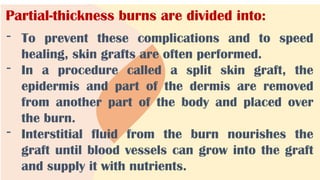
Partial-thickness burns aredivided into:
- To prevent these complications and to speed
healing, skin grafts are often performed.
- In a procedure called a split skin graft, the
epidermis and part of the dermis are removed
from another part of the body and placed over
the burn.
- Interstitial fluid from the burn nourishes the
graft until blood vessels can grow into the graft
and supply it with nutrients.
64.
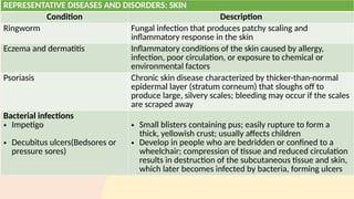
REPRESENTATIVE DISEASES ANDDISORDERS: SKIN
Condition Description
Ringworm Fungal infection that produces patchy scaling and
inflammatory response in the skin
Eczema and dermatitis Inflammatory conditions of the skin caused by allergy,
infection, poor circulation, or exposure to chemical or
environmental factors
Psoriasis Chronic skin disease characterized by thicker-than-normal
epidermal layer (stratum corneum) that sloughs off to
produce large, silvery scales; bleeding may occur if the scales
are scraped away
Bacterial infections
• Impetigo
• Decubitus ulcers(Bedsores or
pressure sores)
• Small blisters containing pus; easily rupture to form a
thick, yellowish crust; usually affects children
• Develop in people who are bedridden or confined to a
wheelchair; compression of tissue and reduced circulation
results in destruction of the subcutaneous tissue and skin,
which later becomes infected by bacteria, forming ulcers
65.
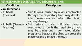
REPRESENTATIVE DISEASES ANDDISORDERS: SKIN
Condition Description
Viral Infections
• Rubeola
(measles)
• Rubella (German
Measles)
• Skin lesions, caused by a virus contracted
through the respiratory tract, may develop
into pneumonia or infect the brain,
causing damage
• Skin lesions; usually mild viral disease
contracted through the respiratory tract;
may be dangerous if contracted during
pregnancy because the virus can cross the
placenta and damage the fetus
66.
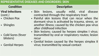
REPRESENTATIVE DISEASES ANDDISORDERS: SKIN
Condition Description
Viral Infections
(cont.)
• Chicken Pox
• Shingles
• Cold Sores (fever
blisters)
• Genital Herpes
• Skin lesions, usually mild, viral disease
contracted through the respiratory tract
• Painful skin lesions that can recur when the
dormant virus is activated by trauma, stress, or
another illness; caused by the chicken pox virus
after childhood infection
• Skin lesions; caused by herpes simplex I virus;
transmitted by oral or respiratory routes; lesion
recur
• Genital lesion caused by the herpes simplex II
virus; transmitted by sexual contact
67.
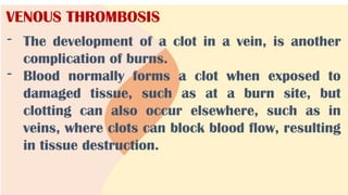
VENOUS THROMBOSIS
- Thedevelopment of a clot in a vein, is another
complication of burns.
- Blood normally forms a clot when exposed to
damaged tissue, such as at a burn site, but
clotting can also occur elsewhere, such as in
veins, where clots can block blood flow, resulting
in tissue destruction.
#2 We are also familiar with this system because we are concerned with the appearance of the integumentary system. Skin without blemishes is considered attractive, whereas acne is a source of embarrassment for many teenagers. The development of wrinkles and the graying or loss of hair are signs of aging. We invest much time, effort, and money on altering and enhancing the appearance of the integumentary system. Many of us apply lotion to our skin, color our hair, and trim our nails. We also try to prevent sweating by using antiperspirants and to reduce or mask body odor by washing and by using deodorants and perfumes.
#3 For example, reduced blood flow through the skin during a heart attack can cause a person to look pale, whereas increased blood flow as a result of increased body temperature can cause a flushed appearance.
Also, some diseases cause skin rashes, such as those characteristic of measles, chicken pox, and allergic reactions.
In addition, the integumentary system and the other systems often interact in complex ways in both healthy and diseased states
#10 To give an analogy, if the subcutaneous tissue is the foundation on which a house rests, the dermis forms most of the house and the epidermis is its roof.
#12 As new cells form, they push older cells to the surface, where they slough, or flake off.
#13 As keratinization proceeds, epithelial cells eventually die and form an outer layer of dead, rigid cells that resists abrasion and acts as a permeability barrier.
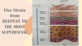
#18 Skin is classified as thick or thin based on the structure of the epidermis. Thick skin has all five strata and is found in areas subject to pressure or friction, such as the palms of the hands, the soles of the feet, and the fingertips. Thin skin lacks the stratum lucidum and covers the rest of the body.
#19 Keratin gives the stratum corneum its structural strength.
These lipids act as waterproofing material, thereby preventing fluid loss through the skin.
Eventually, the desmosomes break apart, and the cells are sloughed from the skin. Excessive sloughing of stratum corneum cells from the surface of the scalp is called dandruff. In skin subjected to fric-tion, the number of layers in the stratum corneum greatly increases, producing a thickened area called a callus (KAL-us; hard skin). Over a bony prominence, the stratum corneum can thicken to form a cone-shaped structure called a corn.
#21 The collagen fibers of the dermis are oriented in many different directions and can resist stretch. However, more collagen fibers are oriented in some directions than in others. This produces cleavage lines, or tension lines, in the skin, and the skin is most resistant to stretch along these lines (figure 5.3). It is important for surgeons to be aware of cleavage lines. An incision made across the cleavage lines is likely to gap and produce considerable scar tissue, but an incision made parallel with the lines tends to gap less and produce less scar tissue.
#22 For example, stretch marks often form on the skin of the abdomen and breasts of a female during pregnancy or on the skin of athletes who have quickly increased muscle size by intense weight training.
Recall that the epidermis lacks blood vessels; however, the dermal papillae contain many blood vessels. Blood flow through these vessels supplies the overlying epidermis with nutrients, removes waste products, and helps regulate body temperature. The dermal papillae in the palms of the hands, the soles of the feet, and the tips of the digits are arranged in parallel, curving ridges that shape the overlying epidermis into patterns called friction ridges. The impressions left on surfaces by these friction ridges are fingerprints and footprints. As the name suggests, the ridges increase friction and improve the grip of the hands and feet.
An injection delivers substances, such as medicines, to the body by puncturing the skin. Substances are administered at different depths in the skin, depending on how quickly the material needs to enter the blood. An intradermal injection delivers material to the blood slowly and is administered by drawing the skin taut and inserting a small needle at a shallow angle into the dermis; an example is the tuberculin skin test. A subcutaneous injection is achieved by pinching the skin to form a "tent" and inserting a short needle into the adipose tissue of the subcutaneous tissue; an example is an insulin injection. An intramuscular injection delivers material to the blood faster than intradermal or subcutaneous injections. An intramuscular injection is accomplished by inserting a long needle at a 90-degree angle to the skin into a muscle deep to the subcutaneous tissue. Intramuscular injections are used for most vaccines and certain antibiotics.
#25 They have many long processes that extend between the epithelial cells of the deep part of the epidermis.
Within melanocytes, the Golgi apparatuses package melanin into vesicles called melanosomes (MEL-ah-no-sohms). Melanosomes move into the cell processes of the melanocytes.
Other areas, such as the lips, palms of the hands, and soles of the feet, contain less melanin. Melanin production is determined by genetic factors, exposure to light, and hormones. Genetic factors are responsible for the amounts of melanin produced resulting in variation in skin color in the human population
#26 Since all humans have about the same number of melanocytes, skin color variations are determined by the amount, kind, and distribution of melanin. Although many genes are responsible for skin color, a single mutation can prevent the production of melanin.
#27 Examples are: darkening the nipples, the pigmented circular areas around the nipples, and the genitalia even more. The cheekbones and forehead can also darken, resulting in "the mask of pregnancy." Also, a dark line of pigmentation can appear on the midline of the abdomen.
Examples include blushing and the redness resulting from the inflammatory response.
#28 Examples include blushing and the redness resulting from the inflammatory response.
The location of pigments and other substances in the skin affects the color produced. If a dark pigment is located in the dermis or subcutaneous tissue, light reflected off the dark pigment can be scattered by collagen fibers of the dermis to produce a blue color. The deeper within the dermis or subcutaneous tissue any dark pigment is located, the bluer the pigment appears because of the light-scattering effect of the overlying tissue. This effect causes the blue color of tattoos, bruises, and some superficial blood vessels.
#29 Just as a house rests on a foundation, the skin rests on the subcutaneous tissue, or hypodermis (high-poh-DER-miss; under the dermis).
The amount and location of adipose tissue vary with age, sex, and diet. Adipose tissue in the subcutaneous tissue functions as padding and insulation, and it is responsible for some of the differences in appearance between males and females as well as between individuals of the same sex. The subcutaneous tissue can be used to estimate total body fat.
#30 The percentage of body fat varies in the population, but on average females have higher total body fat than do males.
A body fat percentage above the acceptable range is an indicator of obesity.
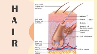
#31 In humans, hair is found everywhere on the skin, except on the palms, the soles, the lips, the nipples, parts of the genitalia, and the distal segments of the fingers and toes. Each hair arises from a hair follicle, an invagination of the epidermis that extends deep into the dermis
#32 Hair within a hair follicle
A helpful analogy for the structure of the hair follicle and hair is a single flower in a vase. The vase is like the hair follicle, and the flower stem is like the hair. The shaft of the hair protrudes above the surface of the skin, whereas the root is below the surface. The hair bulb is the expanded base of the root. A hair has a hard cortex, which surrounds a softer center, the medulla (meh-DULL-ah). The cortex is covered by the cuticle (KEW-tih-cul; skin), a single layer of overlapping cells that holds the hair in the hair follicle. Because the hair follicle is composed of epithelial tissue, hair follicles can play an important role in repair of the skin. If the surface epidermis is damaged, the epithelial cells within the hair follicle can divide and serve as a source of new epithelial cells.
#33 Hair is produced in cycles of growth and rest. During the growth stage, a hair is formed by epithelial cells within the hair bulb. These cells, like the cells of the stratum basale in the skin, divide and undergo keratinization. The hair grows longer as these cells are added to the base of the hair within the hair bulb. Thus, the hair root and shaft consist of columns of dead keratinized epithelial cells. During the resting stage, growth stops and the hair is held in the hair follicle. When the next growth stage begins, a new hair is formed and the old hair falls out.
#34 The loss of hair normally means that the hair is being replaced because the old hair falls out of the hair follicle when the new hair begins to grow. In some individuals, however, a permanent loss of hair results in "pattern baldness." Although many of the hair follicles are lost, some remain and produce a very short, transparent hair, which for practical purposes is invisible. These changes occur when male sex hormones act on the hair follicles of individuals who have the genetic predisposition for pattern baldness.
#35 Gray hair is usually a mixture of unfaded, faded, and white hairs. Associated with each hair follicle are smooth muscle cells called the arrector (ah-REK-tor; that which raises) pili (PIE-lee; hair) (see figure 5.6a). Contraction of the arrector pili causes the hair to become more perpendicular to the skin's surface, or to "stand on end," and it produces a raised area of skin called a "goose bump."
#36 Sebaceous glands are simple, branched acinar glands
#37 Most are connected by a duct to the superficial part of a hair follicle. They produce sebum, an oily, white substance rich in lipids. The sebum is released by holo-crine secretion (see chapter 4) and lubricates the hair and the surface of the skin, which prevents drying and protects against some bacteria.
#38 They produce a secretion that is mostly water with a few salts. Eccrine sweat glands have ducts that open onto the surface of the skin through sweat pores. When the body temperature starts to rise above normal levels, the sweat glands produce sweat, which evaporates and cools the body. Emotional sweating can also occur in the palms, soles, armpits, and other places.
#39 These substances are released primarily by merocrine secretion, though some glands demonstrate holocrine secretion. They open into hair follicles, but only in the armpits and genitalia.
The organic secretion, which is essentially odorless when released, is quickly broken down by bacteria into substances responsible for what is commonly known as body odor.
#40 The visible part of the nail is the nail body, and the part of the nail covered by skin is the nail root
The cuticle, or eponychium (ep-oh-NIK-ee-um), is stratum corneum that extends onto the nail body. The nail root extends distally from the nail matrix. The nail also attaches to the underlying nail bed, which is located distal to the nail matrix. The nail matrix and bed are epithelial tissue with a stratum basale that gives rise to the cells that form the nail. The nail matrix is thicker than the nail bed and produces most of the nail. A small part of the nail matrix, the lunula (LOO-noo-lah; moon), can be seen through the nail body as a whitish, crescent-shaped area at the base of the nail. Cell production within the nail matrix causes the nail to grow. Unlike hair, nails grow continuously and do not have a resting stage.
#46 If exposed to enough ultraviolet light, humans can produce all the vitamin D they need. However, many people need to ingest vitamin D as well because clothing and indoor living reduce their exposure to ultraviolet light.
#47 Fatty fish (and fish oils) and vitamin D-fortified milk are the best sources of vitamin D. Eggs, butter, and liver contain small amounts of vitamin D but are not considered significant sources because too large a serving size is necessary to meet the daily vitamin D requirement.
#48 Even slight changes in temperature can make enzymes operate less efficiently and disrupt the normal rates of chemical changes in the body.
Exercise, fever, and an increase in environmental temperature tend to raise body temperature. In order to maintain homeostasis, the body must rid itself of excess heat.
#54 Can you think of three "illnesses" that are indicated by changes in the skin?
#56 Normally, the liver secretes bile pigments, breakdown products of worn-out red blood cells, into the small intestine. Bile pigments are yellow, and their buildup in the blood and tissues can indicate impaired liver function.
#57 For example, scarlet fever results when bacteria infecting the throat release a toxin into the blood that causes a reddish rash on the skin.
#58 The condition of the skin, hair, and nails is affected by nutritional status.
Example for hair:
For example, lead poisoning results in high levels of lead in the hair. However, the use of hair analysis to determine the general health or nutritional status of an individual is unreliable.
#60 1st degree burn:
They can be caused by sunburn or brief exposure to very hot or very cold objects, and they heal without scarring in about a week.
2nd degree burns:
However, if the burn goes deep into the dermis, the wound appears red, tan, or white; can take several months to heal; and might scar. In all second-degree burns, the epidermis, including the stratum basale where the stem cells are found, is damaged. As a result, the epidermis regenerates from epithelial tissue in hair follicles and sweat glands, as well as from the edges of the wound.
#61 Although the first- and second-degree burn areas are painful, the region of third-degree burn is usually painless because sensory receptors in the epidermis and dermis have been destroyed.
#62 Like third-degree burns, fourth-degree burns are painless due to the destruction of the pain receptors.
Resultant increased susceptibility to infection can cause death.
#63 Meanwhile, the donor tissue produces new epidermis from epithelial tissue in the hair follicles and sweat glands in the same manner as in superficial second-degree burns. When it is not possible or practical to move skin from one part of the body to a burn site, physicians sometimes use artificial skin or grafts from human cadavers. However, these techniques are often unsatisfactory because the body's immune system recognizes the graft as a foreign substance and rejects it. A solution to this problem is to grow some of the burn victim's own skin in the laboratory. A piece of healthy skin from the burn victim is removed and placed in a flask with nutrients and hormones that stimulate rapid growth. The new skin that results consists only of epidermis and does not contain glands or hair.
#67 The concentration of chemicals that cause blood clotting (called clotting factors) increases for two reasons:
Loss of fluid from the burn patient concentrates the chemicals
The liver releases an increased amount of clotting factors


![[TRANS] HES 029 - Lecture 3 (The Integumentary System).pdf](https://cdn.slidesharecdn.com/ss_thumbnails/transhes029-lecture3theintegumentarysystem-221001083441-a0e9cb33-thumbnail.jpg?width=640&height=640&fit=bounds)