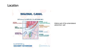
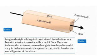
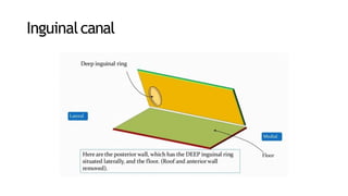
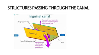
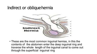
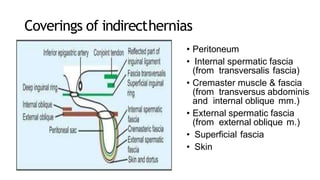
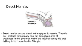
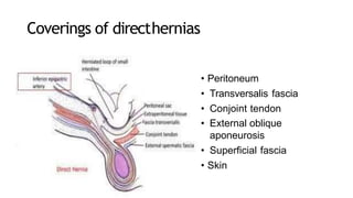
Inguinal canal – Anatomy
InguinalCanal
• This is an oblique intermuscular passage in the lower part of
the anterior abdominal wall , Situated just above the medial
half of the inguinal ligament
Location
Inferior part of the anterolateral
abdominal wall
Length &direction
• It is about 4cm(1.5 inches) long, and is
directed downwards, forwards and medially
• The inguinal canal extends from the deep inguinal
ring to the superficial inguinal ring
Deep inguinalRing
• An oval opening in the fascia
transversalis situated 1.2 cm above the
midinguinal
point, and immediately lateral to the stem
of the inferior epigastric artery
Inguinalcanal
Superficial inguinalring
• Is a triangular gap in the external oblique
aponeurosis . It is shaped like an obtuse angled
triangle . The base of the triangle is formed by the
pubic crest, the two sides of the triangle from the
lateral or lower and the medial or upper margins
of the opening. It is 2.5 cm long and 1.2 cm
broad at the base these margins are referred as
crura. At and beyond the apex of the triangle 2
crura are united by intercrural fibers
BOUNDARIESOF INGUINAL CANAL
THEANTERIORWALL
• .In its whole
extent
• Skin
• Superficial fascia
• External
oblique
aponeurosis
•
• In its lateral one-
third
• The fleshy fibres
of the internal
oblique muscle.
Posterior wall of the inguinalcanal
• In ~ 75 % of individuals ,the posterior wall(floor) of the canal is
formed laterally by the aponeurosis of tranversalis abdominal
muscle and the tranversalis fascia
• The remaining , the Posterior wall is tranversalis fascia only
• Medially , the Posterior wall is reinforced by the internal
oblique aponeurosis
ROOF
• It is formed by the
arched fibres of the
internal oblique and
transverse
abdominis muscles
and aponeurosis
FLOOR
• Is formed by the grooved upper surface of the inguinal ligament;
and at the medial end by the lacunae ligament
STRUCTURESPASSING THROUGHTHE CANAL
• 1.The spermatic cord in males, or the round ligament of the
uterus in females, enters the inguinal canal through the deep
inguinal ring and passes out through the superficial inguinal
ring.
• 2.The ilioinguinal nerve enters the canal through the
interval between the external and internal oblique muscles
and passes out through the superficial inguinalring.
• Allow contents of the scrotum to communicate with intra-
abdominal contents
• Prevent mobile intra-abdominal contents (e.g. Intestine) from
entering the scrotum and possibly becoming damaged, while at
the same time permitting blood vessels, nerves, lymphatics, vas
deferens etc. To supply the scrotal contents
MECHANISMOF INGUINAL CANAL
• The presence of the inguinal canal is
the cause of weakness in the lower
part of the anterior abdominal wall.
This weakness is compensated by
the following factors
Obliquityof the inguinalcanal
• The two inguinal rings do not lie opposite
to each other.
• Therefore, when the intra- abdominal
pressure rises the anterior and posterior
walls of the canal are approximated, thus
obliterating the passage.
• This is known as the flap valve
mechanism.
• The superficial inguinal ring is guarded from behind by the
conjoint tendon and by the reflected part of the inguinal canal.
•
• The deep inguinal ring is guarded from the front by the fleshy
fibres of the internal oblique.
Shutter mechanism of the internal oblique
• This muscle has a triple relation to the inguinal
canal. It forms the anterior wall, the roof, and the
posterior wall of the canal. When it contracts the
roof is approximated to the floor, like a shutter.
Ball valvemechanism
• Contraction of the cremaster helps the
spermatic cord to plug the superficial
inguinal ring
Slit valvemechanism
• Contraction of the external oblique
results in approximation of the two
crura of the superficial inguinal
ring .
• The integrity of the superficial
inguinal ring is greatly increased
by the intercrural fibres.
Whenever, there is a rise in intra abdominal
pressure as in coughing , sneezing, lifting heavy
weights all these mechanisms come to play, so
that the inguinal canal is obliterated, its openings
are closed, and herniation of abdominal viscera is
prevented.
HESSELBACH’STRIANGLE Triangle is an important
structure as it is the site
for direct hernias. The
triangle has the
following borders:
• 1) Medial border of
rectus
abdominus(medially)
• 2) Inguinal
ligament
(inferiorly)
• 3) Inferior
epigastric
vessels(laterally)
Hernias- A briefmention
• Hernias are abnormal outpouchings of the
abdominal contents (such as the small intestine)
from the cavity in which they belong.
• There are two main types of hernias that occur at
the inguinal region. Direct hernia and indirect
hernia.
Indirect or obliquehernia
• These are the most common inguinal hernias, in this the
contents of the abdomen enter the deep inguinal ring and
traverse the whole length of the inguinal canal to come out
through the superficial inguinal ring
Coverings of indirecthernias
• Peritoneum
• Internal spermatic fascia
(from transversalis fascia)
• Cremaster muscle & fascia
(from transversus abdominis
and internal oblique mm.)
• External spermatic fascia
(from external oblique m.)
• Superficial fascia
• Skin
Direct Hernias
• Direct hernias occurs lateral to the epigastric vessels. They do
not protrude through any ring, but through an area of
weakness in the posterior wall of the inguinal canal; this area
is likely to be Hesselbach’s Triangle.
Coverings of directhernias
• Peritoneum
• Transversalis fascia
• Conjoint tendon
• External oblique
aponeurosis
• Superficial fascia
• Skin
inguinal canal anatomy.pptx

inguinal canal anatomy.pptx

  • 1.
  • 2.
    InguinalCanal • This isan oblique intermuscular passage in the lower part of the anterior abdominal wall , Situated just above the medial half of the inguinal ligament
  • 3.
    Location Inferior part ofthe anterolateral abdominal wall
  • 4.
    Length &direction • Itis about 4cm(1.5 inches) long, and is directed downwards, forwards and medially • The inguinal canal extends from the deep inguinal ring to the superficial inguinal ring
  • 6.
    Deep inguinalRing • Anoval opening in the fascia transversalis situated 1.2 cm above the midinguinal point, and immediately lateral to the stem of the inferior epigastric artery
  • 7.
  • 8.
    Superficial inguinalring • Isa triangular gap in the external oblique aponeurosis . It is shaped like an obtuse angled triangle . The base of the triangle is formed by the pubic crest, the two sides of the triangle from the lateral or lower and the medial or upper margins of the opening. It is 2.5 cm long and 1.2 cm broad at the base these margins are referred as crura. At and beyond the apex of the triangle 2 crura are united by intercrural fibers
  • 10.
  • 11.
    THEANTERIORWALL • .In itswhole extent • Skin • Superficial fascia • External oblique aponeurosis • • In its lateral one- third • The fleshy fibres of the internal oblique muscle.
  • 12.
    Posterior wall ofthe inguinalcanal • In ~ 75 % of individuals ,the posterior wall(floor) of the canal is formed laterally by the aponeurosis of tranversalis abdominal muscle and the tranversalis fascia • The remaining , the Posterior wall is tranversalis fascia only • Medially , the Posterior wall is reinforced by the internal oblique aponeurosis
  • 13.
    ROOF • It isformed by the arched fibres of the internal oblique and transverse abdominis muscles and aponeurosis
  • 14.
    FLOOR • Is formedby the grooved upper surface of the inguinal ligament; and at the medial end by the lacunae ligament
  • 15.
  • 16.
    • 1.The spermaticcord in males, or the round ligament of the uterus in females, enters the inguinal canal through the deep inguinal ring and passes out through the superficial inguinal ring. • 2.The ilioinguinal nerve enters the canal through the interval between the external and internal oblique muscles and passes out through the superficial inguinalring.
  • 18.
    • Allow contentsof the scrotum to communicate with intra- abdominal contents • Prevent mobile intra-abdominal contents (e.g. Intestine) from entering the scrotum and possibly becoming damaged, while at the same time permitting blood vessels, nerves, lymphatics, vas deferens etc. To supply the scrotal contents
  • 19.
    MECHANISMOF INGUINAL CANAL •The presence of the inguinal canal is the cause of weakness in the lower part of the anterior abdominal wall. This weakness is compensated by the following factors
  • 20.
    Obliquityof the inguinalcanal •The two inguinal rings do not lie opposite to each other. • Therefore, when the intra- abdominal pressure rises the anterior and posterior walls of the canal are approximated, thus obliterating the passage. • This is known as the flap valve mechanism.
  • 21.
    • The superficialinguinal ring is guarded from behind by the conjoint tendon and by the reflected part of the inguinal canal. • • The deep inguinal ring is guarded from the front by the fleshy fibres of the internal oblique.
  • 22.
    Shutter mechanism ofthe internal oblique • This muscle has a triple relation to the inguinal canal. It forms the anterior wall, the roof, and the posterior wall of the canal. When it contracts the roof is approximated to the floor, like a shutter.
  • 23.
    Ball valvemechanism • Contractionof the cremaster helps the spermatic cord to plug the superficial inguinal ring
  • 24.
    Slit valvemechanism • Contractionof the external oblique results in approximation of the two crura of the superficial inguinal ring . • The integrity of the superficial inguinal ring is greatly increased by the intercrural fibres.
  • 25.
    Whenever, there isa rise in intra abdominal pressure as in coughing , sneezing, lifting heavy weights all these mechanisms come to play, so that the inguinal canal is obliterated, its openings are closed, and herniation of abdominal viscera is prevented.
  • 28.
    HESSELBACH’STRIANGLE Triangle isan important structure as it is the site for direct hernias. The triangle has the following borders: • 1) Medial border of rectus abdominus(medially) • 2) Inguinal ligament (inferiorly) • 3) Inferior epigastric vessels(laterally)
  • 29.
    Hernias- A briefmention •Hernias are abnormal outpouchings of the abdominal contents (such as the small intestine) from the cavity in which they belong. • There are two main types of hernias that occur at the inguinal region. Direct hernia and indirect hernia.
  • 30.
    Indirect or obliquehernia •These are the most common inguinal hernias, in this the contents of the abdomen enter the deep inguinal ring and traverse the whole length of the inguinal canal to come out through the superficial inguinal ring
  • 31.
    Coverings of indirecthernias •Peritoneum • Internal spermatic fascia (from transversalis fascia) • Cremaster muscle & fascia (from transversus abdominis and internal oblique mm.) • External spermatic fascia (from external oblique m.) • Superficial fascia • Skin
  • 32.
    Direct Hernias • Directhernias occurs lateral to the epigastric vessels. They do not protrude through any ring, but through an area of weakness in the posterior wall of the inguinal canal; this area is likely to be Hesselbach’s Triangle.
  • 33.
    Coverings of directhernias •Peritoneum • Transversalis fascia • Conjoint tendon • External oblique aponeurosis • Superficial fascia • Skin