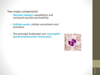
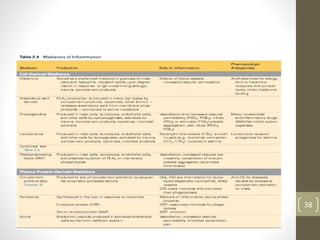
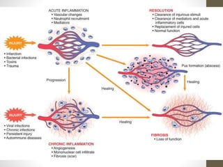
INFLAMMATION
1
Chapter 3
Inflamm…
• A complex reaction to injurious agents( microbes
and damaged ,usually necrotic cells…) that
consists of vascular responses, migration and
activation of leukocytes and systemic reactions
• Inflame-‘to burn’ , ‘set a fire’
2
Inflamm…
Unique feature
• Reaction of blood vessels, leading to accumulation
of fluid and leukocytes in extravascular tissues
3
Inflamm…
Purposes
• Destroy
• Dilute
• Wall off injurious agents
• Brings defense to the area
• Initiates healing
NB: Inflammatory response is closely intertwined
with the process of repair
4
Inflamm…
• Fundamentally a protective response
• Non-specific
Ultimate goal
• To rid the organism of both the initial cause of
injury (e.g toxins, microbes) and the consequences
of such injury (necrotic cells and tissues)
5
Inflamm…
Without inflammation
• Infections would go unchecked
• Wounds would never heal
• Injured organs remain permanent festering sores
6
Inflammation & repair may be potentially harmful
• Rheumatoid arthritis
• Atherosclerosis
• Lung fibrosis
• Hypersensitivity reactions to insect bites, drugs and
toxins
• Repair by fibrosis….disfigured scars and fibrous
bands e.g intestinal obstruction, pericardial fibrous
bands ,joint limitation
7
Two components
• Vascular reaction
• Cellular reaction
8
Many tissues and cells involved
• Fluid and proteins of plasma
• Circulating cells (neutrophils, monocytes,
eosinophils, lymphocytes, basophils and platelets)
• Blood vessels
9
Cellular and extracellular constituents of connective
tissue
• Mast cells, fibroblasts, resident macrophages and
lymphocytes
• Structural proteins (collagen, elastin)
• Adhesive glycoproteins (fibronectin, laminin,
nonfibrillar collagen,tenacin…)
• Proteoglycans
• Basement membrane (esp. ECM with adhesive
glycoproteins & proteoglycans)
10
11
Classification
• Based on duration of occurrence and morphologic
differences
• Acute inflammation
• Chronic inflammation
12
Acute inflammation- short duration (lasting for
minutes, hrs or few days)
• Characterized by exudation of fluid and plasma
proteins and migration of leukocytes( predominant
PMNs)
13
Chronic inflammation- longer duration (weeks,
months, even years)
• Predominant cells- lymphocytes and macrophages
• Proliferation of blood vessels, fibrosis and tissue
necrosis
14
Termination
• When the offending agent is eliminated
• Mediators broken down or dissipated
Anti inflammatory mechanisms
Control response and prevents excessive damage to
the host
15
Naming of inflammatory lesions
The suffix ‘itis’, after the involved organ’s name
• Pancreatitis
• Meningitis
• Arthritis
• Appendicitis …..
16
Historical Highlights
• Description of clinical features of inflammation in
an Egyptian papyrus(~3000BC)
Four cardinal signs
• Rubor-redness
• Tumor-swelling
• Dolor –pain
• Calor-heat
• ‘Functio laesa- loss of function- Virchow ,1882
17
Cornelius Celsus, first century AD,
Roman writer
18
Heat Redness Swelling Pain Loss Of Func.
Calor, Rubor, Dolor, Tumor, Loss of function.
Surgical wound inflammation
19
Acute inflammation
20
21
Acute inflamm…
• a rapid response to injury or microbes and other
foreign substances that is designed to deliver
leukocytes and plasma proteins to sites of injury
Two major components
• Vascular changes: vasodilation and
increased vascular permeability
• Cellular events: cellular recruitment and
activation
• The principal leukocytes are neutrophils
(polymorphonuclear leukocytes)
22
23
Stimuli (causes)
Infections (bacterial, viral, fungal, parasitic)
Trauma (blunt and penetrating)
Physical and chemical agents (thermal injury,
e.g., burns or frostbite; irradiation; chemicals)
Tissue necrosis (from any cause)
Foreign bodies
Immune reactions ( hypersensitivity reactions)
against environmental substances or against self
tissues
24
Vascular changes
• changes in vascular flow and caliber
• Changes in blood vessels begin rapidly after infection or injury and
occur in the following order
Vasodilation – occurs after transient vasoconstriction (lasting only
for seconds),
- results in locally increased blood flow
Vasodilation – Increasesnutrientandcellularsupply,
pressure,fluidfiltration
• Mediators of Vasodilation
• Histamine
• Nitric oxide
• Bradykinin other kinins
• Serotonin
25
26
Increased permeability – leads to outpouring of protein rich fluid to
the extracellular environment
Stasis- as a result of increase in concentration of RBCs , increased
viscosity of blood , flow slows
Margination - leukocytes (principally neutrophils) begin to
accumulate along the vascular endothelial surface
27
28
Cellular events :
leukocyte extravasation and phagocytosis
• Critical function of inflammation is to deliver
leukocytes to the site of injury and to activate them
to perform their function
• The sequence of events in the extravasation of
leukocytes from the vascular lumen to the
extravascular space is divided into
(1) margination and rolling,
(2) adhesion and transmigration between endothelial cells,
and
(3) migration in interstitial tissues toward a chemotactic
stimulus
diapedsis
• The type of emigrating leucocytes depends on the duration of
inflammatory response and type of stimulus
• 6-24hrs-neutrophils predominate
• 24-48hrs-monocytes
• Viral-lymphocytes
• Allergy, parasites-eosinophils, main cells
30
31
Chemotaxis
• After extravasation leukocytes migrate toward sites of
infection or injury along a chemical
gradient…..Chemotaxis
• Chemotactic factors -exogenous and endogenous
substances
(1) bacterial products, particularly peptides with N-
formylmethionine termini;
(2) cytokines, especially those of the chemokine family;
e.g.IL-8
(3) components of the complement system, particularly C5a;
and
(4) products of the lipoxygenase pathway of arachidonic
acid (AA) metabolism, particularly leukotriene B4 (LTB4)
32
Phagocytosis
• Phagocytosis consists of three distinct but interrelated steps :
(1)recognition and attachment of the particle to the ingesting
leukocyte;
(2) engulfment, with subsequent formation of a phagocytic vacuole;
and
(3)killing and degradation of the ingested material
Chemical mediators
of inflammation
33
34
Chemical Mediators…..
Direct the vascular and cellular events
• Mediators may be:
• Plasma derived (typically synthesized by the liver), or
• produced locally by cells at the site of inflammation
• Plasma-derived
• Complement proteins
• kinins,
• coagulation factors
• Circulate as inactive precursors
34
• Cell-derived
• Sequestered in intracellular granules (e.g., histamine in mast
cells) or
• Synthesized de novo in response to a stimulus (e.g.,
prostaglandins, cytokines)
35
Cellular sources
• Platelets
• Mast cells
• Neutrophils
• Monocytes/macrophages
• Lymphocytes
• Mesenchymal cells (endothelium, fibroblasts)
• Some epithelia
36
Once activated & released, most are short lived
• Decay (e.g. AA products)
• Inactivated (e.g. kianase- bradykinin)
• Scavenged (e.g. antioxidants)
• Inhibited (e.g. complement regulatory proteins)
• Most mediators have the potential to cause harmful
effects
37
38
39
Acute inflammation generally has one of three outcomes:
Resolution
Healing by fibrosis
Progression to chronic inflammation
Outcomes of acute
inflammation
40
41
Chronic
inflammation
42
43
Inflammation of prolonged duration
(weeks to months to years) in which
active inflammation, tissue injury, and
healing proceed simultaneously
• Morphologic Features:
• Infiltration with mononuclear cells (macrophages, lymphocytes &
plasma cells)
• indicates persistent reaction to injury
• Tissue destruction
• Done by way of Inflammatory cells
• Repair involving angiogenesis and fibrosis
• Attempt to replace lost tissue
44
45
Causes
Chronic inflammation arises in the following settings:
• Persistent microbial infections,
• mycobacteria , Treponema pallidum , and certain
fungi, viruses, parasites
• These organisms are of low direct pathogenicity,
but typically they evoke an immune response
called delayed hypersensitivity , which may
culminate in a granulomatous reaction
• Intracellular infections of any kind typically
require lymphocytes (and macrophages) to
identify and eradicate infected cells
46
• Prolonged exposure to potentially toxic agents
• Endogenous or exogenous
• e.g. exogenous nondegradable material-inhaled particulate
silica………silicosis
• endogenous agents -chronically elevated plasma lipid
components….. atherosclerosis
• Autoimmune diseases,
• in which an individual develops an immune
response to self-antigens and tissues
• Because the responsible antigens are in most
instances constantly renewed, a self-
perpetuating immune reaction results
e.g. rheumatoid arthritis, systemic lupus
erythematosus
47
48
49
Chronic inflammatorycells and mediators
Macrophages
• Dominant cells
• Mononuclear Phagocyte System
• Circulating blood monocytes →Tissue
macrophages
↓
Kupffer cells (liver)
Sinus Histiocytes (spleen,LN)
Microglia (CNS)
Alveolar Macrophages (lung)
• Activated macrophage products serve to eliminate microbes,
initiate repair process and responsible for tissue injury in
chronic inflammation
• Tissue destruction is one of the hall marks
50
51
Other cells in chronic inflammation
Lymphocytes,
Plasma Cells,
Eosinophils, and Mast Cells
• Though neutrophils are the predominant cells in acute
inflammation they can be seen in some persistent
inflammatory lesions
• E.g. acute osteomyelitis……chronic osteomyelitis
52
53
Granulomatous Inflammation
• a distinctive pattern characterized by formation of
epithelioid granuloma
• Granulomas can form in the setting of persistent T-
cell responses to certain microbes (e.g. M.TB),
where T-cell-derived cytokines are responsible for
persistent macrophage activation
• GRANULOMA – microscopic nodular
collection of Epithelioid macrophages
surrounded by a rim of LYMPHOCYTES,
occasional plasma cells, in long standing
granuloma …fibrosis
• In tuberculosis with central caseous necrosis
• ‘Epithelioid’- epithelial-like, squamous cell-
like activated macrophages
• H&E-With a pale pink granular cytoplasm &
indistinct cell borders 54
55
56
• Frequently epithelioid cells fuse to form giant cells
• The giant cells have a large mass of cytoplasm
containing 20 or more nuclei arranged peripherally
(langhans- type) or haphazardly (foreign body-
type)
57
• Tuberculosis is the archetypal granulomatous
disease
• Granulomas may also develop in response to
relatively inert foreign bodies (e.g., suture, splinter,
breast implant),…..foreign body granulomas
• the formation of a granuloma effectively "walls off"
the offending agent and is therefore a useful
defense mechanism
58
Two types of granulomas which differ in their pathogenesis
Foreign body type
Immune granuloma
Foreign body granulomas
• Are incited by relatively inert foreign bodies (e.g. talc),
• Typically form when material such as sutures and
other fibers are large enough to preclude phagocytosis
• The foreign material can usually be identified in the
center of the granuloma
• Don’t incite any specific inflammatory or immune
responses
59
60
Systemiceffectsof inflammation
61
62
• ‘Acute phase response’ or ‘systemic inflammatory
response syndrome (SIRS)’
• Systemic changes are reactions to cytokines whose
production is stimulated by bacterial products and
inflammatory stimuli
Fever
• an elevation of body temperature, usually by 1° to
4°C,
• one of the most prominent manifestations of the
acute-phase response, especially when inflammation is
caused by infection
• produced in response pyrogens that act by stimulating
prostaglandin (PG) synthesis in the vascular and
perivascular cells of the hypothalamus
63
64
Elevated plasma levels of acute-phase proteins
• mostly synthesized in the liver, whose concentrations
may increase as part of the response to inflammatory
stimuli
• Three of the best-known of these proteins:
C-reactive protein (CRP), fibrinogen, and serum amyloid
A (SAA) protein
• Synthesis of these molecules by hepatocytes is up-
regulated by cytokines, especially IL-6
• Many acute-phase proteins, such as CRP and SAA can act
65
• Fibrinogen binds to erythrocytes and causes them
to form stacks (rouleaux) that sediment more
rapidly than individual erythrocytes
• This is the basis for measuring the erythrocyte
sedimentation rate (ESR) as a simple test for the
systemic inflammatory response, caused by any
number of stimuli, including LPS
66
Leukocytosis
• leukocyte count usually climbs to 15,000 or 20,000 cells/μL, but
sometimes it may reach as high as 40,000 to 100,000 cells/μL
• These extreme elevations are referred to as leukemoid reactions
• leukocytosis occurs initially because of accelerated release of
cells from the bone marrow reserve pool (caused by cytokines,
including TNF and IL-1)
• Prolonged infection also stimulates production of colony-
stimulating factors (CSFs) GM-CSF
67
• Most bacterial infections induce neutrophilia
• Viral infections, such as infectious mononucleosis,
mumps. . .are associated with lymphocytosis
• Bronchial asthma, hay fever, and parasite
infestations – eosinophilia
• Certain infections (typhoid fever and infections
caused by some viruses, rickettsiae, and certain
protozoa) are paradoxically associated with
leukopenia
68
Other acute phase responses include
• increased heart rate and blood pressure;
• decreased sweating,
• rigors (shivering),
• chills,
• anorexia,
• somnolence,
• Malaise…….
• In chronic inflammation a
wasting syndrome called
cachexia
• result of TNF-mediated
appetite suppression and
mobilization of fat stores
69
• In severe bacterial infections (sepsis) , large
quantities of cytokines, notably TNF, as well as IL-
12 and IL-1 are produced
• This is associated with disseminated intravascular
coagulation (DIC), hypoglycemia and hypotensive
shock…..septic shock
70
71

Medical Pathology edited inflammation.pptx

  • 1.
  • 2.
    Inflamm… • A complexreaction to injurious agents( microbes and damaged ,usually necrotic cells…) that consists of vascular responses, migration and activation of leukocytes and systemic reactions • Inflame-‘to burn’ , ‘set a fire’ 2
  • 3.
    Inflamm… Unique feature • Reactionof blood vessels, leading to accumulation of fluid and leukocytes in extravascular tissues 3
  • 4.
    Inflamm… Purposes • Destroy • Dilute •Wall off injurious agents • Brings defense to the area • Initiates healing NB: Inflammatory response is closely intertwined with the process of repair 4
  • 5.
    Inflamm… • Fundamentally aprotective response • Non-specific Ultimate goal • To rid the organism of both the initial cause of injury (e.g toxins, microbes) and the consequences of such injury (necrotic cells and tissues) 5
  • 6.
    Inflamm… Without inflammation • Infectionswould go unchecked • Wounds would never heal • Injured organs remain permanent festering sores 6
  • 7.
    Inflammation & repairmay be potentially harmful • Rheumatoid arthritis • Atherosclerosis • Lung fibrosis • Hypersensitivity reactions to insect bites, drugs and toxins • Repair by fibrosis….disfigured scars and fibrous bands e.g intestinal obstruction, pericardial fibrous bands ,joint limitation 7
  • 8.
    Two components • Vascularreaction • Cellular reaction 8
  • 9.
    Many tissues andcells involved • Fluid and proteins of plasma • Circulating cells (neutrophils, monocytes, eosinophils, lymphocytes, basophils and platelets) • Blood vessels 9
  • 10.
    Cellular and extracellularconstituents of connective tissue • Mast cells, fibroblasts, resident macrophages and lymphocytes • Structural proteins (collagen, elastin) • Adhesive glycoproteins (fibronectin, laminin, nonfibrillar collagen,tenacin…) • Proteoglycans • Basement membrane (esp. ECM with adhesive glycoproteins & proteoglycans) 10
  • 11.
  • 12.
    Classification • Based onduration of occurrence and morphologic differences • Acute inflammation • Chronic inflammation 12
  • 13.
    Acute inflammation- shortduration (lasting for minutes, hrs or few days) • Characterized by exudation of fluid and plasma proteins and migration of leukocytes( predominant PMNs) 13
  • 14.
    Chronic inflammation- longerduration (weeks, months, even years) • Predominant cells- lymphocytes and macrophages • Proliferation of blood vessels, fibrosis and tissue necrosis 14
  • 15.
    Termination • When theoffending agent is eliminated • Mediators broken down or dissipated Anti inflammatory mechanisms Control response and prevents excessive damage to the host 15
  • 16.
    Naming of inflammatorylesions The suffix ‘itis’, after the involved organ’s name • Pancreatitis • Meningitis • Arthritis • Appendicitis ….. 16
  • 17.
    Historical Highlights • Descriptionof clinical features of inflammation in an Egyptian papyrus(~3000BC) Four cardinal signs • Rubor-redness • Tumor-swelling • Dolor –pain • Calor-heat • ‘Functio laesa- loss of function- Virchow ,1882 17 Cornelius Celsus, first century AD, Roman writer
  • 18.
    18 Heat Redness SwellingPain Loss Of Func.
  • 19.
    Calor, Rubor, Dolor,Tumor, Loss of function. Surgical wound inflammation 19
  • 20.
  • 21.
    21 Acute inflamm… • arapid response to injury or microbes and other foreign substances that is designed to deliver leukocytes and plasma proteins to sites of injury
  • 22.
    Two major components •Vascular changes: vasodilation and increased vascular permeability • Cellular events: cellular recruitment and activation • The principal leukocytes are neutrophils (polymorphonuclear leukocytes) 22
  • 23.
    23 Stimuli (causes) Infections (bacterial,viral, fungal, parasitic) Trauma (blunt and penetrating) Physical and chemical agents (thermal injury, e.g., burns or frostbite; irradiation; chemicals) Tissue necrosis (from any cause) Foreign bodies Immune reactions ( hypersensitivity reactions) against environmental substances or against self tissues
  • 24.
    24 Vascular changes • changesin vascular flow and caliber • Changes in blood vessels begin rapidly after infection or injury and occur in the following order Vasodilation – occurs after transient vasoconstriction (lasting only for seconds), - results in locally increased blood flow
  • 25.
    Vasodilation – Increasesnutrientandcellularsupply, pressure,fluidfiltration •Mediators of Vasodilation • Histamine • Nitric oxide • Bradykinin other kinins • Serotonin 25
  • 26.
    26 Increased permeability –leads to outpouring of protein rich fluid to the extracellular environment Stasis- as a result of increase in concentration of RBCs , increased viscosity of blood , flow slows Margination - leukocytes (principally neutrophils) begin to accumulate along the vascular endothelial surface
  • 27.
  • 28.
    28 Cellular events : leukocyteextravasation and phagocytosis • Critical function of inflammation is to deliver leukocytes to the site of injury and to activate them to perform their function • The sequence of events in the extravasation of leukocytes from the vascular lumen to the extravascular space is divided into (1) margination and rolling, (2) adhesion and transmigration between endothelial cells, and (3) migration in interstitial tissues toward a chemotactic stimulus
  • 29.
  • 30.
    • The typeof emigrating leucocytes depends on the duration of inflammatory response and type of stimulus • 6-24hrs-neutrophils predominate • 24-48hrs-monocytes • Viral-lymphocytes • Allergy, parasites-eosinophils, main cells 30
  • 31.
    31 Chemotaxis • After extravasationleukocytes migrate toward sites of infection or injury along a chemical gradient…..Chemotaxis • Chemotactic factors -exogenous and endogenous substances (1) bacterial products, particularly peptides with N- formylmethionine termini; (2) cytokines, especially those of the chemokine family; e.g.IL-8 (3) components of the complement system, particularly C5a; and (4) products of the lipoxygenase pathway of arachidonic acid (AA) metabolism, particularly leukotriene B4 (LTB4)
  • 32.
    32 Phagocytosis • Phagocytosis consistsof three distinct but interrelated steps : (1)recognition and attachment of the particle to the ingesting leukocyte; (2) engulfment, with subsequent formation of a phagocytic vacuole; and (3)killing and degradation of the ingested material
  • 33.
  • 34.
    34 Chemical Mediators….. Direct thevascular and cellular events • Mediators may be: • Plasma derived (typically synthesized by the liver), or • produced locally by cells at the site of inflammation • Plasma-derived • Complement proteins • kinins, • coagulation factors • Circulate as inactive precursors 34
  • 35.
    • Cell-derived • Sequesteredin intracellular granules (e.g., histamine in mast cells) or • Synthesized de novo in response to a stimulus (e.g., prostaglandins, cytokines) 35
  • 36.
    Cellular sources • Platelets •Mast cells • Neutrophils • Monocytes/macrophages • Lymphocytes • Mesenchymal cells (endothelium, fibroblasts) • Some epithelia 36
  • 37.
    Once activated &released, most are short lived • Decay (e.g. AA products) • Inactivated (e.g. kianase- bradykinin) • Scavenged (e.g. antioxidants) • Inhibited (e.g. complement regulatory proteins) • Most mediators have the potential to cause harmful effects 37
  • 38.
  • 39.
    39 Acute inflammation generallyhas one of three outcomes: Resolution Healing by fibrosis Progression to chronic inflammation
  • 40.
  • 41.
  • 42.
  • 43.
    43 Inflammation of prolongedduration (weeks to months to years) in which active inflammation, tissue injury, and healing proceed simultaneously
  • 44.
    • Morphologic Features: •Infiltration with mononuclear cells (macrophages, lymphocytes & plasma cells) • indicates persistent reaction to injury • Tissue destruction • Done by way of Inflammatory cells • Repair involving angiogenesis and fibrosis • Attempt to replace lost tissue 44
  • 45.
    45 Causes Chronic inflammation arisesin the following settings: • Persistent microbial infections, • mycobacteria , Treponema pallidum , and certain fungi, viruses, parasites • These organisms are of low direct pathogenicity, but typically they evoke an immune response called delayed hypersensitivity , which may culminate in a granulomatous reaction • Intracellular infections of any kind typically require lymphocytes (and macrophages) to identify and eradicate infected cells
  • 46.
    46 • Prolonged exposureto potentially toxic agents • Endogenous or exogenous • e.g. exogenous nondegradable material-inhaled particulate silica………silicosis • endogenous agents -chronically elevated plasma lipid components….. atherosclerosis
  • 47.
    • Autoimmune diseases, •in which an individual develops an immune response to self-antigens and tissues • Because the responsible antigens are in most instances constantly renewed, a self- perpetuating immune reaction results e.g. rheumatoid arthritis, systemic lupus erythematosus 47
  • 48.
  • 49.
    49 Chronic inflammatorycells andmediators Macrophages • Dominant cells • Mononuclear Phagocyte System • Circulating blood monocytes →Tissue macrophages ↓ Kupffer cells (liver) Sinus Histiocytes (spleen,LN) Microglia (CNS) Alveolar Macrophages (lung)
  • 50.
    • Activated macrophageproducts serve to eliminate microbes, initiate repair process and responsible for tissue injury in chronic inflammation • Tissue destruction is one of the hall marks 50
  • 51.
    51 Other cells inchronic inflammation Lymphocytes, Plasma Cells, Eosinophils, and Mast Cells
  • 52.
    • Though neutrophilsare the predominant cells in acute inflammation they can be seen in some persistent inflammatory lesions • E.g. acute osteomyelitis……chronic osteomyelitis 52
  • 53.
    53 Granulomatous Inflammation • adistinctive pattern characterized by formation of epithelioid granuloma • Granulomas can form in the setting of persistent T- cell responses to certain microbes (e.g. M.TB), where T-cell-derived cytokines are responsible for persistent macrophage activation
  • 54.
    • GRANULOMA –microscopic nodular collection of Epithelioid macrophages surrounded by a rim of LYMPHOCYTES, occasional plasma cells, in long standing granuloma …fibrosis • In tuberculosis with central caseous necrosis • ‘Epithelioid’- epithelial-like, squamous cell- like activated macrophages • H&E-With a pale pink granular cytoplasm & indistinct cell borders 54
  • 55.
  • 56.
    56 • Frequently epithelioidcells fuse to form giant cells • The giant cells have a large mass of cytoplasm containing 20 or more nuclei arranged peripherally (langhans- type) or haphazardly (foreign body- type)
  • 57.
    57 • Tuberculosis isthe archetypal granulomatous disease • Granulomas may also develop in response to relatively inert foreign bodies (e.g., suture, splinter, breast implant),…..foreign body granulomas • the formation of a granuloma effectively "walls off" the offending agent and is therefore a useful defense mechanism
  • 58.
    58 Two types ofgranulomas which differ in their pathogenesis Foreign body type Immune granuloma
  • 59.
    Foreign body granulomas •Are incited by relatively inert foreign bodies (e.g. talc), • Typically form when material such as sutures and other fibers are large enough to preclude phagocytosis • The foreign material can usually be identified in the center of the granuloma • Don’t incite any specific inflammatory or immune responses 59
  • 60.
  • 61.
  • 62.
    62 • ‘Acute phaseresponse’ or ‘systemic inflammatory response syndrome (SIRS)’ • Systemic changes are reactions to cytokines whose production is stimulated by bacterial products and inflammatory stimuli
  • 63.
    Fever • an elevationof body temperature, usually by 1° to 4°C, • one of the most prominent manifestations of the acute-phase response, especially when inflammation is caused by infection • produced in response pyrogens that act by stimulating prostaglandin (PG) synthesis in the vascular and perivascular cells of the hypothalamus 63
  • 64.
    64 Elevated plasma levelsof acute-phase proteins • mostly synthesized in the liver, whose concentrations may increase as part of the response to inflammatory stimuli • Three of the best-known of these proteins: C-reactive protein (CRP), fibrinogen, and serum amyloid A (SAA) protein • Synthesis of these molecules by hepatocytes is up- regulated by cytokines, especially IL-6 • Many acute-phase proteins, such as CRP and SAA can act
  • 65.
    65 • Fibrinogen bindsto erythrocytes and causes them to form stacks (rouleaux) that sediment more rapidly than individual erythrocytes • This is the basis for measuring the erythrocyte sedimentation rate (ESR) as a simple test for the systemic inflammatory response, caused by any number of stimuli, including LPS
  • 66.
    66 Leukocytosis • leukocyte countusually climbs to 15,000 or 20,000 cells/μL, but sometimes it may reach as high as 40,000 to 100,000 cells/μL • These extreme elevations are referred to as leukemoid reactions • leukocytosis occurs initially because of accelerated release of cells from the bone marrow reserve pool (caused by cytokines, including TNF and IL-1) • Prolonged infection also stimulates production of colony- stimulating factors (CSFs) GM-CSF
  • 67.
    67 • Most bacterialinfections induce neutrophilia • Viral infections, such as infectious mononucleosis, mumps. . .are associated with lymphocytosis • Bronchial asthma, hay fever, and parasite infestations – eosinophilia • Certain infections (typhoid fever and infections caused by some viruses, rickettsiae, and certain protozoa) are paradoxically associated with leukopenia
  • 68.
    68 Other acute phaseresponses include • increased heart rate and blood pressure; • decreased sweating, • rigors (shivering), • chills, • anorexia, • somnolence, • Malaise…….
  • 69.
    • In chronicinflammation a wasting syndrome called cachexia • result of TNF-mediated appetite suppression and mobilization of fat stores 69
  • 70.
    • In severebacterial infections (sepsis) , large quantities of cytokines, notably TNF, as well as IL- 12 and IL-1 are produced • This is associated with disseminated intravascular coagulation (DIC), hypoglycemia and hypotensive shock…..septic shock 70
  • 71.