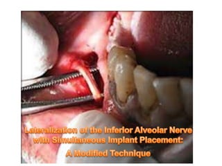
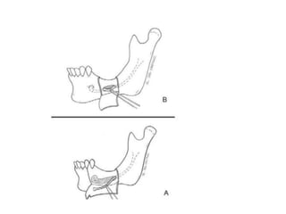
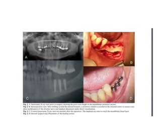
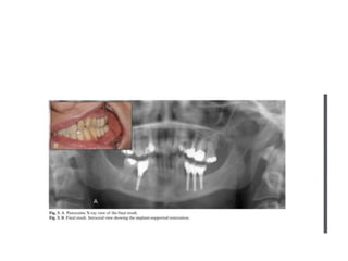
Introduction
•

Progressive bone resorption often occurs following tooth loss or
extraction, resulting in a moderately to severely atrophied
mandible. Often, the bone height posterior to the mental
foramen is inadequate to allow proper placement of endosteal
implants and the use of optimal fixture lengths without
potentially injuring the inferior alveolar nerve. One approach to
avoiding nerve injury when placing implants in these situations
is to reposition the inferior alveolar nerve laterally and then
place the implants medial to the nerve.
•

Several repositioning techniques have been presented in the
literature over the past 10 years, each with limitations
• Some of these techniques involve tranpositioning the nerve by
creating a window that includes the mental foramen as well as
the
area of implant placement, then releasing the nerve from the
mental
foramen and
replacing the nerve distal to its original location
Because this creates
bone segment that must be
• manipulated within the a large nerve area, permanent nerve
mental
damage is a significant risk.
• Other techniques involve lateralizing the nerve by repositioning
it through a posterior cortical window rather than engaging the
mental foramen. This approach, how- ever, requires extensive
stretching of the nerve.
•
•
•

•

To begin the actual repositioning procedure, local anesthesia
was obtained by infiltrating xylocaine 2% containing 1:100,000
epinephrine.
Intra- venous sedation is recommended because of the
procedure’s technique-sensitive nature and the need for patient
cooperation.
A crestal incision with anterior- and posterior- releasing incisions
was made, and a labial mucoperiosteal flap was
reflected, exposing the alveolar ridge and buccal cortex
The incision should extend at least 1 cm beyond the anticipated
site of the osteotomy. .
•
•

•

•

Care was taken during flap reflection to preserve the integrity of
the periosteum and the neurovascular bundle where it exits the
mental foramen and enters the soft tissue
To increase flap relaxation and improve exposure, dissection
was performed below the neurovascular bundle where it exits
the mental foramen. CT was used to locate the approximate
area of the mental foramen, after which blunt dissection was
used to identify and isolate the mental nerve
In the area that is 2 to 3 cm posterior to the mental foramen, a
702-bur was used to create a rectangular osteotomy.
A second circumferential osteotomy around the mental foramen
was then created.
•
•
•
•
•

The posterior osteotomy should extend 1 to 2 cm posteriorly
beyond the intended position of the most distal implant. This
allows for passive positioning of the neurovascular bundle after
implant placement
A small curved osteotome was then used to carefully remove
the posterior rectangular segment of the mandibular cortical
bone overlying the inferior alveolar nerve.
Next, the anterior round segment of mandibular cortical
bone, which included the mental foramen, was gently lifted.
Small curettes were used to carefully remove the medullary
bone lateral to the neurovascular bundle along the entire length
of the bony window.
It is important to remove all sharp edges of bone and any
cancellous spicules along the window that could lacerate the
neurovascular bundle.
•
•

•
•

•

After the neurovascular bundle has been identified, it was
carefully “teased” from the inferior alveolar canal using a nerve
retractor and small curettes.
These instruments should be blunt, and new instruments can be
altered in the laboratory to reduce the possibility of traumatizing
the neurovascular bundle
If possible, the retraction instruments should be left in place
during the procedure because repeatedly introducing and
retracting the instruments increases the risk of trauma to the
neurovascular bundle
While the neurovascular bundle was retracted laterally, the
endosseous implants were placed using standard techniques.
Note that cylindrical nonthreaded implants are recommended as
threaded implants in close contact with the nerve may cause
neurosensory problems.
•
•
•
•
•
•
•

Paralleling pins were used to assess the final implant location.
The paralleling pins were removed while the nerve retractor
held the repositioned nerve in place.
The implants were then placed medial to the inferior alveolar
nerve.
After implant placement, demineralized freeze dried bone
allograft (DFDBA) was placed between the implant and the
nerve to avoid any direct contact between the two
A collagen membrane was placed lateral to the nerve
In situations involving a narrow bucco- lingual width, the cortical
plate is used for ridge augmentation and to cover any
dehiscence on the buccal aspect of the implant
Releasing horizontal incisions were made in the periosteum to
enable a tension-free closure.
•
•

•

It is important to document the new location of the
neurovascular bundle in the medical record in case any future
surgical intervention is required
Postoperative follow-up in this study included radiographic
examinations and assessment of inferior alveolar nerve function
using the pin-prick sensation test
To allow an adequate amount of time for
osseointegration, stage-II surgery was per- formed after 6 to 8
months.
inferior alveolar nerve lateralization

inferior alveolar nerve lateralization

  • 2.
    Introduction • Progressive bone resorptionoften occurs following tooth loss or extraction, resulting in a moderately to severely atrophied mandible. Often, the bone height posterior to the mental foramen is inadequate to allow proper placement of endosteal implants and the use of optimal fixture lengths without potentially injuring the inferior alveolar nerve. One approach to avoiding nerve injury when placing implants in these situations is to reposition the inferior alveolar nerve laterally and then place the implants medial to the nerve.
  • 3.
    • Several repositioning techniqueshave been presented in the literature over the past 10 years, each with limitations • Some of these techniques involve tranpositioning the nerve by creating a window that includes the mental foramen as well as the area of implant placement, then releasing the nerve from the mental foramen and replacing the nerve distal to its original location Because this creates bone segment that must be • manipulated within the a large nerve area, permanent nerve mental damage is a significant risk. • Other techniques involve lateralizing the nerve by repositioning it through a posterior cortical window rather than engaging the mental foramen. This approach, how- ever, requires extensive stretching of the nerve.
  • 5.
    • • • • To begin theactual repositioning procedure, local anesthesia was obtained by infiltrating xylocaine 2% containing 1:100,000 epinephrine. Intra- venous sedation is recommended because of the procedure’s technique-sensitive nature and the need for patient cooperation. A crestal incision with anterior- and posterior- releasing incisions was made, and a labial mucoperiosteal flap was reflected, exposing the alveolar ridge and buccal cortex The incision should extend at least 1 cm beyond the anticipated site of the osteotomy. .
  • 7.
    • • • • Care was takenduring flap reflection to preserve the integrity of the periosteum and the neurovascular bundle where it exits the mental foramen and enters the soft tissue To increase flap relaxation and improve exposure, dissection was performed below the neurovascular bundle where it exits the mental foramen. CT was used to locate the approximate area of the mental foramen, after which blunt dissection was used to identify and isolate the mental nerve In the area that is 2 to 3 cm posterior to the mental foramen, a 702-bur was used to create a rectangular osteotomy. A second circumferential osteotomy around the mental foramen was then created.
  • 8.
    • • • • • The posterior osteotomyshould extend 1 to 2 cm posteriorly beyond the intended position of the most distal implant. This allows for passive positioning of the neurovascular bundle after implant placement A small curved osteotome was then used to carefully remove the posterior rectangular segment of the mandibular cortical bone overlying the inferior alveolar nerve. Next, the anterior round segment of mandibular cortical bone, which included the mental foramen, was gently lifted. Small curettes were used to carefully remove the medullary bone lateral to the neurovascular bundle along the entire length of the bony window. It is important to remove all sharp edges of bone and any cancellous spicules along the window that could lacerate the neurovascular bundle.
  • 10.
    • • • • • After the neurovascularbundle has been identified, it was carefully “teased” from the inferior alveolar canal using a nerve retractor and small curettes. These instruments should be blunt, and new instruments can be altered in the laboratory to reduce the possibility of traumatizing the neurovascular bundle If possible, the retraction instruments should be left in place during the procedure because repeatedly introducing and retracting the instruments increases the risk of trauma to the neurovascular bundle While the neurovascular bundle was retracted laterally, the endosseous implants were placed using standard techniques. Note that cylindrical nonthreaded implants are recommended as threaded implants in close contact with the nerve may cause neurosensory problems.
  • 11.
    • • • • • • • Paralleling pins wereused to assess the final implant location. The paralleling pins were removed while the nerve retractor held the repositioned nerve in place. The implants were then placed medial to the inferior alveolar nerve. After implant placement, demineralized freeze dried bone allograft (DFDBA) was placed between the implant and the nerve to avoid any direct contact between the two A collagen membrane was placed lateral to the nerve In situations involving a narrow bucco- lingual width, the cortical plate is used for ridge augmentation and to cover any dehiscence on the buccal aspect of the implant Releasing horizontal incisions were made in the periosteum to enable a tension-free closure.
  • 12.
    • • • It is importantto document the new location of the neurovascular bundle in the medical record in case any future surgical intervention is required Postoperative follow-up in this study included radiographic examinations and assessment of inferior alveolar nerve function using the pin-prick sensation test To allow an adequate amount of time for osseointegration, stage-II surgery was per- formed after 6 to 8 months.