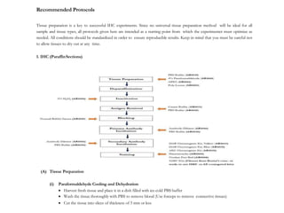
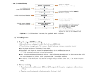
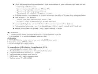
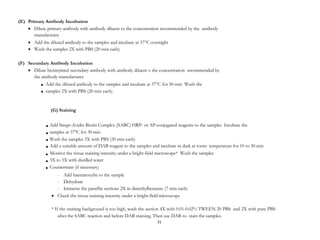
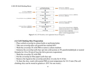
IHC Protocol
By BosterBio
https://www.bosterbio.com/
Recommended Protocols
Tissue preparation is a key to successful IHC experiments. Since no universal tissue preparation method will be ideal for all
sample and tissue types, all protocols given here are intended as a starting point from which the experimenter must optimize as
needed. All conditions should be standardized in order to ensure reproducible results. Keep in mind that you must be careful not
to allow tissues to dry out at any time.
1. IHC (ParaffinSections)
Figure 7. IHC (Paraffin Sections) Workflow with Applicable Boster’s Reagents.
(A) Tissue Preparation
(i) Paraformaldehyde Cooling and Dehydration
 Harvest fresh tissue and place it in a dish filled with ice-cold PBS buffer
 Wash the tissue thoroughly with PBS to remove blood (Use forceps to remove connective tissues)
 Cut the tissue into slices of thickness of 3 mm or less
 Immerse the slices in 4% paraformaldehyde at room temperature for 8 min
 Immerse the slices in 4% paraformaldehyde (pre-cool at 4°C) for 6 to 7 hrs. The paraformaldehyde volume
should be 20X greater than the tissue volume by weight
 Wash the tissue 3X with PBS (1 min each)
 Dehydrate the tissue by immersing the tissue sequentially as follows:
- 1X into 80% ethanol (1 hr at4°C)
- 1X into 90% ethanol (1 hr at4°C)
- 3X into 95% ethanol (1 hr each at 4°C)
- 3X into 100% ethanol (1 hr each at 4°C)
- 3X into dimethylbenzene (0.5 hr each at room temperature)
25
(ii) Liquid Paraffin Section
 Prepare the first portion of liquid paraffin in a suitable bath and allow the paraffin to reach and maintain at
60°C
 Immerse the tissue 2X into the paraffin bath (2 hrseach)
 Prepare the second portion of liquid paraffin in a suitable bath and allow the paraffin to reach and maintain at
60°C
 Pour the second portion of paraffin into a mold
 Quickly transport the tissue from the paraffin bath to the mold with paraffin
 Incubate the tissue at room temperature until it coagulates
 Store the tissue at 4°C
(iii) Section Slicing and Incubation
 Secure the paraffin section on slicer
 Slice one to two pieces of section to adjust the slicer so that the section and blade are parallel
 Slice the remaining section carefully with ~5 µm thick
 Incubate the sliced section in 40 to 50°C water to unfold
 Mount the tissue section onto Poly-Lysine or APES coated glass slides
 Incubate the slides overnight at 37°C
(B) Dewaxing/Deparaffinization
 Prepare the following reagents:
- 90% dimethylbenzene
- 95% dimethylbenzene
- 100% dimethylbenzene
- 90%ethanol
- 95%ethanol
- 100% ethanol
 Sequentially immerse paraffin sections into:
- 90% dimethylbenzene (for 7 min)
- 95% dimethylbenzene (for 7 min)
- 100% dimethylbenzene (for 7 min)
- 90% ethanol (for 7 min)
- 95% ethanol (for 7 min)
- 100% ethanol (for 7 min)
 Wash the slides with water to remove ethanol
Note: The process of dewaxing should be done in a fume hood at room temperature in summer. When the
temperature is lower than 18°C, it is recommended to dewax at 50°C.
(C) Inactivation
 Immerse dewaxed paraffin section into the 3% H2O2 at room temperature for 10min
 Wash the section 3X to 5X with distilled water (total 3 to 5 min)
(D) Antigen Retrieval (Heat Induced Epitope Retrieval: HIER)
 Immerse the paraffin sections in citrate buffer
 Heat the buffer in microwave and turn it off when the buffer has boiled
 Keep the boiled buffer in microwave for 5 to 10 min
 Repeat the heating as outlined above 1X to 2X
 Cool the slides until it reaches room temperature
 Wash the sections 1X to 2X with PBS
(C) Blocking
 Add 5% BSA blocking solution or normal goat serum to the HIER treated samples
 Incubate the samples at 37°C for 30 min
 Discard extra liquid (No washing required)
(D) Primary Antibody Incubation
 Dilute primary antibody with antibody diluent to the concentration recommended by the antibody manufacturer
 Add the diluted antibody to the samples and incubate at 37°C overnight
 Wash the samples 2X with PBS (20 min each)
(E) Secondary Antibody Incubation
 Dilute biotinylated secondary antibody with antibody diluent to the concentration recommended by the antibody manufacturer
 Add the diluted antibody to the samples and incubate at 37°C for 30 min
 Wash the samples 2X with PBS (20 min each)
(F) Staining
 Add Strept-Avidin Biotin Complex (SABC) HRP- or AP-conjugated reagents to the samples
 Incubate the samples at 37°C for 30 min
 Wash the samples 3X with PBS (20 min each)
 Add a suitable amount of DAB reagent to the samples and incubate in dark at room temperature for 10 to 30 min
 Monitor the tissue staining intensity under a bright-field microscope*
 Wash the samples 3X to 5X with distilled water
 Counterstain (if necessary)
- Add haematoxylin to the sample
- Dehydrate
- Immerse the paraffin sections 2X in dimethylbenzene (7 min each)
 Check the tissue staining intensity under a bright-field microscope
* If the staining background is too high, wash the section 4X with 0.01-0.02% TWEEN 20 PBS and 2X with pure PBS after the SABC reaction and before DAB
staining. Then use DAB to stain the samples.
2. IHC (Frozen Sections)
Figure 8. IHC (Frozen Sections) Workflow with Applicable Boster’s Reagents.
(A) Tissue Preparation
(i) Snap Freezing and OCT Embedding
 Harvest fresh tissue and place it in a dish filled with ice-cold PBS buffer
 Wash the tissue thoroughly with PBS to remove blood (Use forceps to remove connective tissues)
 Cut the tissue into slices of thickness of 3 mm or less
 Immediately snap freeze the tissue in iso-pentane cooled in dry ice and keep the tissue at
-70°C (Do not allow frozen tissue to thaw beforecutting)
 Prior to cryostat sectioning, position the tissue in a mold (which can be simply made by using tin foil) and cover
the tissue completely in Optimal Cutting Temperature (OCT) embedding medium
 Use forceps to take the bottom part of mold into liquid nitrogen for 1 to 2 min (The OCT should change to
white)
(i) Cryostat Sectioning
 Pre-cool a slicer box and detector to -22°C and -24°C, respectively (Ensure the completeness and smoothness
of blade)
 Place the tissue from the mold to the detector where the tissue is fixed
 Quickly and carefully slice the cryostat sections at 5-10 µm and mount them on gelatin-coated histological slides. Note that:
- Use coverslip to take sliced tissue
- Cryostat temperature should be between -15°C and -23°C
- The sections will curl up if the specimen is too cold
- The sections will stick to the knife if the specimen is too warm
 Air dry the sections at room temperature for 30 min to prevent them from falling off the slides during antibody incubations
 Store the slides at -70°C. Note that:
- The slides can be stored unfixed for several months at -70°C
- Frozen tissue samples saved for later analysis should be stored intact
 Immediately add 50 µL of ice-cold fixation buffer to each tissuesection upon removal from the freezer
 Fix frozen section by immersing it into 4% paraformaldehyde at 2-8°C for 8 min (Or optimally at -20°C for 20 min)
 Wash the section 3X with PBS and allow it to dry at room temperature for 30 min
(B) Inactivation
• Immerse dewaxed paraffin section into the 3% H2O2 at room temperature for 10 min
• Wash the section 3X to 5X with distilled water (total 3 to 5 min)
(C) Blocking
Add 5% BSA blocking solution or normal goat serum to the HIER treated samples
Incubate the samples at 37°C for 30 min
Discard extra liquid (No washing required)
D) Antigen Retrieval (Heat Induced Epitope Retrieval: HIER)
• Immerse the paraffin sections in citrate buffer
• Heat the buffer in microwave and turn it off when the buffer has boiled
• Keep the boiled buffer in microwave for 5 to 10 min
• Repeat the heating as outlined above 1X to 2X
• Cool the slides until it reaches room temperature
• Wash the sections 1X to 2X with PBS
2


31
Add the diluted antibody to the samples and incubate at 37°C for 30 min Wash the
samples 2X with PBS (20 min each)
(G) Staining







Add Strept-Avidin Biotin Complex (SABC) HRP- or AP-conjugated reagents to the samples Incubate the
samples at 37°C for 30 min
Wash the samples 3X with PBS (20 min each)
Add a suitable amount of DAB reagent to the samples and incubate in dark at room temperature for 10 to 30 min
Monitor the tissue staining intensity under a bright-field microscope* Wash the samples
3X to 5X with distilled water
Counterstain (if necessary)
- Add haematoxylin to the sample
- Dehydrate
- Immerse the paraffin sections 2X in dimethylbenzene (7 min each)
 Check the tissue staining intensity under a bright-field microscope
* If the staining background is too high, wash the section 4X with 0.01-0.02% TWEEN 20 PBS and 2X with pure PBS
after the SABC reaction and before DAB staining. Then use DAB to stain the samples.
(E) Primary Antibody Incubation
 Dilute primary antibody with antibody diluent to the concentration recommended by the antibody
manufacturer
 Add the diluted antibody to the samples and incubate at 37°C overnight
 Wash the samples 2X with PBS (20 min each)
(F) Secondary Antibody Incubation
 Dilute biotinylated secondary antibody with antibody diluent o the concentration recommended by
the antibody manufacturer
3. ICC/IF (Cell Climbing Slices)
Figure 9. ICC/IF Workflow with Applicable Boster’s Reagents.
30
(A) Cell Climbing Slice Preparation
Place settled coverslip in culture bottle or perforated plate
Take out coverslip after cell growth has reached 60%
Wash the coverslip 3X with PBS to remove culture medium
Immerse the coverslip (cells face up) into cold acetone or 4% paraformaldehyde or neutral
formalin for 10 to 20 min (Close the lid to prevent evaporation)
Wash the coverslip 3X with PBS
Put the coverslip on filter paper (cells face up)
Remove the liquid on the coverslip and allow it to dry for 8-10 hrs
To thaw the slice, wash with neutral PBS at room temperature for 10-15 min (The cell
climbing slice can be stored in gelatin at -20°C for one week.)
Note: This fixation procedure using paraformaldehyde and formalin fixatives may cause autofluorescence in the
green spectrum. In this case, you may try fluorophores in the (i) red range or (ii) infrared range if you have an
infrared detection system.
(B) Inactivation
Mix H2O2 with distilled water (v/v: 1:50)
Immerse frozen section or cell climbing slice into the diluted H2O2 at room temperature for 10 min
Wash the section 3X distilled water (1 min each)
(C) Antigen Retrieval (Proteolytic Induced Epitope Retrieval: PIER)
Dry the cell slices with filter paper
Add compound digestion solution (e.g. Trypsin solution or other enzymatic antigen retrieval solution) to the
slices (We recommend the addition of 0.1% Triton to the samples before the digestion. This reduces surface
tension and allows reagents to easily cover the entire sample.)
Incubate the slices at room temperature for 10 min
Wash with 3X PBS (10 min each)
(D) Blocking
Add 5% BSA blocking solution or normal goat serum to the PIER treated samples
Incubate the samples at 37°C for 30 min
Shake off extra liquid and dry the samples with filter paper (No washing required)
(E) Primary Antibody Incubation
Dilute primary antibody with antibody diluent to the concentration recommended by the antibody manufacturer
Add the diluted antibody (Recommended concentration: 0.4 μg to 2 μg) to the samples and incubate at 4°C
overnight
Wash the samples 3X with PBS (15 min each)
(F) Secondary Antibody Incubation
Dilute biotinylated secondary antibody with antibody diluent to the concentration recommended by the antibody
manufacturer
Add the diluted antibody to the samples and incubate at 37°C for 30 min
Wash the samples 3X with PBS (8 min each)
(G) Staining
Add Strept-Avidin Biotin Complex – Fluorescence Iso-Thio-Cyanate (SABC-FITC) or Strept-Avidin Biotin Complex
– Cyanine-3 (SABC-Cy3) reagents to the samples
Incubate the samples at 37°C for 30 min (Avoid light)
Wash the samples 2X with PBS (Total 2 hrs)
Seal the slices with water soluble sealing reagent
Monitor the staining intensity under a fluorescence microscope
Counterstain by adding DAPI staining solution to the sample
Check again the staining intensity under a fluorescence microscope
For slide storage without significant decay in fluorescence signal, add 20 μL of anti-fade solution to the sample
followed by a cover glass (Avoid bubbles)
The End
Thank You.

Immunohistochemistry (IHC) Protocol

  • 1.
  • 2.
    Recommended Protocols Tissue preparationis a key to successful IHC experiments. Since no universal tissue preparation method will be ideal for all sample and tissue types, all protocols given here are intended as a starting point from which the experimenter must optimize as needed. All conditions should be standardized in order to ensure reproducible results. Keep in mind that you must be careful not to allow tissues to dry out at any time. 1. IHC (ParaffinSections) Figure 7. IHC (Paraffin Sections) Workflow with Applicable Boster’s Reagents. (A) Tissue Preparation (i) Paraformaldehyde Cooling and Dehydration  Harvest fresh tissue and place it in a dish filled with ice-cold PBS buffer  Wash the tissue thoroughly with PBS to remove blood (Use forceps to remove connective tissues)  Cut the tissue into slices of thickness of 3 mm or less
  • 3.
     Immerse theslices in 4% paraformaldehyde at room temperature for 8 min  Immerse the slices in 4% paraformaldehyde (pre-cool at 4°C) for 6 to 7 hrs. The paraformaldehyde volume should be 20X greater than the tissue volume by weight  Wash the tissue 3X with PBS (1 min each)  Dehydrate the tissue by immersing the tissue sequentially as follows: - 1X into 80% ethanol (1 hr at4°C) - 1X into 90% ethanol (1 hr at4°C) - 3X into 95% ethanol (1 hr each at 4°C) - 3X into 100% ethanol (1 hr each at 4°C) - 3X into dimethylbenzene (0.5 hr each at room temperature) 25 (ii) Liquid Paraffin Section  Prepare the first portion of liquid paraffin in a suitable bath and allow the paraffin to reach and maintain at 60°C  Immerse the tissue 2X into the paraffin bath (2 hrseach)  Prepare the second portion of liquid paraffin in a suitable bath and allow the paraffin to reach and maintain at 60°C  Pour the second portion of paraffin into a mold  Quickly transport the tissue from the paraffin bath to the mold with paraffin  Incubate the tissue at room temperature until it coagulates  Store the tissue at 4°C (iii) Section Slicing and Incubation  Secure the paraffin section on slicer  Slice one to two pieces of section to adjust the slicer so that the section and blade are parallel  Slice the remaining section carefully with ~5 µm thick  Incubate the sliced section in 40 to 50°C water to unfold  Mount the tissue section onto Poly-Lysine or APES coated glass slides  Incubate the slides overnight at 37°C
  • 4.
    (B) Dewaxing/Deparaffinization  Preparethe following reagents: - 90% dimethylbenzene - 95% dimethylbenzene - 100% dimethylbenzene - 90%ethanol - 95%ethanol - 100% ethanol  Sequentially immerse paraffin sections into: - 90% dimethylbenzene (for 7 min) - 95% dimethylbenzene (for 7 min) - 100% dimethylbenzene (for 7 min) - 90% ethanol (for 7 min) - 95% ethanol (for 7 min) - 100% ethanol (for 7 min)  Wash the slides with water to remove ethanol Note: The process of dewaxing should be done in a fume hood at room temperature in summer. When the temperature is lower than 18°C, it is recommended to dewax at 50°C. (C) Inactivation  Immerse dewaxed paraffin section into the 3% H2O2 at room temperature for 10min  Wash the section 3X to 5X with distilled water (total 3 to 5 min) (D) Antigen Retrieval (Heat Induced Epitope Retrieval: HIER)  Immerse the paraffin sections in citrate buffer  Heat the buffer in microwave and turn it off when the buffer has boiled  Keep the boiled buffer in microwave for 5 to 10 min  Repeat the heating as outlined above 1X to 2X  Cool the slides until it reaches room temperature  Wash the sections 1X to 2X with PBS
  • 5.
    (C) Blocking  Add5% BSA blocking solution or normal goat serum to the HIER treated samples  Incubate the samples at 37°C for 30 min  Discard extra liquid (No washing required) (D) Primary Antibody Incubation  Dilute primary antibody with antibody diluent to the concentration recommended by the antibody manufacturer  Add the diluted antibody to the samples and incubate at 37°C overnight  Wash the samples 2X with PBS (20 min each) (E) Secondary Antibody Incubation  Dilute biotinylated secondary antibody with antibody diluent to the concentration recommended by the antibody manufacturer  Add the diluted antibody to the samples and incubate at 37°C for 30 min  Wash the samples 2X with PBS (20 min each) (F) Staining  Add Strept-Avidin Biotin Complex (SABC) HRP- or AP-conjugated reagents to the samples  Incubate the samples at 37°C for 30 min  Wash the samples 3X with PBS (20 min each)  Add a suitable amount of DAB reagent to the samples and incubate in dark at room temperature for 10 to 30 min  Monitor the tissue staining intensity under a bright-field microscope*  Wash the samples 3X to 5X with distilled water  Counterstain (if necessary) - Add haematoxylin to the sample - Dehydrate - Immerse the paraffin sections 2X in dimethylbenzene (7 min each)  Check the tissue staining intensity under a bright-field microscope * If the staining background is too high, wash the section 4X with 0.01-0.02% TWEEN 20 PBS and 2X with pure PBS after the SABC reaction and before DAB staining. Then use DAB to stain the samples.
  • 6.
    2. IHC (FrozenSections) Figure 8. IHC (Frozen Sections) Workflow with Applicable Boster’s Reagents. (A) Tissue Preparation (i) Snap Freezing and OCT Embedding  Harvest fresh tissue and place it in a dish filled with ice-cold PBS buffer  Wash the tissue thoroughly with PBS to remove blood (Use forceps to remove connective tissues)  Cut the tissue into slices of thickness of 3 mm or less  Immediately snap freeze the tissue in iso-pentane cooled in dry ice and keep the tissue at -70°C (Do not allow frozen tissue to thaw beforecutting)  Prior to cryostat sectioning, position the tissue in a mold (which can be simply made by using tin foil) and cover the tissue completely in Optimal Cutting Temperature (OCT) embedding medium  Use forceps to take the bottom part of mold into liquid nitrogen for 1 to 2 min (The OCT should change to white) (i) Cryostat Sectioning  Pre-cool a slicer box and detector to -22°C and -24°C, respectively (Ensure the completeness and smoothness of blade)  Place the tissue from the mold to the detector where the tissue is fixed
  • 7.
     Quickly andcarefully slice the cryostat sections at 5-10 µm and mount them on gelatin-coated histological slides. Note that: - Use coverslip to take sliced tissue - Cryostat temperature should be between -15°C and -23°C - The sections will curl up if the specimen is too cold - The sections will stick to the knife if the specimen is too warm  Air dry the sections at room temperature for 30 min to prevent them from falling off the slides during antibody incubations  Store the slides at -70°C. Note that: - The slides can be stored unfixed for several months at -70°C - Frozen tissue samples saved for later analysis should be stored intact  Immediately add 50 µL of ice-cold fixation buffer to each tissuesection upon removal from the freezer  Fix frozen section by immersing it into 4% paraformaldehyde at 2-8°C for 8 min (Or optimally at -20°C for 20 min)  Wash the section 3X with PBS and allow it to dry at room temperature for 30 min (B) Inactivation • Immerse dewaxed paraffin section into the 3% H2O2 at room temperature for 10 min • Wash the section 3X to 5X with distilled water (total 3 to 5 min) (C) Blocking Add 5% BSA blocking solution or normal goat serum to the HIER treated samples Incubate the samples at 37°C for 30 min Discard extra liquid (No washing required) D) Antigen Retrieval (Heat Induced Epitope Retrieval: HIER) • Immerse the paraffin sections in citrate buffer • Heat the buffer in microwave and turn it off when the buffer has boiled • Keep the boiled buffer in microwave for 5 to 10 min • Repeat the heating as outlined above 1X to 2X • Cool the slides until it reaches room temperature • Wash the sections 1X to 2X with PBS 2
  • 8.
      31 Add the dilutedantibody to the samples and incubate at 37°C for 30 min Wash the samples 2X with PBS (20 min each) (G) Staining        Add Strept-Avidin Biotin Complex (SABC) HRP- or AP-conjugated reagents to the samples Incubate the samples at 37°C for 30 min Wash the samples 3X with PBS (20 min each) Add a suitable amount of DAB reagent to the samples and incubate in dark at room temperature for 10 to 30 min Monitor the tissue staining intensity under a bright-field microscope* Wash the samples 3X to 5X with distilled water Counterstain (if necessary) - Add haematoxylin to the sample - Dehydrate - Immerse the paraffin sections 2X in dimethylbenzene (7 min each)  Check the tissue staining intensity under a bright-field microscope * If the staining background is too high, wash the section 4X with 0.01-0.02% TWEEN 20 PBS and 2X with pure PBS after the SABC reaction and before DAB staining. Then use DAB to stain the samples. (E) Primary Antibody Incubation  Dilute primary antibody with antibody diluent to the concentration recommended by the antibody manufacturer  Add the diluted antibody to the samples and incubate at 37°C overnight  Wash the samples 2X with PBS (20 min each) (F) Secondary Antibody Incubation  Dilute biotinylated secondary antibody with antibody diluent o the concentration recommended by the antibody manufacturer
  • 9.
    3. ICC/IF (CellClimbing Slices) Figure 9. ICC/IF Workflow with Applicable Boster’s Reagents. 30 (A) Cell Climbing Slice Preparation Place settled coverslip in culture bottle or perforated plate Take out coverslip after cell growth has reached 60% Wash the coverslip 3X with PBS to remove culture medium Immerse the coverslip (cells face up) into cold acetone or 4% paraformaldehyde or neutral formalin for 10 to 20 min (Close the lid to prevent evaporation) Wash the coverslip 3X with PBS Put the coverslip on filter paper (cells face up) Remove the liquid on the coverslip and allow it to dry for 8-10 hrs To thaw the slice, wash with neutral PBS at room temperature for 10-15 min (The cell climbing slice can be stored in gelatin at -20°C for one week.)
  • 10.
    Note: This fixationprocedure using paraformaldehyde and formalin fixatives may cause autofluorescence in the green spectrum. In this case, you may try fluorophores in the (i) red range or (ii) infrared range if you have an infrared detection system. (B) Inactivation Mix H2O2 with distilled water (v/v: 1:50) Immerse frozen section or cell climbing slice into the diluted H2O2 at room temperature for 10 min Wash the section 3X distilled water (1 min each) (C) Antigen Retrieval (Proteolytic Induced Epitope Retrieval: PIER) Dry the cell slices with filter paper Add compound digestion solution (e.g. Trypsin solution or other enzymatic antigen retrieval solution) to the slices (We recommend the addition of 0.1% Triton to the samples before the digestion. This reduces surface tension and allows reagents to easily cover the entire sample.) Incubate the slices at room temperature for 10 min Wash with 3X PBS (10 min each) (D) Blocking Add 5% BSA blocking solution or normal goat serum to the PIER treated samples Incubate the samples at 37°C for 30 min Shake off extra liquid and dry the samples with filter paper (No washing required) (E) Primary Antibody Incubation Dilute primary antibody with antibody diluent to the concentration recommended by the antibody manufacturer Add the diluted antibody (Recommended concentration: 0.4 μg to 2 μg) to the samples and incubate at 4°C overnight Wash the samples 3X with PBS (15 min each)
  • 11.
    (F) Secondary AntibodyIncubation Dilute biotinylated secondary antibody with antibody diluent to the concentration recommended by the antibody manufacturer Add the diluted antibody to the samples and incubate at 37°C for 30 min Wash the samples 3X with PBS (8 min each) (G) Staining Add Strept-Avidin Biotin Complex – Fluorescence Iso-Thio-Cyanate (SABC-FITC) or Strept-Avidin Biotin Complex – Cyanine-3 (SABC-Cy3) reagents to the samples Incubate the samples at 37°C for 30 min (Avoid light) Wash the samples 2X with PBS (Total 2 hrs) Seal the slices with water soluble sealing reagent Monitor the staining intensity under a fluorescence microscope Counterstain by adding DAPI staining solution to the sample Check again the staining intensity under a fluorescence microscope For slide storage without significant decay in fluorescence signal, add 20 μL of anti-fade solution to the sample followed by a cover glass (Avoid bubbles)
  • 12.