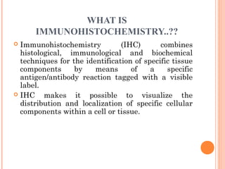
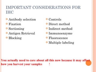
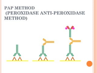
BASICS OF
IMMUNOHISTOCHEMIST
RY
Submtted By
Dr. Aderao Ganesh N.
P-2063
Animal Nutrition Division
Submitted to
Dr. Vikash Chandra
Sr. Scientist
Department of Physiology and Climatology
COMMON METHODS OF PROTEIN
DETECTION
 ELISA
 Gel Electrophoresis
 Western blot
 Immunoprecipitation
 Spectrophotometry
 Enzyme assays
 X-ray crystallography
 NMR
 Immunohistochemistry
WHY
IMMUNOHISTOCHEMISTRY..??
 Antibodies bind to antigen in specific manner
 Gives information about spatial location
 Can be used to locate particular cells and
proteins
 Can be used to identify cellular events – e.g.
apoptosis
WHAT IS
IMMUNOHISTOCHEMISTRY..??
 Immunohistochemistry (IHC) combines
histological, immunological and biochemical
techniques for the identification of specific tissue
components by means of a specific
antigen/antibody reaction tagged with a visible
label.
 IHC makes it possible to visualize the
distribution and localization of specific cellular
components within a cell or tissue.
HISTORY
 The principle has existed since the 1930s.
 Started in 1941 when Coons identified pneumococci using
a direct fluorescent method.
 Indirect method
 Addition of horseradish peroxidase
 Peroxidase anti-peroxidase technique in 1979
 Use of Avidin & Biotin complex in early 1980’s
CELLULAR ANTIGENS CAN
WE TARGET?
 Cytoplasmic
 Nuclear
 Cell membrane
 Lipids
 Proteins
IMPORTANT CONSIDERATIONS FOR
IHC
 Antibody selection
 Fixation
 Sectioning
 Antigen Retrieval
 Blocking
 Controls
 Direct method
 Indirect method
 Immunoenzyme
 Fluorescence
 Multiple labeling
You actually need to care about all this now because it may affect
how you harvest your samples !
MONOCLONAL V. POLYCLONAL
 Monoclonal
 Mouse or rabbit
hybridoma
 Tends to be ‘cleaner’
 Very consistent batch-
to-batch
 More likely to get false
negative results
 Polyclonal
 Many different species
 Tends to have more
non-specific reactivity
 Can have very
different
avidity/affinity batch-
to-batch
 More likely to have
success in an
unknown application
STEPS OF
IMMUNOHISTOCHEMISTRY
FIXATION
 To preserve tissue morphology and retain the
antigenicity
 of the target molecules
 To avoid proteolytic changes
 Helps to prevent
 Elution
 Degradation •
 Modification
 Preserves the position of the Ag
 Preserves the secondary and tertiary structure to a
possible extent
 Provides target for Ab molecules
 Formaldehyde is the preferred fixative as it keeps all
the proteins intact and is cheaper one
FIXATION
 Aldehyde
 10% NBF
 4% formaldehyde with
PBS buffer
 2% formaldehyde with
picric acid and PBS
 The paraformaldehyde
paradox
 Immersion v.
transcardial perfusion
 24-72 hours
 Best for good
architecture
 Frozen
 LN2
 With or without sucrose
 Fix with Alcohol,
acetone or methanol
(fix by coagulation, also
permeabilizes)
 Best for cell membrane
antigens, cytokines
CHEMICAL FIXATIVES
 Simple fixatives
 Formalin
 Mercuric chloride
 Osmic acid
 Picric acid
 Acetone
 Ethyl alcohol
 Osmium tetaoxide
 Campound fixatives
Microanatomical
Formal saline
Neutral buffere
formal saline
Zenkers fluid
Bounes fluid
Cytological
Nuclear
Carnoys fluid
Cytoplasmic-
Chamoys fluid
SECTIONING
 Paraffin
 Must heat and process
through xylenes and
alcohols – ruins some
antigens
 Most commonly used
 BEST if not stored
more than two weeks
– lose antigenicity
after that time
 Frozen
 Better survival of
many antigens
 Poor morphology
 Poor resolution at
higher mag
 Special storage
 Cutting difficulty
Done to have better picture and thin layer of cells for better
processing and better result
ANTIGEN RETRIVAL
 To prepare the tissue to receive the primary antibody
 Ables the partial reversal of formaldehyde induced
confirmational change of Ags
 Increases the accessibility of the Ab to the Ag
 Two methods:
 Heat
 Enzyme digestion
 Choice of Ag retrieval depends on the Ag to be
demonstrated
 Heat Induced Epitope Retrieval (HIER) is widely
used.
ANTIGEN RETRIEVAL
 HIER (Heat induced
epitope retrieval )
 Use
MW/steamer/pressure
cooker ~ 20 minutes,
slow cool
 Citrate 6.0
 Tris-EDTA 9.0
 EDTA 8.0
 Must determine for
each new
antibody/antigen
target
 PIER (Protease
induced epitope
retrieval )
 Proteinase K
 Trypsin
 Pepsin
 Pronase,etc.
 Destroys some
epitopes
 Bad for morphology
IMPROVING ANTIBODY
PENETRATION
 Need this for intracellular (cytoplasmic, nuclear) or
membrane components when epitope is inside cell
membrane
 Detergents most popular
 Triton-X
 Tween
 Also decreases surface tension – better coverage
 Can’t be used for membrane proteins (surface tension
reduced)
 Acetone/Methanol
 Precipitate proteins outside cell membranes- more
accessible
 Saponin
 Punches holes in cell membrane – holes close up when
removed
BLOCKING
 Blocking is done to avoid binding of primary
antibodies to non specific sites and hamper result.
 Done by BSA or 10% serum
 Background staining
 Specific
 Polyclonal antibodies – impure antigen used
 Inadequate fixation – diffusion of antigen – often
worse in center of large block
 Non-specific
 Non-immunologic binding – usually uniform
 Endogenous peroxidases
 Endogenous biotin
NON-SPECIFIC STAINING
Before blocking After blocking
CONTROLS
 Controls should be kept in order to have better
estimate and specificity.
 Positive control
 Best is tissue with known specificity
 Negative control
 Best is IgG from same species immunized against
non-biologic molecule – e.g. BRDU when no BRDU is
present in tissue
 Can also use non-immunized serum from same
species
ANTIBODY BINDING
 Direct Method
Labelled Ab reacts directly with Ag in tissue
sections
Single step method
Short and quick
Insensitive due to little signal amplification
E.g., FITC conjugated Antisera
Herein the antibodies are incubated for 30-60
min.
DIRECT METHOD-
PRIMARY ANTIBODY ONLY
ANTIBODY BINDING - INDIRECT
METHOD
 Unlabelled Primary Ab reacts with Ag and the
labelled secondary Ab reacts with the primary Ab
 Sensitive due to signal amplification
 Economical as single secondary Ab can be used
against many Abs from same species
 Peroxidase Anti-Peroxidase/ Alkaline
Phosphatase Anti-Alkaline Phosphatase (PAP/
APAAP) Method
 Avidin-Biotin Complex (ABC) Method
 Streptavidin – Peroxidase Method
INDIRECT METHOD – PRIMARY AND
SECONDARY ANTIBODIES
Goat anti-actin
Donkey anti-goat
labeled with 488
ENZYME LINKAGE INDIRECT
METHOD
MULTIPLE
IMMUNOFLUORESCENCE
DETECTION METHODS
 Ag-Ab conjugates are visualized by the use of a
label
 Enzymes that produce a colored precipitate in
the presence of a substrate are used as labels
 Labels :
Peroxidase
 Alkaline Phosphatase
 Detection systems:
Direct or Single step Method
Indirect or Two step Method
MULTIPLE LABELLING OF A TISSUE SECTION
ENZYMATIC DETECTION
METHODS
 Brightfield microscope sufficient for analysis of
specimens
 Suitable for tissue analysis at low magnification
 Resolution of subcellular structures not as good as with
fluorescence methods, but can be combined with electron
microscopy
 Unimited shelf life of labelled specimens
 Substrate reagents often toxic/carcinogenic
PAP METHOD
(PEROXIDASE ANTI-PEROXIDASE
METHOD)
ABC METHOD
SUMMARY
 IHC = immunology +histology + chemistry
 Has strengths and weaknesses
 Think about your planned assay before acquiring
tissue
 Good block, appropriately fixed and sectioned can
give you great data
 Bad block, inappropriately fixed and sectioned,
can give you misleading data and waste money
SUMMARY
 Very efficient and sensitive method for localizing
of antigens and their identification
 Although, advances has been occurred for protein
identification but this is used till now due to its
benifit that it identifies the protein in intact cell.
 Use of secondary antibody is always good as it
will be less expensive and yield good results.
 IHC should can be used in the cells in their
natural state.
Immunohistochemistry

Immunohistochemistry

  • 1.
    BASICS OF IMMUNOHISTOCHEMIST RY Submtted By Dr.Aderao Ganesh N. P-2063 Animal Nutrition Division Submitted to Dr. Vikash Chandra Sr. Scientist Department of Physiology and Climatology
  • 2.
    COMMON METHODS OFPROTEIN DETECTION  ELISA  Gel Electrophoresis  Western blot  Immunoprecipitation  Spectrophotometry  Enzyme assays  X-ray crystallography  NMR  Immunohistochemistry
  • 3.
    WHY IMMUNOHISTOCHEMISTRY..??  Antibodies bindto antigen in specific manner  Gives information about spatial location  Can be used to locate particular cells and proteins  Can be used to identify cellular events – e.g. apoptosis
  • 4.
    WHAT IS IMMUNOHISTOCHEMISTRY..??  Immunohistochemistry(IHC) combines histological, immunological and biochemical techniques for the identification of specific tissue components by means of a specific antigen/antibody reaction tagged with a visible label.  IHC makes it possible to visualize the distribution and localization of specific cellular components within a cell or tissue.
  • 5.
    HISTORY  The principlehas existed since the 1930s.  Started in 1941 when Coons identified pneumococci using a direct fluorescent method.  Indirect method  Addition of horseradish peroxidase  Peroxidase anti-peroxidase technique in 1979  Use of Avidin & Biotin complex in early 1980’s
  • 6.
    CELLULAR ANTIGENS CAN WETARGET?  Cytoplasmic  Nuclear  Cell membrane  Lipids  Proteins
  • 7.
    IMPORTANT CONSIDERATIONS FOR IHC Antibody selection  Fixation  Sectioning  Antigen Retrieval  Blocking  Controls  Direct method  Indirect method  Immunoenzyme  Fluorescence  Multiple labeling You actually need to care about all this now because it may affect how you harvest your samples !
  • 8.
    MONOCLONAL V. POLYCLONAL Monoclonal  Mouse or rabbit hybridoma  Tends to be ‘cleaner’  Very consistent batch- to-batch  More likely to get false negative results  Polyclonal  Many different species  Tends to have more non-specific reactivity  Can have very different avidity/affinity batch- to-batch  More likely to have success in an unknown application
  • 9.
  • 10.
    FIXATION  To preservetissue morphology and retain the antigenicity  of the target molecules  To avoid proteolytic changes  Helps to prevent  Elution  Degradation •  Modification  Preserves the position of the Ag  Preserves the secondary and tertiary structure to a possible extent  Provides target for Ab molecules  Formaldehyde is the preferred fixative as it keeps all the proteins intact and is cheaper one
  • 11.
    FIXATION  Aldehyde  10%NBF  4% formaldehyde with PBS buffer  2% formaldehyde with picric acid and PBS  The paraformaldehyde paradox  Immersion v. transcardial perfusion  24-72 hours  Best for good architecture  Frozen  LN2  With or without sucrose  Fix with Alcohol, acetone or methanol (fix by coagulation, also permeabilizes)  Best for cell membrane antigens, cytokines
  • 12.
    CHEMICAL FIXATIVES  Simplefixatives  Formalin  Mercuric chloride  Osmic acid  Picric acid  Acetone  Ethyl alcohol  Osmium tetaoxide  Campound fixatives Microanatomical Formal saline Neutral buffere formal saline Zenkers fluid Bounes fluid Cytological Nuclear Carnoys fluid Cytoplasmic- Chamoys fluid
  • 13.
    SECTIONING  Paraffin  Mustheat and process through xylenes and alcohols – ruins some antigens  Most commonly used  BEST if not stored more than two weeks – lose antigenicity after that time  Frozen  Better survival of many antigens  Poor morphology  Poor resolution at higher mag  Special storage  Cutting difficulty Done to have better picture and thin layer of cells for better processing and better result
  • 14.
    ANTIGEN RETRIVAL  Toprepare the tissue to receive the primary antibody  Ables the partial reversal of formaldehyde induced confirmational change of Ags  Increases the accessibility of the Ab to the Ag  Two methods:  Heat  Enzyme digestion  Choice of Ag retrieval depends on the Ag to be demonstrated  Heat Induced Epitope Retrieval (HIER) is widely used.
  • 15.
    ANTIGEN RETRIEVAL  HIER(Heat induced epitope retrieval )  Use MW/steamer/pressure cooker ~ 20 minutes, slow cool  Citrate 6.0  Tris-EDTA 9.0  EDTA 8.0  Must determine for each new antibody/antigen target  PIER (Protease induced epitope retrieval )  Proteinase K  Trypsin  Pepsin  Pronase,etc.  Destroys some epitopes  Bad for morphology
  • 16.
    IMPROVING ANTIBODY PENETRATION  Needthis for intracellular (cytoplasmic, nuclear) or membrane components when epitope is inside cell membrane  Detergents most popular  Triton-X  Tween  Also decreases surface tension – better coverage  Can’t be used for membrane proteins (surface tension reduced)  Acetone/Methanol  Precipitate proteins outside cell membranes- more accessible  Saponin  Punches holes in cell membrane – holes close up when removed
  • 17.
    BLOCKING  Blocking isdone to avoid binding of primary antibodies to non specific sites and hamper result.  Done by BSA or 10% serum  Background staining  Specific  Polyclonal antibodies – impure antigen used  Inadequate fixation – diffusion of antigen – often worse in center of large block  Non-specific  Non-immunologic binding – usually uniform  Endogenous peroxidases  Endogenous biotin
  • 18.
  • 19.
    CONTROLS  Controls shouldbe kept in order to have better estimate and specificity.  Positive control  Best is tissue with known specificity  Negative control  Best is IgG from same species immunized against non-biologic molecule – e.g. BRDU when no BRDU is present in tissue  Can also use non-immunized serum from same species
  • 20.
    ANTIBODY BINDING  DirectMethod Labelled Ab reacts directly with Ag in tissue sections Single step method Short and quick Insensitive due to little signal amplification E.g., FITC conjugated Antisera Herein the antibodies are incubated for 30-60 min.
  • 21.
  • 22.
    ANTIBODY BINDING -INDIRECT METHOD  Unlabelled Primary Ab reacts with Ag and the labelled secondary Ab reacts with the primary Ab  Sensitive due to signal amplification  Economical as single secondary Ab can be used against many Abs from same species  Peroxidase Anti-Peroxidase/ Alkaline Phosphatase Anti-Alkaline Phosphatase (PAP/ APAAP) Method  Avidin-Biotin Complex (ABC) Method  Streptavidin – Peroxidase Method
  • 23.
    INDIRECT METHOD –PRIMARY AND SECONDARY ANTIBODIES Goat anti-actin Donkey anti-goat labeled with 488
  • 24.
  • 25.
  • 26.
    DETECTION METHODS  Ag-Abconjugates are visualized by the use of a label  Enzymes that produce a colored precipitate in the presence of a substrate are used as labels  Labels : Peroxidase  Alkaline Phosphatase  Detection systems: Direct or Single step Method Indirect or Two step Method
  • 27.
    MULTIPLE LABELLING OFA TISSUE SECTION
  • 28.
    ENZYMATIC DETECTION METHODS  Brightfieldmicroscope sufficient for analysis of specimens  Suitable for tissue analysis at low magnification  Resolution of subcellular structures not as good as with fluorescence methods, but can be combined with electron microscopy  Unimited shelf life of labelled specimens  Substrate reagents often toxic/carcinogenic
  • 29.
  • 30.
  • 31.
    SUMMARY  IHC =immunology +histology + chemistry  Has strengths and weaknesses  Think about your planned assay before acquiring tissue  Good block, appropriately fixed and sectioned can give you great data  Bad block, inappropriately fixed and sectioned, can give you misleading data and waste money
  • 32.
    SUMMARY  Very efficientand sensitive method for localizing of antigens and their identification  Although, advances has been occurred for protein identification but this is used till now due to its benifit that it identifies the protein in intact cell.  Use of secondary antibody is always good as it will be less expensive and yield good results.  IHC should can be used in the cells in their natural state.

Editor's Notes

  • #12 Formaldehyde fixes by formation of hydroxymethyl adducts on reactive side chains of proteins. Once sufficient adducts are formed, they slowly cross-link to stabilize the proteins in a gel-like formation. At room temperature, it takes approximately 24 hours for maximal binding of formaldehyde to occur and hence all the adducts to form. These initial adducts, and any initial cross-links formed, are unstable and easily reversed. For tissues fixed for 24 hours, the cross-links are largely reversed by washing ( in water or 70% EtOH) after a few hours. It probably takes at least 5-7 days for most of the cross-links to form, and a small amount of cross-linking still continues over time. Even after fixation for 7 days or more, the cross-links can still be reversed by prolonged washing. That's great for IHC, since cross-links can be reversed by HIER (which is just washing at elevated temperatures!).
  • #22 Fluorochrome conjugated primary antibody