Western Blot Customer Review Anti-Endophilin I Antibody (STJ92924)
•
0 likes•102 views

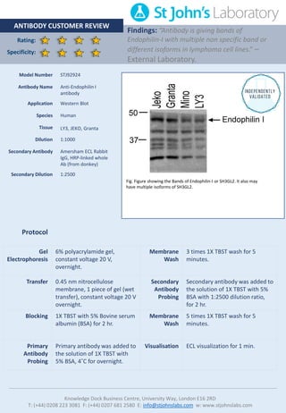
Implicated in synaptic vesicle endocytosis. May recruit other proteins to membranes with high curvature. Brain, mostly in frontal cortex. Expressed at high level in fetal cerebellum. Anti-Endophilin I -http://www.stjohnslabs.com/endophilin-i-antibody Join our Antibody Validation Project - http://www.stjohnslabs.com/services/antibody-validation
Report
Share
Report
Share
Download to read offline
Recommended
Western Blot Customer Review Anti-β-tubulin Antibody (STJ97037)
Tubulin is the major constituent of microtubules. It binds two moles of GTP, one at an exchangeable site on the beta chain and one at a non-exchangeable site on the alpha chain. TUBB3 plays a critical role in proper axon guidance and mantainance.
Anti-β-tubulin -http://www.stjohnslabs.com/b-tubulin-antibody-p-98672
Join our Antibody Validation Project - http://www.stjohnslabs.com/services/antibody-validation
Western Blot Customer Review Anti Glucocorticoid Receptor antibody (STJ97101)
Western Blot Customer Review Anti Glucocorticoid Receptor antibody (STJ97101)St John's Laboratory Ltd
Receptor for glucocorticoids (GC). Has a dual mode of action: as a transcription factor that binds to glucocorticoid response elements (GRE), both for nuclear and mitochondrial DNA, and as a modulator of other transcription factors. Affects inflammatory responses, cellular proliferation and differentiation in target tissues. Involved in chromatin remodeling . Plays a role in rapid mRNA degradation by binding to the 5' UTR of target mRNAs and interacting with PNRC2 in a ligand-dependent manner. Could act as a coactivator for STAT5-dependent transcription upon growth hormone (GH) stimulation and could reveal an essential role of hepatic GR in the control of body growth (By similarity). Has transcriptional activation and repression activity . Mediates glucocorticoid-induced apoptosis . Promotes accurate chromosome segregation during mitosis . May act as a tumor suppressor . May play a negative role in adipogenesis through the regulation of lipolytic and antilipogenic gene expression (By similarity). / Isoform Beta: Acts as a dominant negative inhibitor of isoform Alpha . Has intrinsic transcriptional activity independent of isoform Alpha when both isoforms are coexpressed.
Join our Antibody Validation Project: http://www.stjohnslabs.com/services/antibody-validation
Anti glucocorticoid receptor antibody (STJ97101):
http://www.stjohnslabs.com/glucocorticoid-receptor-antibody-p-98736?filter_name=STJ97101Western Blot Antibody Customer Review for Anti-Histone H3 (Phospho-Tyr41) An...
Western Blot Antibody Customer Review for Anti-Histone H3 (Phospho-Tyr41) An...St John's Laboratory Ltd
Core component of nucleosome. Nucleosomes wrap and compact DNA into chromatin, limiting DNA accessibility to the cellular machineries which require DNA as a template. Histones thereby play a central role in transcription regulation, DNA repair, DNA replication and chromosomal stability. DNA accessibility is regulated via a complex set of post-translational modifications of histones, also called histone code, and nucleosome remodeling.
Anti-Histone H3 (Phospho-Tyr41) - http://www.stjohnslabs.com/histone-h3-phospho-tyr41-antibody?filter_name=STJ97138
Join our Antibody Validation Project - http://www.stjohnslabs.com/services/antibody-validationWestern Blot Antibody Customer Review for Anti-Phospho-ALK (Y1507) Polyclonal...
Western Blot Antibody Customer Review for Anti-Phospho-ALK (Y1507) Polyclonal...St John's Laboratory Ltd
Anaplastic lymphoma kinase (ALK) also known as ALK tyrosine kinase receptor or CD246 (cluster of differentiation 246) is an enzyme that in humans is encoded by the ALK gene.
ALK plays an important role in the development of the brain and exerts its effects on specific neurons in the nervous system. Transduces signals from ligands at the cell surface, through specific activation of the mitogen-activated protein kinase (MAPK) pathway. Phospho-ALK (Y1507) Polyclonal Antibody detects endogenous levels of ALK protein only when phosphorylated at Y1507.
Anti-Phospho-ALK - http://www.stjohnslabs.com/phospho-alk-y1507-antibody?filter_name=STJ90845
Join our Antibody Validation Project - http://www.stjohnslabs.com/services/antibody-validation
Western Blot Customer Review Anti-Phospho-Cofilin (S3) Antibody (STJ90230)
Binds to F-actin and exhibits pH-sensitive F-actin depolymerizing activity. Regulates actin cytoskeleton dynamics. Important for normal progress through mitosis and normal cytokinesis. Plays a role in the regulation of cell morphology and cytoskeletal organization. Required for the up-regulation of atypical chemokine receptor ACKR2 from endosomal compartment to cell membrane, increasing its efficiency in chemokine uptake and degradation.
Anti-Phospho-Cofilin (S3) -http://www.stjohnslabs.com/phospho-cofilin-s3-antibody
Join our Antibody Validation Project - http://www.stjohnslabs.com/services/antibody-validation
Customer Review For LPCAT1 Polyclonal Antibody (STJ27005)
LPCAT1 antibody possess both acyltransferase and acetyltransferase activities. Activity is calcium-independent. Mediates the conversion of 1-acyl-sn-glycero-3-phosphocholine (LPC) into phosphatidylcholine (PC). Displays a clear preference for saturated fatty acyl-CoAs, and 1-myristoyl or 1-palmitoyl LPC as acyl donors and acceptors, respectively. May synthesize phosphatidylcholine in pulmonary surfactant, thereby playing a pivotal role in respiratory physiology.
Publication:
http://www.ncbi.nlm.nih.gov/pubmed/16864775
http://www.ncbi.nlm.nih.gov/pubmed/21498505
To purchase this antibody, use the following link: http://www.stjohnslabs.com/lpcat1-antibody-p-68679?filter_name=STJ27005
Customer Review For GSK3β Polyclonal Antibody (STJ93447)
GSK3β Polyclonal Antibody is an active protein kinase that acts as a negative regulator in the hormonal control of glucose homeostasis, Wnt signaling and regulation of transcription factors and microtubules, by phosphorylating and inactivating glycogen synthase (GYS1 or GYS2), EIF2B, CTNNB1/beta-catenin, APC, AXIN1, DPYSL2/CRMP2, JUN, NFATC1/NFATC, MAPT/TAU and MACF1. Requires primed phosphorylation of the majority of its substrates.
To purchase this antibody, use the following link: http://www.stjohnslabs.com/gsk3b-antibody-p-92568?filter_name=STJ93447
Customer Review for Histone H4 (TriMethyl Lys20) Polyclonal Antibody (STJ97154)
Customer Review for Histone H4 (TriMethyl Lys20) Polyclonal Antibody (STJ97154)St John's Laboratory Ltd
Histones are nuclear proteins that form octameric structures which bind DNA to form units of chromatin called nucleosomes. The family of histones—H2A, H2B, H3, and H4—are key players in gene regulation. They undergo a number of post-translational modifications (PTM) in response to various stimuli, including phosphorylation on serine and threonine residues and methylation on lysine residues. PTMs produce configural changes in histone proteins that may induce nucleosome remodeling and expose or hide DNA sequences from transcriptional complexes. Histone H4 lysine 20 (H4K20) may undergo mono-, di-, or trimethylation, which is catalyzed by the methyltransferase PR-Set7 (Set8 or KMT5a). Methylated H4K20 plays a role in regulating DNA damage responses, mitosis, DNA replication, and gene expression. Trimethylation of H4K20 contributes to gene silencing, and is a mark of the repressive heterochromatin state.
To purchase this antibody use the following link: http://www.stjohnslabs.com/histone-h4-tri-methyl-lys20-antibody?filter_name=STJ97154Recommended
Western Blot Customer Review Anti-β-tubulin Antibody (STJ97037)
Tubulin is the major constituent of microtubules. It binds two moles of GTP, one at an exchangeable site on the beta chain and one at a non-exchangeable site on the alpha chain. TUBB3 plays a critical role in proper axon guidance and mantainance.
Anti-β-tubulin -http://www.stjohnslabs.com/b-tubulin-antibody-p-98672
Join our Antibody Validation Project - http://www.stjohnslabs.com/services/antibody-validation
Western Blot Customer Review Anti Glucocorticoid Receptor antibody (STJ97101)
Western Blot Customer Review Anti Glucocorticoid Receptor antibody (STJ97101)St John's Laboratory Ltd
Receptor for glucocorticoids (GC). Has a dual mode of action: as a transcription factor that binds to glucocorticoid response elements (GRE), both for nuclear and mitochondrial DNA, and as a modulator of other transcription factors. Affects inflammatory responses, cellular proliferation and differentiation in target tissues. Involved in chromatin remodeling . Plays a role in rapid mRNA degradation by binding to the 5' UTR of target mRNAs and interacting with PNRC2 in a ligand-dependent manner. Could act as a coactivator for STAT5-dependent transcription upon growth hormone (GH) stimulation and could reveal an essential role of hepatic GR in the control of body growth (By similarity). Has transcriptional activation and repression activity . Mediates glucocorticoid-induced apoptosis . Promotes accurate chromosome segregation during mitosis . May act as a tumor suppressor . May play a negative role in adipogenesis through the regulation of lipolytic and antilipogenic gene expression (By similarity). / Isoform Beta: Acts as a dominant negative inhibitor of isoform Alpha . Has intrinsic transcriptional activity independent of isoform Alpha when both isoforms are coexpressed.
Join our Antibody Validation Project: http://www.stjohnslabs.com/services/antibody-validation
Anti glucocorticoid receptor antibody (STJ97101):
http://www.stjohnslabs.com/glucocorticoid-receptor-antibody-p-98736?filter_name=STJ97101Western Blot Antibody Customer Review for Anti-Histone H3 (Phospho-Tyr41) An...
Western Blot Antibody Customer Review for Anti-Histone H3 (Phospho-Tyr41) An...St John's Laboratory Ltd
Core component of nucleosome. Nucleosomes wrap and compact DNA into chromatin, limiting DNA accessibility to the cellular machineries which require DNA as a template. Histones thereby play a central role in transcription regulation, DNA repair, DNA replication and chromosomal stability. DNA accessibility is regulated via a complex set of post-translational modifications of histones, also called histone code, and nucleosome remodeling.
Anti-Histone H3 (Phospho-Tyr41) - http://www.stjohnslabs.com/histone-h3-phospho-tyr41-antibody?filter_name=STJ97138
Join our Antibody Validation Project - http://www.stjohnslabs.com/services/antibody-validationWestern Blot Antibody Customer Review for Anti-Phospho-ALK (Y1507) Polyclonal...
Western Blot Antibody Customer Review for Anti-Phospho-ALK (Y1507) Polyclonal...St John's Laboratory Ltd
Anaplastic lymphoma kinase (ALK) also known as ALK tyrosine kinase receptor or CD246 (cluster of differentiation 246) is an enzyme that in humans is encoded by the ALK gene.
ALK plays an important role in the development of the brain and exerts its effects on specific neurons in the nervous system. Transduces signals from ligands at the cell surface, through specific activation of the mitogen-activated protein kinase (MAPK) pathway. Phospho-ALK (Y1507) Polyclonal Antibody detects endogenous levels of ALK protein only when phosphorylated at Y1507.
Anti-Phospho-ALK - http://www.stjohnslabs.com/phospho-alk-y1507-antibody?filter_name=STJ90845
Join our Antibody Validation Project - http://www.stjohnslabs.com/services/antibody-validation
Western Blot Customer Review Anti-Phospho-Cofilin (S3) Antibody (STJ90230)
Binds to F-actin and exhibits pH-sensitive F-actin depolymerizing activity. Regulates actin cytoskeleton dynamics. Important for normal progress through mitosis and normal cytokinesis. Plays a role in the regulation of cell morphology and cytoskeletal organization. Required for the up-regulation of atypical chemokine receptor ACKR2 from endosomal compartment to cell membrane, increasing its efficiency in chemokine uptake and degradation.
Anti-Phospho-Cofilin (S3) -http://www.stjohnslabs.com/phospho-cofilin-s3-antibody
Join our Antibody Validation Project - http://www.stjohnslabs.com/services/antibody-validation
Customer Review For LPCAT1 Polyclonal Antibody (STJ27005)
LPCAT1 antibody possess both acyltransferase and acetyltransferase activities. Activity is calcium-independent. Mediates the conversion of 1-acyl-sn-glycero-3-phosphocholine (LPC) into phosphatidylcholine (PC). Displays a clear preference for saturated fatty acyl-CoAs, and 1-myristoyl or 1-palmitoyl LPC as acyl donors and acceptors, respectively. May synthesize phosphatidylcholine in pulmonary surfactant, thereby playing a pivotal role in respiratory physiology.
Publication:
http://www.ncbi.nlm.nih.gov/pubmed/16864775
http://www.ncbi.nlm.nih.gov/pubmed/21498505
To purchase this antibody, use the following link: http://www.stjohnslabs.com/lpcat1-antibody-p-68679?filter_name=STJ27005
Customer Review For GSK3β Polyclonal Antibody (STJ93447)
GSK3β Polyclonal Antibody is an active protein kinase that acts as a negative regulator in the hormonal control of glucose homeostasis, Wnt signaling and regulation of transcription factors and microtubules, by phosphorylating and inactivating glycogen synthase (GYS1 or GYS2), EIF2B, CTNNB1/beta-catenin, APC, AXIN1, DPYSL2/CRMP2, JUN, NFATC1/NFATC, MAPT/TAU and MACF1. Requires primed phosphorylation of the majority of its substrates.
To purchase this antibody, use the following link: http://www.stjohnslabs.com/gsk3b-antibody-p-92568?filter_name=STJ93447
Customer Review for Histone H4 (TriMethyl Lys20) Polyclonal Antibody (STJ97154)
Customer Review for Histone H4 (TriMethyl Lys20) Polyclonal Antibody (STJ97154)St John's Laboratory Ltd
Histones are nuclear proteins that form octameric structures which bind DNA to form units of chromatin called nucleosomes. The family of histones—H2A, H2B, H3, and H4—are key players in gene regulation. They undergo a number of post-translational modifications (PTM) in response to various stimuli, including phosphorylation on serine and threonine residues and methylation on lysine residues. PTMs produce configural changes in histone proteins that may induce nucleosome remodeling and expose or hide DNA sequences from transcriptional complexes. Histone H4 lysine 20 (H4K20) may undergo mono-, di-, or trimethylation, which is catalyzed by the methyltransferase PR-Set7 (Set8 or KMT5a). Methylated H4K20 plays a role in regulating DNA damage responses, mitosis, DNA replication, and gene expression. Trimethylation of H4K20 contributes to gene silencing, and is a mark of the repressive heterochromatin state.
To purchase this antibody use the following link: http://www.stjohnslabs.com/histone-h4-tri-methyl-lys20-antibody?filter_name=STJ97154Customer Review For CAR Polyclonal Antibody (STJ91996)
Component of the epithelial apical junction complex that may function as an homophilic cell adhesion molecule and is essential for tight junction integrity. Also involved in transepithelial migration of leukocytes through adhesive interactions with AMICA1/JAML a transmembrane protein of the plasma membrane of leukocytes. The interaction between both receptors also mediates the activation of gamma-delta T-cells, a subpopulation of T-cells residing in epithelia and involved in tissue homeostasis and repair. Upon epithelial CXADR-binding, AMICA1 induces downstream cell signaling events in gamma-delta T-cells through PI3-kinase and MAP kinases. It results in proliferation and production of cytokines and growth factors by T-cells that in turn stimulate epithelial tissues repair.
To purchase this antibody, use the following link: http://www.stjohnslabs.com/car-antibody?filter_name=STJ91996
Customer Review for TriMethyl-Histone H3-K56 Polyclonal Antibody (STJ29403)
Histones are basic nuclear proteins that are responsible for the nucleosome structure of the chromosomal fiber in eukaryotes. Nucleosomes consist of approximately 146 bp of DNA wrapped around a histone octamer composed of pairs of each of the four core histones (H2A, H2B, H3, and H4). The chromatin fiber is further compacted through the interaction of a linker histone, H1, with the DNA between the nucleosomes to form higher order chromatin structures. This gene is intronless and encodes a member of the histone H3 family. Transcripts from this gene lack polyA tails; instead, they contain a palindromic termination element. This gene is located separately from the other H3 genes that are in the histone gene cluster on chromosome 6p22-p21.3.
To purchase this antibody use the following link: http://www.stjohnslabs.com/trimethyl-histone-h3-k56?filter_name=STJ29403
Western Blot Protocol - St John's Laboratory
The western blot technique uses gel electrophoresis to separate proteins in a tissue homogenate or extract by molecular weight. The separated proteins on the gel are then transferred to a membrane (usually nitrocellulose or PVDF) which is then incubated with an antibody specific for a target protein. The protein of interest can be visualised using conjugated secondary antibodies and detection reagents.
Do you have a technical question? Get in touch: info@stjohnslabs.com
Protocol series - http://www.stjohnslabs.com/services/video-protocol-series
Customer Review For ERBB4 Polyclonal Antibody (STJ27886)
Tyrosine-protein kinase that plays an essential role as cell surface receptor for neuregulins and EGF family members and regulates development of the heart, the central nervous system and the mammary gland, gene transcription, cell proliferation, differentiation, migration and apoptosis. Required for normal cardiac muscle differentiation during embryonic development, and for postnatal cardiomyocyte proliferation. Required for normal development of the embryonic central nervous system, especially for normal neural crest cell migration and normal axon guidance. Required for mammary gland differentiation, induction of milk proteins and lactation. Acts as cell-surface receptor for the neuregulins NRG1, NRG2, NRG3 and NRG4 and the EGF family members BTC, EREG and HBEGF. Ligand binding triggers receptor dimerization and autophosphorylation at specific tyrosine residues that then serve as binding sites for scaffold proteins and effectors. Ligand specificity and signaling is modulated by alternative splicing, proteolytic processing, and by the formation of heterodimers with other ERBB family members, thereby creating multiple combinations of intracellular phosphotyrosines that trigger ligand- and context-specific cellular responses. Mediates phosphorylation of SHC1 and activation of the MAP kinases MAPK1/ERK2 and MAPK3/ERK1. Isoform JM-A CYT-1 and isoform JM-B CYT-1 phosphorylate PIK3R1, leading to the activation of phosphatidylinositol 3-kinase and AKT1 and protect cells against apoptosis. Isoform JM-A CYT-1 and isoform JM-B CYT-1 mediate reorganization of the actin cytoskeleton and promote cell migration in response to NRG1. Isoform JM-A CYT-2 and isoform JM-B CYT-2 lack the phosphotyrosine that mediates interaction with PIK3R1, and hence do not phosphorylate PIK3R1, do not protect cells against apoptosis, and do not promote reorganization of the actin cytoskeleton and cell migration. Proteolytic processing of isoform JM-A CYT-1 and isoform JM-A CYT-2 gives rise to the corresponding soluble intracellular domains (4ICD) that translocate to the nucleus, promote nuclear import of STAT5A, activation of STAT5A, mammary epithelium differentiation, cell proliferation and activation of gene expression. The ERBB4 soluble intracellular domains (4ICD) colocalize with STAT5A at the CSN2 promoter to regulate transcription of milk proteins during lactation. The ERBB4 soluble intracellular domains can also translocate to mitochondria and promote apoptosis.
To purchase this antibody, use the following link: http://www.stjohnslabs.com/erbb4?filter_name=STJ27886
Customer Review For EphB4 Polyclonal Antibody (STJ23550)
Ephrin receptors and their ligands, the ephrins, mediate numerous developmental processes, particularly in the nervous system. Based on their structures and sequence relationships, ephrins are divided into the ephrin-A (EFNA) class, which are anchored to the membrane by a glycosylphosphatidylinositol linkage, and the ephrin-B (EFNB) class, which are transmembrane proteins. The Eph family of receptors are divided into 2 groups based on the similarity of their extracellular domain sequences and their affinities for binding ephrin-A and ephrin-B ligands. Ephrin receptors make up the largest subgroup of the receptor tyrosine kinase (RTK) family. The protein encoded by this gene binds to ephrin-B2 and plays an essential role in vascular development.
To purchase this antibody, use the following link: http://www.stjohnslabs.com/ephb4-antibody-p-65270?filter_name=STJ23550
Independent Antibody Validation For mCherry Tag Monoclonal Antibody (STJ34373)
Independent Antibody Validation For mCherry Tag Monoclonal Antibody (STJ34373)St John's Laboratory Ltd
St John’s Laboratory suppliers a highly photostable mCherry tag antibody, which is resistant to photobleaching. Independent validation reviews provide feedback on real-experimental use:
https://www.scienceexchange.com/validations/29688
https://www.scienceexchange.com/validations/29733
https://www.scienceexchange.com/validations/29708
To purchase this antibody, use the following link: http://www.stjohnslabs.com/mcherry-tag-antibody?filter_name=STJ34373Customer Review For FAAH Polyclonal Antibody (STJ23602)
FAAH antibody degrades bioactive fatty acid amides like oleamide, the endogenous cannabinoid, anandamide and myristic amide to their corresponding acids, thereby serving to terminate the signaling functions of these molecules. Hydrolyzes polyunsaturated substrate anandamide preferentially as compared to monounsaturated substrates.
To purchase this antibody, use the following link: http://www.stjohnslabs.com/faah-antibody-p-65320?filter_name=STJ23602
Customer Review For NFκB-p65 Polyclonal Antibody (STJ94468)
NF-kappa-B is a pleiotropic transcription factor present in almost all cell types and is the endpoint of a series of signal transduction events that are initiated by a vast array of stimuli related to many biological processes such as inflammation, immunity, differentiation, cell growth, tumorigenesis and apoptosis. NF-kappa-B is a homo- or heterodimeric complex formed by the Rel-like domain-containing proteins RELA/p65, RELB, NFKB1/p105, NFKB1/p50, REL and NFKB2/p52 and the heterodimeric p65-p50 complex appears to be most abundant one. The dimers bind at kappa-B sites in the DNA of their target genes and the individual dimers have distinct preferences for different kappa-B sites that they can bind with distinguishable affinity and specificity. Different dimer combinations act as transcriptional activators or repressors, respectively.
To purchase this antibody, use the following link: http://www.stjohnslabs.com/nfkb-p65-antibody-p-93367?filter_name=STJ94468
Customer Review For ENT1 Polyclonal Antibody (STJ96396)
Equilibrative nucleoside transporter 1 (ENT1) is a protein that in humans is encoded by the SLC29A1 gene. Multiple alternatively spliced variants, encoding the same protein, have been found for this gene.
This gene is a member of the equilibrative nucleoside transporter family. The gene encodes a transmembrane glycoprotein that localizes to the plasma and mitochondrial membranes and mediates the cellular uptake of nucleosides from the surrounding medium. The protein is categorized as an equilibrative (as opposed to concentrative) transporter that is sensitive to inhibition by nitrobenzylmercaptopurine ribonucleoside (NBMPR). Nucleoside transporters are required for nucleotide synthesis in cells that lack de novo nucleoside synthesis pathways, and are also necessary for the uptake of cytotoxic nucleosides used for cancer and viral chemotherapies.
To purchase this antibody, use the following link: http://www.stjohnslabs.com/ent1-antibody?filter_name=STJ96396
Antibody Customer Review For VAPB Polyclonal Antibody (STJ47066)
Vesicle-associated membrane protein-associated protein B (VAPB) is a type IV membrane protein found in plasma and intracellular vesicle membranes. The protein is found as a homodimer and as a heterodimer with VAPA. It also can interact with VAMP1 and VAMP2 and may be involved in vesicle trafficking. Like VAPA, VAPB binds to proteins that contain a FFAT motif. Considerable interest in VAPB has arisen because mutations in this protein are associated with rare, familial forms of Motor Neurone Disease (also called Amyotrophic Lateral Sclerosis and Lou Gehrig's disease).
To purchase this antibody use the following link: http://www.stjohnslabs.com/vapb-antibody-c-term-p-79486?filter_name=STJ47066
Customer Review For Actin β Polyclonal Antibody (STJ91464)
Beta-actin (human gene and protein symbol ACTB/ACTB) is one of six different actin isoforms which have been identified in humans. This is one of the two nonmuscle cytoskeletal actins. Actins are highly conserved proteins that are involved in cell motility, structure and integrity. Alpha actins are a major constituent of the contractile apparatus.
To purchase this antibody, use the following link: http://www.stjohnslabs.com/actin-b-antibody?filter_name=STJ91464
Customer Review for His-tag Polyclonal Antibody (STJ90106)
A polyhistidine-tag is an amino acid motif in proteins that consists of at least six histidine (His) residues, often at the N- or C-terminus of the protein. It is also known as hexa histidine-tag, 6xHis-tag, His6 tag and by the trademarked name His-tag. Polyhistidine-tags are often used for affinity purification of polyhistidine-tagged recombinant proteins expressed in Escherichia coli and other prokaryotic expression systems.
To purchase this antibody, use the following link: http://www.stjohnslabs.com/his-tag-antibody-p-95792?filter_name=STJ90106
Customer Review For PIWIL4 Polyclonal Antibody (STJ25009)
PIWIL4 belongs to the Argonaute family of proteins, which function in development and maintenance of germline stem cells. Plays a central role during spermatogenesis by repressing transposable elements and prevent their mobilization, which is essential for the germline integrity. Acts via the piRNA metabolic process, which mediates the repression of transposable elements during meiosis by forming complexes composed of piRNAs and Piwi proteins and govern the methylation and subsequent repression of transposons. Directly binds piRNAs, a class of 24 to 30 nucleotide RNAs that are generated by a Dicer-independent mechanism and are primarily derived from transposons and other repeated sequence elements. Associates with secondary piRNAs antisense and PIWIL2/MILI is required for such association.
To purchase this antibody, use the following link: http://www.stjohnslabs.com/piwil4-antibody-p-66699?filter_name=STJ25009
Customer Review For GAPDH Polyclonal Antibody (STJ96417)
Has both glyceraldehyde-3-phosphate dehydrogenase and nitrosylase activities, thereby playing a role in glycolysis and nuclear functions, respectively. Participates in nuclear events including transcription, RNA transport, DNA replication and apoptosis. Nuclear functions are probably due to the nitrosylase activity that mediates cysteine S-nitrosylation of nuclear target proteins such as SIRT1, HDAC2 and PRKDC. Modulates the organization and assembly of the cytoskeleton. Facilitates the CHP1-dependent microtubule and membrane associations through its ability to stimulate the binding of CHP1 to microtubules (By similarity). Glyceraldehyde-3-phosphate dehydrogenase is a key enzyme in glycolysis that catalyzes the first step of the pathway by converting D-glyceraldehyde 3-phosphate (G3P) into 3-phospho-D-glyceroyl phosphate. Component of the GAIT (gamma interferon-activated inhibitor of translation) complex which mediates interferon-gamma-induced transcript-selective translation inhibition in inflammation processes. Upon interferon-gamma treatment assembles into the GAIT complex which binds to stem loop-containing GAIT elements in the 3'-UTR of diverse inflammatory mRNAs (such as ceruplasmin) and suppresses their translation.
To purchase this antibody, use the following link: http://www.stjohnslabs.com/gapdh-antibody-p-94882?filter_name=STJ96417
Customer Review For mTOR Polyclonal Antibody (STJ94280)
Serine/threonine protein kinase which is a central regulator of cellular metabolism, growth and survival in response to hormones, growth factors, nutrients, energy and stress signals. MTOR directly or indirectly regulates the phosphorylation of at least 800 proteins. Functions as part of 2 structurally and functionally distinct signaling complexes mTORC1 and mTORC2 (mTOR complex 1 and 2). Activated mTORC1 up-regulates protein synthesis by phosphorylating key regulators of mRNA translation and ribosome synthesis. This includes phosphorylation of EIF4EBP1 and release of its inhibition toward the elongation initiation factor 4E (eiF4E). Moreover, phosphorylates and activates RPS6KB1 and RPS6KB2 that promote protein synthesis by modulating the activity of their downstream targets including ribosomal protein S6, eukaryotic translation initiation factor EIF4B, and the inhibitor of translation initiation PDCD4. Stimulates the pyrimidine biosynthesis pathway, both by acute regulation through RPS6KB1-mediated phosphorylation of the biosynthetic enzyme CAD, and delayed regulation, through transcriptional enhancement of the pentose phosphate pathway which produces 5-phosphoribosyl-1-pyrophosphate (PRPP), an allosteric activator of CAD at a later step in synthesis, this function is dependent on the mTORC1 complex. Regulates ribosome synthesis by activating RNA polymerase III-dependent transcription through phosphorylation and inhibition of MAF1 an RNA polymerase III-repressor.
To purchase this antibody, use the following link: http://www.stjohnslabs.com/mtor-antibody-p-93228?filter_name=STJ94280
Customer Review For Alpha-synuclein Polyclonal Antibody (STJ95862)
Alpha-synuclein is involved in the regulation of dopamine release and transport. Induces fibrillization of microtubule-associated protein tau. Reduces neuronal responsiveness to various apoptotic stimuli, leading to a decreased caspase-3 activation.
To purchase this antibody, use the following link: http://www.stjohnslabs.com/synuclein-a-antibody-p-94486?filter_name=STJ95862
Customer Review For Tubulin-beta Polyclonal Antibody- IF Application (STJ31562)
Customer Review For Tubulin-beta Polyclonal Antibody- IF Application (STJ31562)St John's Laboratory Ltd
Tubulin is the major constituent of microtubules. It binds two moles of GTP, one at an exchangeable site on the beta chain and one at a non-exchangeable site on the alpha chain. TUBB3 plays a critical role in proper axon guidance and maintenance.
To purchase this antibody use the following link: http://www.stjohnslabs.com/tubulin-beta-antibody?filter_name=STJ31562 Customer Review For Tubulin-beta Polyclonal Antibody - IF&ICC Application (ST...
Customer Review For Tubulin-beta Polyclonal Antibody - IF&ICC Application (ST...St John's Laboratory Ltd
Tubulin is the major constituent of microtubules. It binds two molecules of GTP, one at an exchangeable site on the beta chain and one at a non-exchangeable site on the alpha chain. TUBB3 plays a critical role in proper axon guidance and mantainance.
To purchase this antibody, use the following link: http://www.stjohnslabs.com/tubulin-beta-antibody?filter_name=STJ31562 Customer Review For D2HGDH Polyclonal Antibody (STJ27209)
This gene encodes D-2hydroxyglutarate dehydrogenase, a mitochondrial enzyme belonging to the FAD-binding oxidoreductase/transferase type 4 family. This enzyme, which is most active in liver and kidney but also active in heart and brain, converts D-2-hydroxyglutarate to 2-ketoglutarate. Mutations in this gene are present in D-2-hydroxyglutaric aciduria, a rare recessive neurometabolic disorder causing developmental delay, epilepsy, hypotonia, and dysmorphic features.
To purchase this antibody, use the following link: http://www.stjohnslabs.com/d2hgdh-antibody-p-68883?filter_name=STJ27209
Antibody Customer Review For PHF19/hPCL3 Polyclonal Antibody (STJ110368)
PHD Finger Protein 19 is coded for by the PHF19 gene (also known as hPCL3). The Polycomb Group (PcG) is a key binding site within the protein that targets trimethylated Histone H3 at Lys-36, where it recruits the PRC2 complex. It is thought to be involved in the transition of embryonic stems cells from an active to repressed state, and research has indicated its overexpression in many types of cancers.
Western Blot Customer Review Anti-WAVE2 Antibody (STJ96263)
Downstream effector molecule involved in the transmission of signals from tyrosine kinase receptors and small GTPases to the actin cytoskeleton. Promotes formation of actin filaments. Part of the WAVE complex that regulates lamellipodia formation. The WAVE complex regulates actin filament reorganization via its interaction with the Arp2/3 complex.
Anti-WAVE2 -http://www.stjohnslabs.com/wave2-antibody
Join our Antibody Validation Project - http://www.stjohnslabs.com/services/antibody-validation
Western Blot Antibody Customer Review for Anti-Peroxin 2 Antibody (STJ95040)
Western Blot Antibody Customer Review for Anti-Peroxin 2 Antibody (STJ95040)St John's Laboratory Ltd
PEX2 is gene on chromosome 8q21.1 that encodes a peroxin, an integral membrane protein involved in peroxisome biosynthesis and integrity, which assembles membrane vesicles before translocation of matrix proteins.
Defects of PEX2 cause peroxisome biogenesis disorder complementation group 5, Zelleger syndrome and a form of Refsum disease.
Anti-Peroxin 2 - http://www.stjohnslabs.com/peroxin-2-antibody?filter_name=STJ95040
Join our Antibody Validation Project - http://www.stjohnslabs.com/services/antibody-validationMore Related Content
What's hot
Customer Review For CAR Polyclonal Antibody (STJ91996)
Component of the epithelial apical junction complex that may function as an homophilic cell adhesion molecule and is essential for tight junction integrity. Also involved in transepithelial migration of leukocytes through adhesive interactions with AMICA1/JAML a transmembrane protein of the plasma membrane of leukocytes. The interaction between both receptors also mediates the activation of gamma-delta T-cells, a subpopulation of T-cells residing in epithelia and involved in tissue homeostasis and repair. Upon epithelial CXADR-binding, AMICA1 induces downstream cell signaling events in gamma-delta T-cells through PI3-kinase and MAP kinases. It results in proliferation and production of cytokines and growth factors by T-cells that in turn stimulate epithelial tissues repair.
To purchase this antibody, use the following link: http://www.stjohnslabs.com/car-antibody?filter_name=STJ91996
Customer Review for TriMethyl-Histone H3-K56 Polyclonal Antibody (STJ29403)
Histones are basic nuclear proteins that are responsible for the nucleosome structure of the chromosomal fiber in eukaryotes. Nucleosomes consist of approximately 146 bp of DNA wrapped around a histone octamer composed of pairs of each of the four core histones (H2A, H2B, H3, and H4). The chromatin fiber is further compacted through the interaction of a linker histone, H1, with the DNA between the nucleosomes to form higher order chromatin structures. This gene is intronless and encodes a member of the histone H3 family. Transcripts from this gene lack polyA tails; instead, they contain a palindromic termination element. This gene is located separately from the other H3 genes that are in the histone gene cluster on chromosome 6p22-p21.3.
To purchase this antibody use the following link: http://www.stjohnslabs.com/trimethyl-histone-h3-k56?filter_name=STJ29403
Western Blot Protocol - St John's Laboratory
The western blot technique uses gel electrophoresis to separate proteins in a tissue homogenate or extract by molecular weight. The separated proteins on the gel are then transferred to a membrane (usually nitrocellulose or PVDF) which is then incubated with an antibody specific for a target protein. The protein of interest can be visualised using conjugated secondary antibodies and detection reagents.
Do you have a technical question? Get in touch: info@stjohnslabs.com
Protocol series - http://www.stjohnslabs.com/services/video-protocol-series
Customer Review For ERBB4 Polyclonal Antibody (STJ27886)
Tyrosine-protein kinase that plays an essential role as cell surface receptor for neuregulins and EGF family members and regulates development of the heart, the central nervous system and the mammary gland, gene transcription, cell proliferation, differentiation, migration and apoptosis. Required for normal cardiac muscle differentiation during embryonic development, and for postnatal cardiomyocyte proliferation. Required for normal development of the embryonic central nervous system, especially for normal neural crest cell migration and normal axon guidance. Required for mammary gland differentiation, induction of milk proteins and lactation. Acts as cell-surface receptor for the neuregulins NRG1, NRG2, NRG3 and NRG4 and the EGF family members BTC, EREG and HBEGF. Ligand binding triggers receptor dimerization and autophosphorylation at specific tyrosine residues that then serve as binding sites for scaffold proteins and effectors. Ligand specificity and signaling is modulated by alternative splicing, proteolytic processing, and by the formation of heterodimers with other ERBB family members, thereby creating multiple combinations of intracellular phosphotyrosines that trigger ligand- and context-specific cellular responses. Mediates phosphorylation of SHC1 and activation of the MAP kinases MAPK1/ERK2 and MAPK3/ERK1. Isoform JM-A CYT-1 and isoform JM-B CYT-1 phosphorylate PIK3R1, leading to the activation of phosphatidylinositol 3-kinase and AKT1 and protect cells against apoptosis. Isoform JM-A CYT-1 and isoform JM-B CYT-1 mediate reorganization of the actin cytoskeleton and promote cell migration in response to NRG1. Isoform JM-A CYT-2 and isoform JM-B CYT-2 lack the phosphotyrosine that mediates interaction with PIK3R1, and hence do not phosphorylate PIK3R1, do not protect cells against apoptosis, and do not promote reorganization of the actin cytoskeleton and cell migration. Proteolytic processing of isoform JM-A CYT-1 and isoform JM-A CYT-2 gives rise to the corresponding soluble intracellular domains (4ICD) that translocate to the nucleus, promote nuclear import of STAT5A, activation of STAT5A, mammary epithelium differentiation, cell proliferation and activation of gene expression. The ERBB4 soluble intracellular domains (4ICD) colocalize with STAT5A at the CSN2 promoter to regulate transcription of milk proteins during lactation. The ERBB4 soluble intracellular domains can also translocate to mitochondria and promote apoptosis.
To purchase this antibody, use the following link: http://www.stjohnslabs.com/erbb4?filter_name=STJ27886
Customer Review For EphB4 Polyclonal Antibody (STJ23550)
Ephrin receptors and their ligands, the ephrins, mediate numerous developmental processes, particularly in the nervous system. Based on their structures and sequence relationships, ephrins are divided into the ephrin-A (EFNA) class, which are anchored to the membrane by a glycosylphosphatidylinositol linkage, and the ephrin-B (EFNB) class, which are transmembrane proteins. The Eph family of receptors are divided into 2 groups based on the similarity of their extracellular domain sequences and their affinities for binding ephrin-A and ephrin-B ligands. Ephrin receptors make up the largest subgroup of the receptor tyrosine kinase (RTK) family. The protein encoded by this gene binds to ephrin-B2 and plays an essential role in vascular development.
To purchase this antibody, use the following link: http://www.stjohnslabs.com/ephb4-antibody-p-65270?filter_name=STJ23550
Independent Antibody Validation For mCherry Tag Monoclonal Antibody (STJ34373)
Independent Antibody Validation For mCherry Tag Monoclonal Antibody (STJ34373)St John's Laboratory Ltd
St John’s Laboratory suppliers a highly photostable mCherry tag antibody, which is resistant to photobleaching. Independent validation reviews provide feedback on real-experimental use:
https://www.scienceexchange.com/validations/29688
https://www.scienceexchange.com/validations/29733
https://www.scienceexchange.com/validations/29708
To purchase this antibody, use the following link: http://www.stjohnslabs.com/mcherry-tag-antibody?filter_name=STJ34373Customer Review For FAAH Polyclonal Antibody (STJ23602)
FAAH antibody degrades bioactive fatty acid amides like oleamide, the endogenous cannabinoid, anandamide and myristic amide to their corresponding acids, thereby serving to terminate the signaling functions of these molecules. Hydrolyzes polyunsaturated substrate anandamide preferentially as compared to monounsaturated substrates.
To purchase this antibody, use the following link: http://www.stjohnslabs.com/faah-antibody-p-65320?filter_name=STJ23602
Customer Review For NFκB-p65 Polyclonal Antibody (STJ94468)
NF-kappa-B is a pleiotropic transcription factor present in almost all cell types and is the endpoint of a series of signal transduction events that are initiated by a vast array of stimuli related to many biological processes such as inflammation, immunity, differentiation, cell growth, tumorigenesis and apoptosis. NF-kappa-B is a homo- or heterodimeric complex formed by the Rel-like domain-containing proteins RELA/p65, RELB, NFKB1/p105, NFKB1/p50, REL and NFKB2/p52 and the heterodimeric p65-p50 complex appears to be most abundant one. The dimers bind at kappa-B sites in the DNA of their target genes and the individual dimers have distinct preferences for different kappa-B sites that they can bind with distinguishable affinity and specificity. Different dimer combinations act as transcriptional activators or repressors, respectively.
To purchase this antibody, use the following link: http://www.stjohnslabs.com/nfkb-p65-antibody-p-93367?filter_name=STJ94468
Customer Review For ENT1 Polyclonal Antibody (STJ96396)
Equilibrative nucleoside transporter 1 (ENT1) is a protein that in humans is encoded by the SLC29A1 gene. Multiple alternatively spliced variants, encoding the same protein, have been found for this gene.
This gene is a member of the equilibrative nucleoside transporter family. The gene encodes a transmembrane glycoprotein that localizes to the plasma and mitochondrial membranes and mediates the cellular uptake of nucleosides from the surrounding medium. The protein is categorized as an equilibrative (as opposed to concentrative) transporter that is sensitive to inhibition by nitrobenzylmercaptopurine ribonucleoside (NBMPR). Nucleoside transporters are required for nucleotide synthesis in cells that lack de novo nucleoside synthesis pathways, and are also necessary for the uptake of cytotoxic nucleosides used for cancer and viral chemotherapies.
To purchase this antibody, use the following link: http://www.stjohnslabs.com/ent1-antibody?filter_name=STJ96396
Antibody Customer Review For VAPB Polyclonal Antibody (STJ47066)
Vesicle-associated membrane protein-associated protein B (VAPB) is a type IV membrane protein found in plasma and intracellular vesicle membranes. The protein is found as a homodimer and as a heterodimer with VAPA. It also can interact with VAMP1 and VAMP2 and may be involved in vesicle trafficking. Like VAPA, VAPB binds to proteins that contain a FFAT motif. Considerable interest in VAPB has arisen because mutations in this protein are associated with rare, familial forms of Motor Neurone Disease (also called Amyotrophic Lateral Sclerosis and Lou Gehrig's disease).
To purchase this antibody use the following link: http://www.stjohnslabs.com/vapb-antibody-c-term-p-79486?filter_name=STJ47066
Customer Review For Actin β Polyclonal Antibody (STJ91464)
Beta-actin (human gene and protein symbol ACTB/ACTB) is one of six different actin isoforms which have been identified in humans. This is one of the two nonmuscle cytoskeletal actins. Actins are highly conserved proteins that are involved in cell motility, structure and integrity. Alpha actins are a major constituent of the contractile apparatus.
To purchase this antibody, use the following link: http://www.stjohnslabs.com/actin-b-antibody?filter_name=STJ91464
Customer Review for His-tag Polyclonal Antibody (STJ90106)
A polyhistidine-tag is an amino acid motif in proteins that consists of at least six histidine (His) residues, often at the N- or C-terminus of the protein. It is also known as hexa histidine-tag, 6xHis-tag, His6 tag and by the trademarked name His-tag. Polyhistidine-tags are often used for affinity purification of polyhistidine-tagged recombinant proteins expressed in Escherichia coli and other prokaryotic expression systems.
To purchase this antibody, use the following link: http://www.stjohnslabs.com/his-tag-antibody-p-95792?filter_name=STJ90106
Customer Review For PIWIL4 Polyclonal Antibody (STJ25009)
PIWIL4 belongs to the Argonaute family of proteins, which function in development and maintenance of germline stem cells. Plays a central role during spermatogenesis by repressing transposable elements and prevent their mobilization, which is essential for the germline integrity. Acts via the piRNA metabolic process, which mediates the repression of transposable elements during meiosis by forming complexes composed of piRNAs and Piwi proteins and govern the methylation and subsequent repression of transposons. Directly binds piRNAs, a class of 24 to 30 nucleotide RNAs that are generated by a Dicer-independent mechanism and are primarily derived from transposons and other repeated sequence elements. Associates with secondary piRNAs antisense and PIWIL2/MILI is required for such association.
To purchase this antibody, use the following link: http://www.stjohnslabs.com/piwil4-antibody-p-66699?filter_name=STJ25009
Customer Review For GAPDH Polyclonal Antibody (STJ96417)
Has both glyceraldehyde-3-phosphate dehydrogenase and nitrosylase activities, thereby playing a role in glycolysis and nuclear functions, respectively. Participates in nuclear events including transcription, RNA transport, DNA replication and apoptosis. Nuclear functions are probably due to the nitrosylase activity that mediates cysteine S-nitrosylation of nuclear target proteins such as SIRT1, HDAC2 and PRKDC. Modulates the organization and assembly of the cytoskeleton. Facilitates the CHP1-dependent microtubule and membrane associations through its ability to stimulate the binding of CHP1 to microtubules (By similarity). Glyceraldehyde-3-phosphate dehydrogenase is a key enzyme in glycolysis that catalyzes the first step of the pathway by converting D-glyceraldehyde 3-phosphate (G3P) into 3-phospho-D-glyceroyl phosphate. Component of the GAIT (gamma interferon-activated inhibitor of translation) complex which mediates interferon-gamma-induced transcript-selective translation inhibition in inflammation processes. Upon interferon-gamma treatment assembles into the GAIT complex which binds to stem loop-containing GAIT elements in the 3'-UTR of diverse inflammatory mRNAs (such as ceruplasmin) and suppresses their translation.
To purchase this antibody, use the following link: http://www.stjohnslabs.com/gapdh-antibody-p-94882?filter_name=STJ96417
Customer Review For mTOR Polyclonal Antibody (STJ94280)
Serine/threonine protein kinase which is a central regulator of cellular metabolism, growth and survival in response to hormones, growth factors, nutrients, energy and stress signals. MTOR directly or indirectly regulates the phosphorylation of at least 800 proteins. Functions as part of 2 structurally and functionally distinct signaling complexes mTORC1 and mTORC2 (mTOR complex 1 and 2). Activated mTORC1 up-regulates protein synthesis by phosphorylating key regulators of mRNA translation and ribosome synthesis. This includes phosphorylation of EIF4EBP1 and release of its inhibition toward the elongation initiation factor 4E (eiF4E). Moreover, phosphorylates and activates RPS6KB1 and RPS6KB2 that promote protein synthesis by modulating the activity of their downstream targets including ribosomal protein S6, eukaryotic translation initiation factor EIF4B, and the inhibitor of translation initiation PDCD4. Stimulates the pyrimidine biosynthesis pathway, both by acute regulation through RPS6KB1-mediated phosphorylation of the biosynthetic enzyme CAD, and delayed regulation, through transcriptional enhancement of the pentose phosphate pathway which produces 5-phosphoribosyl-1-pyrophosphate (PRPP), an allosteric activator of CAD at a later step in synthesis, this function is dependent on the mTORC1 complex. Regulates ribosome synthesis by activating RNA polymerase III-dependent transcription through phosphorylation and inhibition of MAF1 an RNA polymerase III-repressor.
To purchase this antibody, use the following link: http://www.stjohnslabs.com/mtor-antibody-p-93228?filter_name=STJ94280
Customer Review For Alpha-synuclein Polyclonal Antibody (STJ95862)
Alpha-synuclein is involved in the regulation of dopamine release and transport. Induces fibrillization of microtubule-associated protein tau. Reduces neuronal responsiveness to various apoptotic stimuli, leading to a decreased caspase-3 activation.
To purchase this antibody, use the following link: http://www.stjohnslabs.com/synuclein-a-antibody-p-94486?filter_name=STJ95862
Customer Review For Tubulin-beta Polyclonal Antibody- IF Application (STJ31562)
Customer Review For Tubulin-beta Polyclonal Antibody- IF Application (STJ31562)St John's Laboratory Ltd
Tubulin is the major constituent of microtubules. It binds two moles of GTP, one at an exchangeable site on the beta chain and one at a non-exchangeable site on the alpha chain. TUBB3 plays a critical role in proper axon guidance and maintenance.
To purchase this antibody use the following link: http://www.stjohnslabs.com/tubulin-beta-antibody?filter_name=STJ31562 Customer Review For Tubulin-beta Polyclonal Antibody - IF&ICC Application (ST...
Customer Review For Tubulin-beta Polyclonal Antibody - IF&ICC Application (ST...St John's Laboratory Ltd
Tubulin is the major constituent of microtubules. It binds two molecules of GTP, one at an exchangeable site on the beta chain and one at a non-exchangeable site on the alpha chain. TUBB3 plays a critical role in proper axon guidance and mantainance.
To purchase this antibody, use the following link: http://www.stjohnslabs.com/tubulin-beta-antibody?filter_name=STJ31562 Customer Review For D2HGDH Polyclonal Antibody (STJ27209)
This gene encodes D-2hydroxyglutarate dehydrogenase, a mitochondrial enzyme belonging to the FAD-binding oxidoreductase/transferase type 4 family. This enzyme, which is most active in liver and kidney but also active in heart and brain, converts D-2-hydroxyglutarate to 2-ketoglutarate. Mutations in this gene are present in D-2-hydroxyglutaric aciduria, a rare recessive neurometabolic disorder causing developmental delay, epilepsy, hypotonia, and dysmorphic features.
To purchase this antibody, use the following link: http://www.stjohnslabs.com/d2hgdh-antibody-p-68883?filter_name=STJ27209
Antibody Customer Review For PHF19/hPCL3 Polyclonal Antibody (STJ110368)
PHD Finger Protein 19 is coded for by the PHF19 gene (also known as hPCL3). The Polycomb Group (PcG) is a key binding site within the protein that targets trimethylated Histone H3 at Lys-36, where it recruits the PRC2 complex. It is thought to be involved in the transition of embryonic stems cells from an active to repressed state, and research has indicated its overexpression in many types of cancers.
What's hot (20)
Customer Review For CAR Polyclonal Antibody (STJ91996)
Customer Review For CAR Polyclonal Antibody (STJ91996)
Customer Review for TriMethyl-Histone H3-K56 Polyclonal Antibody (STJ29403)
Customer Review for TriMethyl-Histone H3-K56 Polyclonal Antibody (STJ29403)
Customer Review For ERBB4 Polyclonal Antibody (STJ27886)
Customer Review For ERBB4 Polyclonal Antibody (STJ27886)
Customer Review For EphB4 Polyclonal Antibody (STJ23550)
Customer Review For EphB4 Polyclonal Antibody (STJ23550)
Independent Antibody Validation For mCherry Tag Monoclonal Antibody (STJ34373)
Independent Antibody Validation For mCherry Tag Monoclonal Antibody (STJ34373)
Customer Review For FAAH Polyclonal Antibody (STJ23602)
Customer Review For FAAH Polyclonal Antibody (STJ23602)
Customer Review For NFκB-p65 Polyclonal Antibody (STJ94468)
Customer Review For NFκB-p65 Polyclonal Antibody (STJ94468)
Customer Review For ENT1 Polyclonal Antibody (STJ96396)
Customer Review For ENT1 Polyclonal Antibody (STJ96396)
Antibody Customer Review For VAPB Polyclonal Antibody (STJ47066)
Antibody Customer Review For VAPB Polyclonal Antibody (STJ47066)
Customer Review For Actin β Polyclonal Antibody (STJ91464)
Customer Review For Actin β Polyclonal Antibody (STJ91464)
Customer Review for His-tag Polyclonal Antibody (STJ90106)
Customer Review for His-tag Polyclonal Antibody (STJ90106)
Customer Review For PIWIL4 Polyclonal Antibody (STJ25009)
Customer Review For PIWIL4 Polyclonal Antibody (STJ25009)
Customer Review For GAPDH Polyclonal Antibody (STJ96417)
Customer Review For GAPDH Polyclonal Antibody (STJ96417)
Customer Review For mTOR Polyclonal Antibody (STJ94280)
Customer Review For mTOR Polyclonal Antibody (STJ94280)
Customer Review For Alpha-synuclein Polyclonal Antibody (STJ95862)
Customer Review For Alpha-synuclein Polyclonal Antibody (STJ95862)
Customer Review For Tubulin-beta Polyclonal Antibody- IF Application (STJ31562)
Customer Review For Tubulin-beta Polyclonal Antibody- IF Application (STJ31562)
Customer Review For Tubulin-beta Polyclonal Antibody - IF&ICC Application (ST...
Customer Review For Tubulin-beta Polyclonal Antibody - IF&ICC Application (ST...
Customer Review For D2HGDH Polyclonal Antibody (STJ27209)
Customer Review For D2HGDH Polyclonal Antibody (STJ27209)
Antibody Customer Review For PHF19/hPCL3 Polyclonal Antibody (STJ110368)
Antibody Customer Review For PHF19/hPCL3 Polyclonal Antibody (STJ110368)
Similar to Western Blot Customer Review Anti-Endophilin I Antibody (STJ92924)
Western Blot Customer Review Anti-WAVE2 Antibody (STJ96263)
Downstream effector molecule involved in the transmission of signals from tyrosine kinase receptors and small GTPases to the actin cytoskeleton. Promotes formation of actin filaments. Part of the WAVE complex that regulates lamellipodia formation. The WAVE complex regulates actin filament reorganization via its interaction with the Arp2/3 complex.
Anti-WAVE2 -http://www.stjohnslabs.com/wave2-antibody
Join our Antibody Validation Project - http://www.stjohnslabs.com/services/antibody-validation
Western Blot Antibody Customer Review for Anti-Peroxin 2 Antibody (STJ95040)
Western Blot Antibody Customer Review for Anti-Peroxin 2 Antibody (STJ95040)St John's Laboratory Ltd
PEX2 is gene on chromosome 8q21.1 that encodes a peroxin, an integral membrane protein involved in peroxisome biosynthesis and integrity, which assembles membrane vesicles before translocation of matrix proteins.
Defects of PEX2 cause peroxisome biogenesis disorder complementation group 5, Zelleger syndrome and a form of Refsum disease.
Anti-Peroxin 2 - http://www.stjohnslabs.com/peroxin-2-antibody?filter_name=STJ95040
Join our Antibody Validation Project - http://www.stjohnslabs.com/services/antibody-validationCustomer Review For Cdk2 Polyclonal Antibody (STJ92197)
Activity of CDK2 is maximal during S phase and G2; activated by interaction with cyclin E during the early stages of DNA synthesis to permit G1-S transition, and subsequently activated by cyclin A2 (cyclin A1 in germ cells) during the late stages of DNA replication to drive the transition from S phase to mitosis, the G2 phase. EZH2 phosphorylation promotes H3K27me3 maintenance and epigenetic gene silencing. Phosphorylates CABLES1 (By similarity). Cyclin E/CDK2 prevents oxidative stress-mediated Ras-induced senescence by phosphorylating MYC. Involved in G1-S phase DNA damage checkpoint that prevents cells with damaged DNA from initiating mitosis; regulates homologous recombination-dependent repair by phosphorylating BRCA2, this phosphorylation is low in S phase when recombination is active, but increases as cells progress towards mitosis. In response to DNA damage, double-strand break repair by homologous recombination a reduction of CDK2-mediated BRCA2 phosphorylation. Phosphorylation of RB1 disturbs its interaction with E2F1. NPM1 phosphorylation by cyclin E/CDK2 promotes its dissociates from unduplicated centrosomes, thus initiating centrosome duplication. Cyclin E/CDK2-mediated phosphorylation of NPAT at G1-S transition and until prophase stimulates the NPAT-mediated activation of histone gene transcription during S phase. Required for vitamin D-mediated growth inhibition by being itself inactivated. Involved in the nitric oxide- (NO) mediated signaling in a nitrosylation/activation-dependent manner. USP37 is activated by phosphorylation and thus triggers G1-S transition.
To purchase this antibody, use the following link: http://www.stjohnslabs.com/cdk2-antibody-p-91630?filter_name=STJ92197
Customer Review For EphA2 Polyclonal Antibody (STJ92940)
Receptor tyrosine kinase which binds promiscuously membrane-bound ephrin-A family ligands residing on adjacent cells, leading to contact-dependent bidirectional signaling into neighboring cells. The signaling pathway downstream of the receptor is referred to as forward signaling while the signaling pathway downstream of the ephrin ligand is referred to as reverse signaling. Activated by the ligand ephrin-A1/EFNA1 regulates migration, integrin-mediated adhesion, proliferation and differentiation of cells. Regulates cell adhesion and differentiation through DSG1/desmoglein-1 and inhibition of the ERK1/ERK2 (MAPK3/MAPK1, respectively) signaling pathway. May also participate in UV radiation-induced apoptosis and have a ligand-independent stimulatory effect on chemotactic cell migration. During development, may function in distinctive aspects of pattern formation and subsequently in development of several fetal tissues. Involved for instance in angiogenesis, in early hindbrain development and epithelial proliferation and branching morphogenesis during mammary gland development. Engaged by the ligand ephrin-A5/EFNA5 may regulate lens fiber cells shape and interactions and be important for lens transparency development and maintenance. With ephrin-A2/EFNA2 may play a role in bone remodeling through regulation of osteoclastogenesis and osteoblastogenesis.
To purchase this antibody, use the following link: http://www.stjohnslabs.com/epha2-antibody-p-96094?filter_name=STJ92940
Western Blot Antibody Customer Review for Anti-Bad Antibody (STJ91797)
The Bcl-2-associated death promoter (BAD) protein is a pro-apoptotic member of the Bcl-2 gene family which is involved in initiating apoptosis. BAD is a member of the BH3-only family, a subfamily of the Bcl-2 family. After activation, it is able to form a heterodimer with anti-apoptotic proteins and prevent them from stopping apoptosis.
Anti-Bad - http://www.stjohnslabs.com/bad-antibody-p-91310?filter_name=STJ91797
Join our Antibody Validation Project - http://www.stjohnslabs.com/services/antibody-validation
Western Blot Antibody Customer Review for p63 Polyclonal Antibody (STJ94912)
Western Blot Antibody Customer Review for p63 Polyclonal Antibody (STJ94912)St John's Laboratory Ltd
TP63 (tumour protein p63) or transformation-related protein 63 acts as a sequence specific DNA binding transcriptional activator or repressor. It is encoded by the TP63 gene in humans. The p63 polyclonal antibody detects endogenous levels of p63 protein.Customer Review For Cdc2 Polyclonal Antibody (STJ92154)
Cdc2 Polyclonal Antibody plays a key role in the control of the eukaryotic cell cycle by modulating the centrosome cycle as well as mitotic onset; promotes G2-M transition, and regulates G1 progress and G1-S transition via association with multiple interphase cyclins.
To purchase this antibody, use the following link: http://www.stjohnslabs.com/cdc2-antibody-p-91597?filter_name=STJ92154
Antibody Customer Review For Tau Polyclonal Antibody (STJ95911)
Tau are stabilising proteins for axonal microtubules within the Central Nervous System, and as such are also recognised as Mictrotubule-associated Protein (MAP). Tau is predominantly active in the distal portions of axons to carry out its function.
Customer Review For AXIN2 Polyclonal Antibody (STJ26209)
The Axin-related protein, Axin2, presumably plays an important role in the regulation of the stability of beta-catenin in the Wnt signaling pathway, like its rodent homologs, mouse conductin/rat axil. In mouse, conductin organizes a multiprotein complex of APC (adenomatous polyposis of the colon), beta-catenin, glycogen synthase kinase 3-beta, and conductin, which leads to the degradation of beta-catenin.
To purchase this antibody, use the following link: http://www.stjohnslabs.com/axin2-antibody-p-67883?filter_name=STJ26209
St John's Laboratory Ltd Booklet 2015
St John's Laboratory Booklet 2015, includes company story, customer reviews, antibody validation project.
Antibody Customer Review For FKRP Polyclonal Antibody (STJ93080)
Fukutin-related protein (FKRP) is targeted to the medial Golgi apparatus and is necessary for post-translational modification of dystroglycan. Mutations in the gene encoding this protein have been associated with congenital muscular dystrophy, mental retardation, and cerebellar cysts. Several alternatively spliced transcript variants of this gene have been described, but the full-length nature of some of these variants has not been determined.
To purchase this antibody use the following link: http://www.stjohnslabs.com/fkrp-antibody-p-95246?filter_name=FKRP
Antibody Customer Review For HIF-1α Polyclonal Antibody (STJ93498)
Hypoxia-inducible factor 1-alpha, also known as HIF-1α, is a subunit of a heterodimeric transcription factor hypoxia-inducible factor 1 (HIF-1) that is encoded by the HIF1α gene. It is a basic helix-loop-helix PAS domain containing protein, and is considered as the master transcriptional regulator of cellular and developmental response to hypoxia. The dysregulation and overexpression of HIF1α by either hypoxia or genetic alternations have been heavily implicated in cancer biology, as well as a number of other pathophysiologies, specifically in areas of vascularization and angiogenesis, energy metabolism, cell survival and tumour invasion.
To purchase this antibody use the following link: http://www.stjohnslabs.com/hif-1a-antibody-p-92614?filter_name=STJ93498
Antibody Customer Review For Phospho-eIF2α (S51) Polyclonal Antibody (STJ90253)
Antibody Customer Review For Phospho-eIF2α (S51) Polyclonal Antibody (STJ90253) St John's Laboratory Ltd
eIF2 is an initiation factor found within eukaryotes. It mediates binding of tRNA to the ribosome in a GTP dependent manner. The alpha subunit is the main target for phosphorylation at Serine-51, and is considered the regulatory subunit of this trimeric (formed of eIF2a, eIF2b and eIF2y).
Visit www.stjohnslabs.comM2-Cinical Immunology
Following is my journal documentation during Master's in Biotechnology completed in 2015. I do understand many changes would've occurred in the curriculum since then, but the basics seldom change. Kindly absorb as per your need.
WESTERN BLOTTING ADYA.pptx
BLO: Transferring the macromolecule from gel to membrane followed by detection on the membrane using antibody is k/a blotting
molecular methods used to identify and measure specific DNA, RNA and protein in complex biological mixtures.
It is the technique för
transferring DNA, RNA and proteins onto a carrier so they can be separated, and often follows the use of a gel electrophoresis.
IMMUNO BLOTTING:
Immunoblotting techniques use antibodies to identify target proteins .
They involve identification of protein target via antigen-antibody (or protein-ligand) specific reactions.
The Southern blot is used for transferring DNA,.
The Northern blot for RNA
The western blot for PROTEIN.
The Eastern blot for PROTEIN, post-translational modifications (PTMS) .
WESTERN BLOTTING:
Principle:
Western blotting technique is used for identification of particular protein from the mixture of protein.
In this method labelled antibody against particular protein is used identify the desired protein, so it is a specific test.
Western blotting is also known as immunoblotting because it uses antibodies to detect the protein.
METHODOLOGY:
Extraction of protein
2. Gel electrophoresis: SDS PAGE
3. Blotting: electrical or capillary blotting
4. Blocking: BSA
5. Treatment with primary antibody
6. Treatment with secondary antibody( enzyme labelled anti Ab)
7. Treatment with specific substrate; if enzyme is alkaline phosphatase, substrate is p-nitro phenyl phosphate which give color.
Western Blot Antibody Customer Review for ADNP Polyclonal Antibody (STJ91502)
Western Blot Antibody Customer Review for ADNP Polyclonal Antibody (STJ91502)St John's Laboratory Ltd
ADNP (Activity-Dependent Neuroprotector Homeobox) is expressed through the ADNP gene in humans. It is a possible transcription factor through having a homeobox and 9 zinc finger domains. It is said to have both stimulatory and inhibitory growth effects on different tumour cells. The ADNP Polyclonal Antibody detects endogenous levels of ADNP protein.Similar to Western Blot Customer Review Anti-Endophilin I Antibody (STJ92924) (18)
Western Blot Customer Review Anti-WAVE2 Antibody (STJ96263)
Western Blot Customer Review Anti-WAVE2 Antibody (STJ96263)
Western Blot Antibody Customer Review for Anti-Peroxin 2 Antibody (STJ95040)
Western Blot Antibody Customer Review for Anti-Peroxin 2 Antibody (STJ95040)
Customer Review For Cdk2 Polyclonal Antibody (STJ92197)
Customer Review For Cdk2 Polyclonal Antibody (STJ92197)
Customer Review For EphA2 Polyclonal Antibody (STJ92940)
Customer Review For EphA2 Polyclonal Antibody (STJ92940)
Western Blot Antibody Customer Review for Anti-Bad Antibody (STJ91797)
Western Blot Antibody Customer Review for Anti-Bad Antibody (STJ91797)
Western Blot Antibody Customer Review for p63 Polyclonal Antibody (STJ94912)
Western Blot Antibody Customer Review for p63 Polyclonal Antibody (STJ94912)
Customer Review For Cdc2 Polyclonal Antibody (STJ92154)
Customer Review For Cdc2 Polyclonal Antibody (STJ92154)
Antibody Customer Review For Tau Polyclonal Antibody (STJ95911)
Antibody Customer Review For Tau Polyclonal Antibody (STJ95911)
Customer Review For AXIN2 Polyclonal Antibody (STJ26209)
Customer Review For AXIN2 Polyclonal Antibody (STJ26209)
Antibody Customer Review For FKRP Polyclonal Antibody (STJ93080)
Antibody Customer Review For FKRP Polyclonal Antibody (STJ93080)
Antibody Customer Review For HIF-1α Polyclonal Antibody (STJ93498)
Antibody Customer Review For HIF-1α Polyclonal Antibody (STJ93498)
Antibody Customer Review For Phospho-eIF2α (S51) Polyclonal Antibody (STJ90253)
Antibody Customer Review For Phospho-eIF2α (S51) Polyclonal Antibody (STJ90253)
Western Blot Antibody Customer Review for ADNP Polyclonal Antibody (STJ91502)
Western Blot Antibody Customer Review for ADNP Polyclonal Antibody (STJ91502)
More from St John's Laboratory Ltd
Immunohistochemistry Customer Review Anti-Smo antibody (STJ95710)
G protein-coupled receptor that probably associates with the patched protein (PTCH) to transduce the hedgehog's proteins signal. Binding of sonic hedgehog (SHH) to its receptor patched is thought to prevent normal inhibition by patched of smoothened (SMO). Required for the accumulation of KIF7 and GLI3 in the cilia.
Anti-Smo antibody (STJ95710): http://www.stjohnslabs.com/smo-antibody-p-94371?filter_name=STJ95710
Join our Antibody Validation Project - http://www.stjohnslabs.com/services/antibody-validation
Scientist of the Month - May Byron Baron
This June, Dr. Byron Baron from the University of Malta, Malta, is our Scientist of the Month! He's shared with us his research highlights, his current projects and some comments on the biotechnology industry.
Want to be our Scientist of the Month? Contact info@stjohnslabs.com
Immunohistochemistry Antibody Validation Report for Anti-Caspase-8 Antibody (...
Immunohistochemistry Antibody Validation Report for Anti-Caspase-8 Antibody (...St John's Laboratory Ltd
Most upstream protease of the activation cascade of caspases responsible for the TNFRSF6/FAS mediated and TNFRSF1A induced cell death. Binding to the adapter molecule FADD recruits it to either receptor. The resulting aggregate called death-inducing signaling complex (DISC) performs CASP8 proteolytic activation. The active dimeric enzyme is then liberated from the DISC and free to activate downstream apoptotic proteases. Proteolytic fragments of the N-terminal propeptide (termed CAP3, CAP5 and CAP6) are likely retained in the DISC. Cleaves and activates CASP3, CASP4, CASP6, CASP7, CASP9 and CASP10. May participate in the GZMB apoptotic pathways. Cleaves ADPRT. Hydrolyzes the small-molecule substrate, Ac-Asp-Glu-Val-Asp-|-AMC. Likely target for the cowpox virus CRMA death inhibitory protein. Isoform 5, isoform 6, isoform 7 and isoform 8 lack the catalytic site and may interfere with the pro-apoptotic activity of the complex. / Strict requirement for Asp at position P1 and has a preferred cleavage sequence of (Leu/Asp/Val)-Glu-Thr-Asp-|-(Gly/Ser/Ala). / Inhibited by the effector protein NleF that is produced by pathogenic E.coli; this inhibits apoptosis.
Anti-Caspase-8-http://www.stjohnslabs.com/caspase-8-antibody-p-99045
Join our Antibody Validation Project - http://www.stjohnslabs.com/services/antibody-validationImmunohistochemistry Antibody Validation Report for Anti-Gamma Tubulin Antibo...
Immunohistochemistry Antibody Validation Report for Anti-Gamma Tubulin Antibo...St John's Laboratory Ltd
Tubulin is the major constituent of microtubules. The gamma chain is found at microtubule organizing centers (MTOC) such as the spindle poles or the centrosome. Pericentriolar matrix component that regulates alpha/beta chain minus-end nucleation, centrosome duplication and spindle formation.
Anti-Gamma Tubulin-http://www.stjohnslabs.com/gamma-tubulin-antibody
Join our Antibody Validation Project - http://www.stjohnslabs.com/services/antibody-validation
Immunohistochemistry Antibody Validation Report for Anti-Epsilon Tubulin Anti...
Immunohistochemistry Antibody Validation Report for Anti-Epsilon Tubulin Anti...St John's Laboratory Ltd
Tubulin in molecular biology can refer either to the tubulin protein superfamily of globular proteins, or one of the member proteins of that superfamily. α- and β-tubulins polymerize into microtubules, a major component of the eukaryotic cytoskeleton. Microtubules function in many essential cellular processes, including mitosis. Tubulin-binding drugs kill cancerous cells by inhibiting microtubule dynamics, which are required for DNA segregation and therefore cell division. In eukaryotes there are six members of the tubulin superfamily, although not all are present in all species (see below). Both α and β tubulins have a mass of around 50 kDa and are thus in a similar range compared to actin with ~42 kDa. In contrast, tubulin polymers (microtubules) tend to be much bigger than actin filaments due to their cylindrical nature. Tubulin was long thought to be specific to eukaryotes. More recently, however, several prokaryotic proteins have been shown to be related to tubulin.
Anti-Epsilon Tubulin -http://www.stjohnslabs.com/epsilon-tubulin-antibody-2
Join our Antibody Validation Project - http://www.stjohnslabs.com/services/antibody-validationImmunohistochemistry Antibody Validation Report for Anti-Luciferase Antibody ...
Immunohistochemistry Antibody Validation Report for Anti-Luciferase Antibody ...St John's Laboratory Ltd
Produces green light with a wavelength of 562 nm. / Photinus luciferin + O2 + ATP = oxidized Photinus luciferin + CO2 + AMP + diphosphate + light.
Anti-Luciferase -http://www.stjohnslabs.com/luciferase-antibody-2
Join our Antibody Validation Project - http://www.stjohnslabs.com/services/antibody-validationImmunohistochemistry Antibody Validation Report for Anti-LC3A Antibody (STJ97...
Immunohistochemistry Antibody Validation Report for Anti-LC3A Antibody (STJ97...St John's Laboratory Ltd
Ubiquitin-like modifier involved in formation of autophagosomal vacuoles (autophagosomes) . Whereas LC3s are involved in elongation of the phagophore membrane, the GABARAP/GATE-16 subfamily is essential for a later stage in autophagosome maturation .
Anti-LC3A-http://www.stjohnslabs.com/lc3a-antibody
Join our Antibody Validation Project - http://www.stjohnslabs.com/services/antibody-validationImmunohistochemistry Antibody Validation Report for Anti-CHOP Antibody (STJ97...
Immunohistochemistry Antibody Validation Report for Anti-CHOP Antibody (STJ97...St John's Laboratory Ltd
Multifunctional transcription factor in ER stress response. Plays an essential role in the response to a wide variety of cell stresses and induces cell cycle arrest and apoptosis in response to ER stress. Plays a dual role both as an inhibitor of CCAAT/enhancer-binding protein (C/EBP) function and as an activator of other genes. Acts as a dominant-negative regulator of C/EBP-induced transcription: dimerizes with members of the C/EBP family, impairs their association with C/EBP binding sites in the promoter regions, and inhibits the expression of C/EBP regulated genes. Positively regulates the transcription of TRIB3, IL6, IL8, IL23, TNFRSF10B/DR5, PPP1R15A/GADD34, BBC3/PUMA, BCL2L11/BIM and ERO1L. Negatively regulates; expression of BCL2 and MYOD1, ATF4-dependent transcriptional activation of asparagine synthetase (ASNS), CEBPA-dependent transcriptional activation of hepcidin (HAMP) and CEBPB-mediated expression of peroxisome proliferator-activated receptor gamma (PPARG).
Anti-CHOP-http://www.stjohnslabs.com/chop-antibody-2
Join our Antibody Validation Project - http://www.stjohnslabs.com/services/antibody-validationImmunohistochemistry Antibody Validation Report for Anti-phospho-MLKL (S358) ...
Immunohistochemistry Antibody Validation Report for Anti-phospho-MLKL (S358) ...St John's Laboratory Ltd
Pseudokinase that plays a key role in TNF-induced necroptosis, a programmed cell death process. Activated following phosphorylation by RIPK3, leading to homotrimerization, localization to the plasma membrane and execution of programmed necrosis characterized by calcium influx and plasma membrane damage. Does not have protein kinase activity.
Anti-phospho-MLKL (S358)-http://www.stjohnslabs.com/phospho-mlkl-s358-antibody-1
Join our Antibody Validation Project - http://www.stjohnslabs.com/services/antibody-validationImmunohistochemistry Antibody Validation Report for Anti-ERK1 Antibody (STJ97...
Immunohistochemistry Antibody Validation Report for Anti-ERK1 Antibody (STJ97...St John's Laboratory Ltd
Serine/threonine kinase which acts as an essential component of the MAP kinase signal transduction pathway. MAPK1/ERK2 and MAPK3/ERK1 are the 2 MAPKs which play an important role in the MAPK/ERK cascade. They participate also in a signaling cascade initiated by activated KIT and KITLG/SCF. Depending on the cellular context, the MAPK/ERK cascade mediates diverse biological functions such as cell growth, adhesion, survival and differentiation through the regulation of transcription, translation, cytoskeletal rearrangements. The MAPK/ERK cascade plays also a role in initiation and regulation of meiosis, mitosis, and postmitotic functions in differentiated cells by phosphorylating a number of transcription factors. About 160 substrates have already been discovered for ERKs. Many of these substrates are localized in the nucleus, and seem to participate in the regulation of transcription upon stimulation.
Anti-ERK1-http://www.stjohnslabs.com/erk1-antibody-3
Join our Antibody Validation Project - http://www.stjohnslabs.com/services/antibody-validation
Immunohistochemistry Antibody Validation Report for Anti-PDGFRα Antibody (STJ...
Immunohistochemistry Antibody Validation Report for Anti-PDGFRα Antibody (STJ...St John's Laboratory Ltd
Tyrosine-protein kinase that acts as a cell-surface receptor for PDGFA, PDGFB and PDGFC and plays an essential role in the regulation of embryonic development, cell proliferation, survival and chemotaxis. Depending on the context, promotes or inhibits cell proliferation and cell migration. Plays an important role in the differentiation of bone marrow-derived mesenchymal stem cells. Required for normal skeleton development and cephalic closure during embryonic development. Required for normal development of the mucosa lining the gastrointestinal tract, and for recruitment of mesenchymal cells and normal development of intestinal villi. Plays a role in cell migration and chemotaxis in wound healing. Plays a role in platelet activation, secretion of agonists from platelet granules, and in thrombin-induced platelet aggregation.
Anti-PDGFRα-http://www.stjohnslabs.com/pdgfra-antibody-2
Join our Antibody Validation Project - http://www.stjohnslabs.com/services/antibody-validationImmunohistochemistry Antibody Validation Report for Anti-Caspase-1 Antibody (...
Immunohistochemistry Antibody Validation Report for Anti-Caspase-1 Antibody (...St John's Laboratory Ltd
Thiol protease that cleaves IL-1 beta between an Asp and an Ala, releasing the mature cytokine which is involved in a variety of inflammatory processes. Important for defense against pathogens. Cleaves and activates sterol regulatory element binding proteins (SREBPs). Can also promote apoptosis. / Strict requirement for an Asp residue at position P1 and has a preferred cleavage sequence of Tyr-Val-Ala-Asp-|-. / Specifically inhibited by the cowpox virus Crma protein.
Anti-Caspase-1-http://www.stjohnslabs.com/caspase-1-antibody-1
Join our Antibody Validation Project - http://www.stjohnslabs.com/services/antibody-validationImmunohistochemistry Antibody Validation Report for Anti-Amyloid-β Antibody (...
Immunohistochemistry Antibody Validation Report for Anti-Amyloid-β Antibody (...St John's Laboratory Ltd
Functions as a cell surface receptor and performs physiological functions on the surface of neurons relevant to neurite growth, neuronal adhesion and axonogenesis. Involved in cell mobility and transcription regulation through protein-protein interactions. Can promote transcription activation through binding to APBB1-KAT5 and inhibits Notch signaling through interaction with Numb. Couples to apoptosis-inducing pathways such as those mediated by G(O) and JIP. Inhibits G(o) alpha ATPase activity (By similarity). Acts as a kinesin I membrane receptor, mediating the axonal transport of beta-secretase and presenilin 1. Involved in copper homeostasis/oxidative stress through copper ion reduction. In vitro, copper-metallated APP induces neuronal death directly or is potentiated through Cu2+-mediated low-density lipoprotein oxidation.
Anti-Amyloid-β-http://www.stjohnslabs.com/amyloid-v-antibody
Join our Antibody Validation Project - http://www.stjohnslabs.com/services/antibody-validationImmunohistochemistry Antibody Validation Report for Anti-Bcl-2 Antibody (STJ9...
Immunohistochemistry Antibody Validation Report for Anti-Bcl-2 Antibody (STJ9...St John's Laboratory Ltd
Suppresses apoptosis in a variety of cell systems including factor-dependent lymphohematopoietic and neural cells. Regulates cell death by controlling the mitochondrial membrane permeability. Appears to function in a feedback loop system with caspases. Inhibits caspase activity either by preventing the release of cytochrome c from the mitochondria and/or by binding to the apoptosis-activating factor (APAF-1). May attenuate inflammation by impairing NLRP1-inflammasome activation, hence CASP1 activation and IL1B release .
Anti-Bcl-2-http://www.stjohnslabs.com/bcl-2-antibody-1
Join our Antibody Validation Project - http://www.stjohnslabs.com/services/antibody-validationImmunohistochemistry Antibody Validation Report for Anti-α-SMA Antibody (STJ9...
Immunohistochemistry Antibody Validation Report for Anti-α-SMA Antibody (STJ9...St John's Laboratory Ltd
Alpha-actin-2 also known as actin, aortic smooth muscle or alpha smooth muscle actin (α-SMA, SMactin, alpha-SM-actin, ASMA) is a protein that in humans is encoded by the ACTA2 gene located on 10q22-q24. Actin alpha 2, the human aortic smooth muscle actin gene, is one of six different actin isoforms which have been identified. Actins are highly conserved proteins that are involved in cell motility, structure and integrity. Alpha actins are a major constituent of the contractile apparatus. Alpha-smooth muscle actin (α-SMA) is commonly used as a marker of myofibroblast formation.
Anti-α-SMA -http://www.stjohnslabs.com/a-sma-antibody-1
Join our Antibody Validation Project - http://www.stjohnslabs.com/services/antibody-validationImmunohistochemistry Antibody Validation Report for Anti-E2F-1 Antibody (STJ9...
Immunohistochemistry Antibody Validation Report for Anti-E2F-1 Antibody (STJ9...St John's Laboratory Ltd
Transcription activator that binds DNA cooperatively with DP proteins through the E2 recognition site, 5'-TTTC[CG]CGC-3' found in the promoter region of a number of genes whose products are involved in cell cycle regulation or in DNA replication. The DRTF1/E2F complex functions in the control of cell-cycle progression from G1 to S phase. E2F1 binds preferentially RB1 in a cell-cycle dependent manner. It can mediate both cell proliferation and TP53/p53-dependent apoptosis. Blocks adipocyte differentiation by binding to specific promoters repressing CEBPA binding to its target gene promoters . / BIRC2/c-IAP1 stimulates its transcriptional activity.
Anti-E2F-1-http://www.stjohnslabs.com/e2f-1-antibody-1
Join our Antibody Validation Project - http://www.stjohnslabs.com/services/antibody-validationImmunohistochemistry Antibody Validation Report for Anti-Annexin I Antibody (...
Immunohistochemistry Antibody Validation Report for Anti-Annexin I Antibody (...St John's Laboratory Ltd
Plays important roles in the innate immune response as effector of glucocorticoid-mediated responses and regulator of the inflammatory process. Has anti-inflammatory activity. Plays a role in glucocorticoid-mediated down-regulation of the early phase of the inflammatory response (By similarity). Promotes resolution of inflammation and wound healing. Functions at least in part by activating the formyl peptide receptors and downstream signaling cascades. Promotes chemotaxis of granulocytes and monocytes via activation of the formyl peptide receptors.
Anti-Annexin I - http://www.stjohnslabs.com/anti-annexin-i-antibody?filter_name=STJ98699
Join our Antibody Validation Project - http://www.stjohnslabs.com/services/antibody-validation
Immunohistochemistry Antibody Validation Report for Anti-Tau Antibody (STJ98827)
Immunohistochemistry Antibody Validation Report for Anti-Tau Antibody (STJ98827)St John's Laboratory Ltd
Promotes microtubule assembly and stability, and might be involved in the establishment and maintenance of neuronal polarity. The C-terminus binds axonal microtubules while the N-terminus binds neural plasma membrane components, suggesting that tau functions as a linker protein between both. Axonal polarity is predetermined by TAU/MAPT localization (in the neuronal cell) in the domain of the cell body defined by the centrosome. The short isoforms allow plasticity of the cytoskeleton whereas the longer isoforms may preferentially play a role in its stabilization.
Anti-Tau- http://www.stjohnslabs.com/anti-tau-antibody?filter_name=STJ98827
Join our Antibody Validation Project - http://www.stjohnslabs.com/services/antibody-validationImmunohistochemistry Antibody Validation Report for Anti-TTR Antibody (STJ98876)
Immunohistochemistry Antibody Validation Report for Anti-TTR Antibody (STJ98876)St John's Laboratory Ltd
Transthyretin (TTR) is a transport protein in the serum and cerebrospinal fluid that carries the thyroid hormone thyroxine (T4) and retinol-binding protein bound to retinol. This is how transthyretin gained its name: transports thyroxine and retinol. The liver secretes transthyretin into the blood, and the choroid plexus secretes TTR into the cerebrospinal fluid.
TTR was originally called prealbumin(or thyroxine-binding prealbumin) because it ran faster than albumin on electrophoresis gels.
Anti-TTR - http://www.stjohnslabs.com/anti-ttr-antibody?filter_name=STJ98876
Join our Antibody Validation Project - http://www.stjohnslabs.com/services/antibody-validation
Immunohistochemistry Antibody Validation Report for Anti-HP-1γ Antibody (STJ9...
Immunohistochemistry Antibody Validation Report for Anti-HP-1γ Antibody (STJ9...St John's Laboratory Ltd
Seems to be involved in transcriptional silencing in heterochromatin-like complexes. Recognizes and binds histone H3 tails methylated at 'Lys-9', leading to epigenetic repression. May contribute to the association of the heterochromatin with the inner nuclear membrane through its interaction with lamin B receptor (LBR). Involved in the formation of functional kinetochore through interaction with MIS12 complex proteins. Contributes to the conversion of local chromatin to a heterochromatin-like repressive state through H3 'Lys-9' trimethylation, mediates the recruitment of the methyltransferases SUV39H1 and/or SUV39H2 by the PER complex to the E-box elements of the circadian target genes such as PER2 itself or PER1.
Anti-HP-1γ-http://www.stjohnslabs.com/anti-hp-1g-antibody?filter_name=STJ98892
Join our Antibody Validation Project - http://www.stjohnslabs.com/services/antibody-validationMore from St John's Laboratory Ltd (20)
Immunohistochemistry Customer Review Anti-Smo antibody (STJ95710)
Immunohistochemistry Customer Review Anti-Smo antibody (STJ95710)
Immunohistochemistry Antibody Validation Report for Anti-Caspase-8 Antibody (...
Immunohistochemistry Antibody Validation Report for Anti-Caspase-8 Antibody (...
Immunohistochemistry Antibody Validation Report for Anti-Gamma Tubulin Antibo...
Immunohistochemistry Antibody Validation Report for Anti-Gamma Tubulin Antibo...
Immunohistochemistry Antibody Validation Report for Anti-Epsilon Tubulin Anti...
Immunohistochemistry Antibody Validation Report for Anti-Epsilon Tubulin Anti...
Immunohistochemistry Antibody Validation Report for Anti-Luciferase Antibody ...
Immunohistochemistry Antibody Validation Report for Anti-Luciferase Antibody ...
Immunohistochemistry Antibody Validation Report for Anti-LC3A Antibody (STJ97...
Immunohistochemistry Antibody Validation Report for Anti-LC3A Antibody (STJ97...
Immunohistochemistry Antibody Validation Report for Anti-CHOP Antibody (STJ97...
Immunohistochemistry Antibody Validation Report for Anti-CHOP Antibody (STJ97...
Immunohistochemistry Antibody Validation Report for Anti-phospho-MLKL (S358) ...
Immunohistochemistry Antibody Validation Report for Anti-phospho-MLKL (S358) ...
Immunohistochemistry Antibody Validation Report for Anti-ERK1 Antibody (STJ97...
Immunohistochemistry Antibody Validation Report for Anti-ERK1 Antibody (STJ97...
Immunohistochemistry Antibody Validation Report for Anti-PDGFRα Antibody (STJ...
Immunohistochemistry Antibody Validation Report for Anti-PDGFRα Antibody (STJ...
Immunohistochemistry Antibody Validation Report for Anti-Caspase-1 Antibody (...
Immunohistochemistry Antibody Validation Report for Anti-Caspase-1 Antibody (...
Immunohistochemistry Antibody Validation Report for Anti-Amyloid-β Antibody (...
Immunohistochemistry Antibody Validation Report for Anti-Amyloid-β Antibody (...
Immunohistochemistry Antibody Validation Report for Anti-Bcl-2 Antibody (STJ9...
Immunohistochemistry Antibody Validation Report for Anti-Bcl-2 Antibody (STJ9...
Immunohistochemistry Antibody Validation Report for Anti-α-SMA Antibody (STJ9...
Immunohistochemistry Antibody Validation Report for Anti-α-SMA Antibody (STJ9...
Immunohistochemistry Antibody Validation Report for Anti-E2F-1 Antibody (STJ9...
Immunohistochemistry Antibody Validation Report for Anti-E2F-1 Antibody (STJ9...
Immunohistochemistry Antibody Validation Report for Anti-Annexin I Antibody (...
Immunohistochemistry Antibody Validation Report for Anti-Annexin I Antibody (...
Immunohistochemistry Antibody Validation Report for Anti-Tau Antibody (STJ98827)
Immunohistochemistry Antibody Validation Report for Anti-Tau Antibody (STJ98827)
Immunohistochemistry Antibody Validation Report for Anti-TTR Antibody (STJ98876)
Immunohistochemistry Antibody Validation Report for Anti-TTR Antibody (STJ98876)
Immunohistochemistry Antibody Validation Report for Anti-HP-1γ Antibody (STJ9...
Immunohistochemistry Antibody Validation Report for Anti-HP-1γ Antibody (STJ9...
Recently uploaded
Structures and textures of metamorphic rocks
It is useful for the Under Graduating students for easy understanding and it's useful for the exam preparations.
insect morphology and physiology of insect
the documents provide complete information of insect morphology and their structure
Richard's aventures in two entangled wonderlands
Since the loophole-free Bell experiments of 2020 and the Nobel prizes in physics of 2022, critics of Bell's work have retreated to the fortress of super-determinism. Now, super-determinism is a derogatory word - it just means "determinism". Palmer, Hance and Hossenfelder argue that quantum mechanics and determinism are not incompatible, using a sophisticated mathematical construction based on a subtle thinning of allowed states and measurements in quantum mechanics, such that what is left appears to make Bell's argument fail, without altering the empirical predictions of quantum mechanics. I think however that it is a smoke screen, and the slogan "lost in math" comes to my mind. I will discuss some other recent disproofs of Bell's theorem using the language of causality based on causal graphs. Causal thinking is also central to law and justice. I will mention surprising connections to my work on serial killer nurse cases, in particular the Dutch case of Lucia de Berk and the current UK case of Lucy Letby.
Predicting property prices with machine learning algorithms.pdf
This is a report about predicting property prices using SVM, random forest and gradient boosting machine
Richard's entangled aventures in wonderland
Since the loophole-free Bell experiments of 2020 and the Nobel prizes in physics of 2022, critics of Bell's work have retreated to the fortress of super-determinism. Now, super-determinism is a derogatory word - it just means "determinism". Palmer, Hance and Hossenfelder argue that quantum mechanics and determinism are not incompatible, using a sophisticated mathematical construction based on a subtle thinning of allowed states and measurements in quantum mechanics, such that what is left appears to make Bell's argument fail, without altering the empirical predictions of quantum mechanics. I think however that it is a smoke screen, and the slogan "lost in math" comes to my mind. I will discuss some other recent disproofs of Bell's theorem using the language of causality based on causal graphs. Causal thinking is also central to law and justice. I will mention surprising connections to my work on serial killer nurse cases, in particular the Dutch case of Lucia de Berk and the current UK case of Lucy Letby.
Seminar of U.V. Spectroscopy by SAMIR PANDA
Spectroscopy is a branch of science dealing the study of interaction of electromagnetic radiation with matter.
Ultraviolet-visible spectroscopy refers to absorption spectroscopy or reflect spectroscopy in the UV-VIS spectral region.
Ultraviolet-visible spectroscopy is an analytical method that can measure the amount of light received by the analyte.
filosofia boliviana introducción jsjdjd.pptx
La filosofía boliviana y la búsqueda por construir pensamientos propios
(May 29th, 2024) Advancements in Intravital Microscopy- Insights for Preclini...
(May 29th, 2024) Advancements in Intravital Microscopy- Insights for Preclini...Scintica Instrumentation
Intravital microscopy (IVM) is a powerful tool utilized to study cellular behavior over time and space in vivo. Much of our understanding of cell biology has been accomplished using various in vitro and ex vivo methods; however, these studies do not necessarily reflect the natural dynamics of biological processes. Unlike traditional cell culture or fixed tissue imaging, IVM allows for the ultra-fast high-resolution imaging of cellular processes over time and space and were studied in its natural environment. Real-time visualization of biological processes in the context of an intact organism helps maintain physiological relevance and provide insights into the progression of disease, response to treatments or developmental processes.
In this webinar we give an overview of advanced applications of the IVM system in preclinical research. IVIM technology is a provider of all-in-one intravital microscopy systems and solutions optimized for in vivo imaging of live animal models at sub-micron resolution. The system’s unique features and user-friendly software enables researchers to probe fast dynamic biological processes such as immune cell tracking, cell-cell interaction as well as vascularization and tumor metastasis with exceptional detail. This webinar will also give an overview of IVM being utilized in drug development, offering a view into the intricate interaction between drugs/nanoparticles and tissues in vivo and allows for the evaluation of therapeutic intervention in a variety of tissues and organs. This interdisciplinary collaboration continues to drive the advancements of novel therapeutic strategies.
insect taxonomy importance systematics and classification
documents provide information about insect classification and taxonomy of insect
Structural Classification Of Protein (SCOP)
A brief information about the SCOP protein database used in bioinformatics.
The Structural Classification of Proteins (SCOP) database is a comprehensive and authoritative resource for the structural and evolutionary relationships of proteins. It provides a detailed and curated classification of protein structures, grouping them into families, superfamilies, and folds based on their structural and sequence similarities.
Nutraceutical market, scope and growth: Herbal drug technology
As consumer awareness of health and wellness rises, the nutraceutical market—which includes goods like functional meals, drinks, and dietary supplements that provide health advantages beyond basic nutrition—is growing significantly. As healthcare expenses rise, the population ages, and people want natural and preventative health solutions more and more, this industry is increasing quickly. Further driving market expansion are product formulation innovations and the use of cutting-edge technology for customized nutrition. With its worldwide reach, the nutraceutical industry is expected to keep growing and provide significant chances for research and investment in a number of categories, including vitamins, minerals, probiotics, and herbal supplements.
Comparative structure of adrenal gland in vertebrates
Adrenal gland comparative structures in vertebrates
Recently uploaded (20)
Predicting property prices with machine learning algorithms.pdf
Predicting property prices with machine learning algorithms.pdf
(May 29th, 2024) Advancements in Intravital Microscopy- Insights for Preclini...
(May 29th, 2024) Advancements in Intravital Microscopy- Insights for Preclini...
insect taxonomy importance systematics and classification
insect taxonomy importance systematics and classification
Nutraceutical market, scope and growth: Herbal drug technology
Nutraceutical market, scope and growth: Herbal drug technology
Comparative structure of adrenal gland in vertebrates
Comparative structure of adrenal gland in vertebrates
In silico drugs analogue design: novobiocin analogues.pptx
In silico drugs analogue design: novobiocin analogues.pptx
Western Blot Customer Review Anti-Endophilin I Antibody (STJ92924)
- 1. Rating: Specificity: ANTIBODY CUSTOMER REVIEW Findings: “Antibody is giving bands of Endophilin-I with multiple non specific band or different isoforms in lymphoma cell lines.” – External Laboratory. Knowledge Dock Business Centre, University Way, London E16 2RD T: (+44) 0208 223 3081 F: (+44) 0207 681 2580 E: info@stjohnslabs.com w: www.stjohnslabs.com Protocol Gel Electrophoresis 6% polyacrylamide gel, constant voltage 20 V, overnight. Membrane Wash 3 times 1X TBST wash for 5 minutes. Transfer 0.45 nm nitrocellulose membrane, 1 piece of gel (wet transfer), constant voltage 20 V overnight. Secondary Antibody Probing Secondary antibody was added to the solution of 1X TBST with 5% BSA with 1:2500 dilution ratio, for 2 hr. Blocking 1X TBST with 5% Bovine serum albumin (BSA) for 2 hr. Membrane Wash 5 times 1X TBST wash for 5 minutes. Primary Antibody Probing Primary antibody was added to the solution of 1X TBST with 5% BSA, 4˚C for overnight. Visualisation ECL visualization for 1 min. Model Number STJ92924 Antibody Name Anti-Endophilin I antibody Application Western Blot Species Human Tissue LY3, JEKO, Granta Dilution 1:1000 Secondary Antibody Amersham ECL Rabbit IgG, HRP-linked whole Ab (from donkey) Secondary Dilution 1:2500 Fig. Figure showing the Bands of Endophilin-I or SH3GL2. It also may have multiple isoforms of SH3GL2.