Peritoneal dialysis involves exchanging dialysis fluid into the peritoneal cavity through a catheter to remove waste and excess fluid. There are different types of peritoneal dialysis including continuous ambulatory peritoneal dialysis (CAPD) and continuous cycling peritoneal dialysis (CCPD). A peritoneal equilibration test (PET) determines a patient's transporter status which guides the best dialysis regimen. High transporters have faster fluid and waste removal but poorer fluid status. New dialysis solutions aim to reduce glucose absorption and improve fluid management.
This document discusses complications of peritoneal dialysis (PD) therapy, including infectious and non-infectious complications. It provides guidance on diagnosing and treating peritonitis, the major infectious complication of PD. It recommends empiric antibiotic therapy for peritonitis including cefazolin/cephalothin and vancomycin or teicoplanin initially. Therapy should be adjusted based on culture results and patient response. Non-infectious complications discussed include mechanical issues like pain or hernias as well as metabolic disturbances like ultrafiltration failure or encapsulating peritoneal sclerosis. Overall the document provides clinical guidelines for managing common complications of peritoneal dialysis.
This document provides an overview of anticoagulation options for hemodialysis. It discusses conventional anticoagulants like unfractionated heparin and low molecular weight heparins. It also covers newer direct thrombin inhibitors and regional anticoagulation methods using citrate or prostacyclin. The risks and benefits of each option are evaluated based on bleeding risks, reversibility, cost, and ability to prevent clotting during hemodialysis procedures. Monitoring requirements and dosing protocols are also reviewed for different anticoagulant regimens.
Causes of hypotension during dialysis by aniqa attaaniqaatta1
title; hypotension and causes of hypotension
this lecture will cover the hemodialysis related hypotension and causes of hypotension during dialysis. like cardiac related factors, idh related volume changes..
This document discusses anticoagulation during hemodialysis. It begins by explaining coagulation and anticoagulants. Unfractionated heparin is the most commonly used anticoagulant for hemodialysis as it can be administered via infusion pumps and has a short half life. The maintenance dose is monitored via aPTT or ACT tests. Low molecular weight heparins are also used as they have fewer side effects compared to unfractionated heparin. Regional anticoagulation was used in the past but has been replaced by heparin-free techniques due to risks of rebound bleeding.
This document summarizes some of the metabolic complications that can occur with peritoneal dialysis (PD). It notes that while PD is generally well-tolerated, it can lead to issues like glucose absorption, protein loss, and lipid abnormalities. Glucose is used as the osmotic agent in PD fluid and is readily absorbed, which can cause hyperinsulinemia and hypertriglyceridemia in patients. Protein is also lost across the peritoneum in PD. Electrolyte imbalances like hyponatremia, hypernatremia, hypokalemia, hyperkalemia, hypocalcemia, and hypermagnesemia can arise depending on the composition of the dialysate fluid and
This document discusses exit site infections in peritoneal dialysis patients. It defines acute and chronic exit site infections and notes that approximately one fifth of peritonitis episodes are associated with exit or tunnel infections. Exit site infections are commonly caused by Staphylococcus aureus or Gram-negative bacteria like Pseudomonas. Treatment depends on the severity and causative organism but may include antibiotics, changing the exit site dressing, or catheter removal in severe cases. Preventing exit site infections through good catheter care and possibly antibiotic prophylaxis can help reduce risks of peritonitis and catheter loss.
This document provides an overview of principles of haemodialysis. It describes the components of haemodialysis including the blood circuit, dialysate circuit and dialyzer. It explains how diffusion and convection work to remove solutes and fluid across the dialyzer membrane. High water purity standards are required for patient safety. Haemodiafiltration combines diffusive and convective clearances and may provide benefits over standard haemodialysis.
This document discusses the prescription of peritoneal dialysis, including the choice of modality (CAPD vs APD), clearance targets, and measurement of clearance through Kt/V and creatinine clearance. It also covers factors that determine clearance like residual renal function, body size, and transport characteristics. For CAPD and APD, prescription factors include exchange frequency and volume, and dwell times. Nutritional monitoring for PD patients includes nPNA, serum albumin, subjective global assessment, and lean body mass. Treatment of malnutrition may include dietitian support, supplements, promotility agents, steroids, and amino acids.
Dialysis dose prescription (the basics) dr ujjawalUjjawal Roy
The document discusses key aspects of dialysis dose prescription, including:
1) Components of the dialysis prescription include dialyzer choice, time, blood and dialysate flow rates, ultrafiltration rate, dialysate composition, temperature, and anticoagulation.
2) Prescription goals are to restore the body's fluid and electrolyte balance and remove waste and excess water from patients with end-stage renal disease.
3) Important considerations for dialysis prescription include a patient's dry weight and risk of intradialytic hypotension.
Catheter related infections atmeda final (1)FarragBahbah
This document discusses catheter-related infections in hemodialysis patients. It covers definitions of different types of infections, pathogenesis, epidemiology, diagnosis, treatment, and prevention strategies. The most common causative pathogens are coagulase-negative staphylococci, S. aureus, enterococci, and Candida species. Antibiotic lock solutions can be used to reduce infections and allow catheter salvage in some cases. Strict adherence to infection control practices and prioritizing arteriovenous fistulas can help reduce catheter-related infections.
- Short-term catheters should only be used for acute dialysis or limited hospital use. Non-cuffed femoral catheters are only for bed-bound patients.
- Long-term catheters should be used with a plan for permanent access and prefer those capable of high flow rates. Choice depends on local experience and goals.
- Long-term catheters should avoid the same side as a maturing arteriovenous access, if possible.
The document provides historical background on the development of peritoneal dialysis (PD) and outlines its use in acute kidney injury (AKI). It discusses:
1. The first experiments using the peritoneal cavity for uremia removal in the 1920s.
2. The development of intermittent PD in the 1960s and continuous ambulatory PD in the 1970s.
3. Evidence that high doses of continuous PD can provide appropriate metabolic control in AKI, with survival and renal recovery rates similar to other renal replacement therapies.
4. Indications for acute PD include hemodynamic instability and bleeding risks, while contraindications include recent abdominal surgery and severe peritonitis.
The document discusses guidelines for reusing dialyzers, including labeling dialyzers with patient names, testing dialyzers after each use, and monitoring patients for reactions. It outlines requirements for reprocessing dialyzers, including using ultrapure water and specific cleaning/disinfecting agents like sodium hypochlorite, hydrogen peroxide, formaldehyde, glutaraldehyde, and peracetic acid. It also covers reprocessing blood tubings and testing their performance.
This document discusses fluid balance, dry weight, and intradialytic hypotension in hemodialysis patients. It defines dry weight as the ideal post-dialysis weight that allows a patient to remain normotensive without medications until their next dialysis session. Achieving the correct dry weight is important for controlling blood pressure. Intradialytic hypotension can occur if ultrafiltration is too rapid or the patient's dry weight is too low. Preventive measures include monitoring blood pressure during dialysis and pausing ultrafiltration if needed.
Dialysis without anticoagulation (Heparin Free Dialysis)Mahmoud Eid
This document discusses techniques for performing dialysis without anticoagulation. It describes indications for heparin-free dialysis such as recent surgery or bleeding risks. Techniques mentioned include regional citrate anticoagulation, saline flushes, heparin-coated membranes, and citrasate dialysate. Signs of clotting and scoring systems are provided. Tips for priming, high blood flows, and alternatives to heparin locking are also outlined. The key recommendations are to prime properly, have no rushing, follow a written protocol, and focus on patient safety above all else.
This document discusses relative blood volume (RBV) monitoring and its potential applications in dialysis. It provides an overview of RBV monitoring principles, compartmental fluid shifts, and RBV profiles in relation to intradialytic hypotension. While RBV monitoring shows promise for fluid management and blood pressure control, evidence from studies is mixed. The largest study to date found RBV monitoring increased mortality and hospitalizations. Further research is still needed to fully understand the clinical utility and appropriate applications of RBV monitoring.
This document discusses the history and physiology of peritoneal dialysis. It describes how peritoneal dialysis evolved from early experiments in the 19th century to the development of continuous ambulatory peritoneal dialysis in the late 20th century. It also summarizes the anatomy of the peritoneum and various models that have been used to describe solute and fluid transport across the peritoneal membrane during dialysis.
1. Anticoagulation is required for hemodialysis due to the activation of coagulation pathways from turbulent blood flow through the dialysis circuit.
2. Standard-risk patients are typically treated with unfractionated heparin or low molecular weight heparin to prevent clotting during the procedure.
3. High bleeding risk patients and those with heparin-induced thrombocytopenia are treated using a "no-heparin" method to avoid systemic anticoagulation.
The document summarizes potential complications of peritoneal dialysis catheters including malfunctioning catheters, early and late non-functioning, and causes such as constipation, intra-abdominal adhesions from previous surgery or peritonitis, catheter migration, blood or fibrin blocking the catheter, and hernias. It describes methods for investigating malfunctioning catheters including abdominal x-rays, x-rays with contrast dye, and CT scans. It provides guidance on managing different causes through measures like laxatives, re-siting the catheter, adding heparin to dialysate, or removing the catheter.
Indications of renal replacement therapyBimal khadka
1. The document discusses the history of hemodialysis and kidney transplantation, beginning with the first hemodialysis treatment in 1924 and the first documented kidney transplant in 1950 in the US.
2. It then covers the development of dialysis therapy and renal replacement therapy, including the first successful kidney transplant between identical twins in 1954.
3. Key aspects of dialysis therapy methods like hemodialysis and peritoneal dialysis are described, along with indications for starting renal replacement therapy and complications of transplantation.
This document discusses permanent vascular access for hemodialysis. It describes the formation and types of arteriovenous fistulae (AVF) and synthetic grafts. AVFs involve surgically connecting an artery and vein and are the preferred permanent access. Synthetic grafts are used when vessels are unsuitable for an AVF. Complications of access include stenosis, thrombosis, ischemia, pseudoaneurysms and infection. Care of the access involves monitoring for complications, proper needle placement and infection prevention.
Ultrafiltration is the process of removing water from the bloodstream during hemodialysis to achieve solute clearance and adequate fluid removal. There are two main methods of controlling ultrafiltration: pressure control, where blood compartment pressure is increased to remove fluid, and volumetric control, which directly measures ultrafiltrate volume for more accurate control. High efficiency dialysis aims for high urea and solute clearance rates using large surface area membranes and optimized blood and dialysate flows. High flux dialysis uses more permeable membranes to improve clearance of middle and large molecules. Hemofiltration provides solute clearance solely through convection while hemodiafiltration combines dialysis with large volume ultrafiltration for combined con
The document outlines CDC core interventions for preventing dialysis bloodstream infections, including catheter reduction, staff education, surveillance and feedback, hand hygiene observations, catheter/vascular access care observations, catheter hub disinfection, patient education, chlorhexidine for skin antisepsis, and antimicrobial ointment for hemodialysis catheter exit sites. It also provides definitions and treatment protocols for exit site infections, tunnel infections, and hemodialysis catheter-related bloodstream infections. Empiric antibiotic and antifungal regimens, treatment durations, and criteria for catheter salvage or removal are discussed in detail.
This document discusses evaluating the adequacy of hemodialysis treatment. It states that numerous studies have shown a correlation between the delivered dose of hemodialysis and patient mortality and morbidity. The urea reduction ratio (URR), which measures the percentage reduction of urea levels pre- and post-dialysis, is one way to evaluate adequacy, with a URR over 60% generally associated with better outcomes. Equilibrated Kt/V is also discussed as a standard measure of dialysis dose, with a minimum of 1.4 recommended. The document outlines factors that influence adequacy, including treatment time and frequency, dialyzer characteristics, blood and dialysate flow rates, and dialysis solution composition
Vascular access for hemodialysis( AVF )Irfan Elahi
There are three main types of vascular access for hemodialysis: arteriovenous fistulae (AVF), arteriovenous grafts, and catheters. AVFs have the lowest rate of failures and complications and are the preferred type of access.
For an AVF to be suitable for cannulation and dialysis, it must undergo a maturation process where the fistula develops adequate blood flow, wall thickness and diameter. A properly matured fistula will have a minimum diameter of 6mm, be less than 6mm deep, have a blood flow over 600ml/min, and be evaluated at 4-6 weeks after creation.
The physical exam is the best tool to determine if an AV
This document discusses anticoagulation during hemodialysis. It begins by explaining coagulation and anticoagulants. It then discusses how hemodialysis can activate coagulation pathways due to interactions with artificial surfaces. Unfractionated heparin is most commonly used for anticoagulation during dialysis due to its low cost and short half-life, though it carries bleeding risks. Low molecular weight heparins are also used and provide benefits like less bleeding risk. Anticoagulation is monitored during dialysis through tests like aPTT and ACT to prevent clotting while limiting bleeding risks.
This document discusses encapsulating peritoneal sclerosis (EPS), a serious complication of long-term peritoneal dialysis where the peritoneal membrane becomes thickened and fibrotic, potentially causing partial or complete intestinal obstruction. Risk factors for EPS include longer duration of PD therapy, especially over 10 years, younger age of PD initiation, and no association with peritonitis episodes. EPS involves an early inflammatory phase with vague abdominal symptoms followed by a sclerosing phase where the membrane thickens and restricts intestinal movement. Diagnosis involves clinical features and abdominal CT or MRI showing thickened, calcified peritoneum and dilated bowel loops. Treatment depends on the phase, using corticosteroids and tamoxifen in inflammation or
Peritoneal dialysis can be performed manually in the hospital or using cycler equipment at home as directed by a physician. Licensed nursing associates carry out peritoneal dialysis according to a physician's orders regarding fluid type and number of exchanges. Any changes to orders are immediately communicated to pharmacy. Nursing staff reinforce aseptic technique and infection prevention education. Intake and output are determined by weighing bags unless direct measurement is ordered.
Peritoneal dialysis is a treatment for kidney failure that uses the peritoneal membrane in the abdomen as a filter. It involves infusing dialysate fluid into the abdomen through a catheter for diffusion and osmosis to occur. There are various types of peritoneal dialysis including continuous ambulatory peritoneal dialysis, automated peritoneal dialysis, and intermittent peritoneal dialysis. Nursing management focuses on preventing infections, monitoring for fluid overload, managing pain, and providing education on catheter care and lifestyle adjustments. Peritoneal dialysis offers patients greater independence compared to hemodialysis.
This document provides an overview of peritoneal dialysis, including:
1. The principles of peritoneal dialysis, which uses the peritoneum as a semipermeable membrane for diffusion and convection of fluids and solutes.
2. The types of peritoneal dialysis solutions and catheters used, as well as factors that influence ultrafiltration.
3. Methods for assessing peritoneal function and dialysis adequacy, along with the complications that can arise with long-term peritoneal dialysis.
The document discusses complications of peritoneal dialysis, specifically peritonitis. It describes the typical presentation of peritonitis as abdominal pain and cloudy dialysate fluid. Causes include breaks in sterile technique or recent infections. Diagnosis requires abdominal pain and cloudy fluid with leukocytosis. Treatment involves empiric antibiotics targeting gram positive and negative organisms. Outcomes depend on causative organisms and whether the peritoneal catheter is infected.
This document provides an overview of peritoneal dialysis, including:
1. Peritoneal dialysis removes waste and fluid from the blood through diffusion and ultrafiltration across the peritoneal membrane in the abdomen.
2. The peritoneal membrane contains pores that allow transport of water, small solutes, and macromolecules. Transport is assessed through the peritoneal equilibration test.
3. Prescriptions are tailored based on membrane transport characteristics, with more frequent exchanges for high transporters to optimize fluid removal and clearance of waste.
Dialysis is a process that removes waste and excess water from the blood when the kidneys fail. It uses the peritoneum as a natural membrane to remove wastes and water from the blood into a dialysate solution in the abdominal cavity. There are two main types of peritoneal dialysis - continuous ambulatory peritoneal dialysis (CAPD) which is done manually throughout the day, and automated peritoneal dialysis (APD) which uses a machine at night. The peritoneal dialysis procedure involves infusing dialysate into the abdominal cavity, allowing it to dwell to enable diffusion and osmosis, then draining the used fluid and repeating the cycles. Nursing care focuses on infection prevention, assessing
Dialysis is a treatment that replaces kidney function by removing waste and excess fluid from the blood. It is commonly prescribed for those with temporary or permanent kidney failure, as their kidneys cannot remove fluids from the body. There are two main types of dialysis - hemodialysis, which circulates blood outside the body through a machine, and peritoneal dialysis, which uses the lining of the abdomen as a filter through a surgically implanted catheter. Dialysis is the primary treatment option for end stage renal disease besides kidney transplantation.
Dialysis is a method of removing waste and toxins from the blood when the kidneys fail. There are two main types: hemodialysis which uses a machine to filter blood outside the body through a semipermeable membrane, and peritoneal dialysis which uses the peritoneal membrane in the abdomen. Hemodialysis treatments typically last 4 hours and occur 3 times per week to cleanse the blood and maintain electrolyte and fluid balance for patients with kidney failure. Access points for hemodialysis include catheters, arteriovenous shunts, and arteriovenous fistulas or grafts.
Pleural effusion occurs when fluid accumulates in the pleural space between the lungs and chest wall due to an imbalance of fluid filtration and reabsorption. It can be caused by conditions that increase hydrostatic pressure or permeability of pulmonary capillaries such as heart failure, or conditions involving the pleura like infections, malignancies, and trauma. Diagnosis involves chest x-ray, CT, or ultrasound imaging to detect fluid levels, with thoracentesis of opaque or symptomatic effusions to analyze appearance, cell count, chemistries and cytology to determine if the effusion is an exudate or transudate and guide treatment of the underlying condition.
Haemodialysis is a medical procedure that removes waste and fluid from the blood using an artificial kidney machine and dialyzer. It was pioneered by Dr. Willem Kolff in 1943. Conditions like acute renal failure and chronic renal failure can necessitate haemodialysis when they lead to acid-base imbalances, electrolyte abnormalities, fluid overload, or uremia. The goals of haemodialysis are solute clearance and fluid removal through diffusion and ultrafiltration across a semi-permeable membrane. It requires a dialyzer, dialysis solution, blood tubing, and a machine. Access points like arteriovenous fistulas and catheters are used to connect the patient's blood to the
This document discusses common complications that can occur during hemodialysis, including intradialytic hypotension, muscle cramps, nausea/vomiting, headaches, chest/back pain, itching, disequilibrium syndrome, dialyzer reactions, hemolysis, and air embolism. For each complication, the document outlines the potential causes, management strategies, and ways to prevent the complications from occurring.
The document discusses several potential complications of hemodialysis, including intradialytic hypotension, dialyzer reactions, disequilibrium syndrome, cramping, air embolism, hemolysis, cardiac arrhythmias, hemorrhage, pruritus, febrile reactions, and hypokalemia. For each complication, the document outlines the etiology, diagnosis, treatment, and prevention strategies.
1. Tubulointerstitial diseases involve the tubules and interstitium of the kidney to a greater degree than the glomeruli and vasculature. They can be primary or secondary due to progressive glomerular or vascular injury.
2. The tubulointerstitial compartment consists of everything other than the glomeruli and makes up the majority of the mature kidney. Injury can result from toxic insults, infections, drugs, or immunological processes.
3. Acute interstitial nephritis is characterized by infiltration of inflammatory cells like T cells and monocytes into the interstitium, sparing the glomeruli. It is commonly caused by drugs but can also result
Renal function, or glomerular filtration rate (GFR), indicates the state of the kidneys and is measured by the volume of fluid filtered from the glomerular capillaries into the Bowman's capsule per unit of time. GFR can be estimated by measuring creatinine or other substances that are freely filtered but not reabsorbed or secreted by the kidneys. While creatinine clearance is commonly used to estimate GFR, it may overestimate by 10-20% due to minimal creatinine secretion. Alternative methods to more directly measure GFR include collecting urine over 24 hours or injecting inulin, though these have limitations as well.
1. Hemodialysis can have acute complications such as hypotension, muscle cramps, headaches, nausea and vomiting, and dialysis disequilibrium syndrome.
2. Hypotension during hemodialysis can be caused by plasma volume contraction and is treated with normal saline or D10%.
3. Muscle cramps are also common and usually involve the lower extremities, occurring near the end of treatments. High serum CPK is a frequent finding.
This document provides an overview of obstructive uropathy. It begins by defining obstructive uropathy as the functional or anatomic obstruction of urine flow at any level of the urinary tract. It then discusses the prevalence of obstructive uropathy and how it can be classified based on factors like duration and site of obstruction. Potential causes of obstructive uropathy are then reviewed for different parts of the urinary tract. The pathophysiology and hemodynamic changes that occur with obstruction are explained. Cellular and molecular changes that can lead to fibrosis and tubular cell death are described. Management of patients is discussed including diagnostic imaging, issues in patient care like hypertension and pain management, and considerations for surgical intervention.
This document discusses the use of peritoneal dialysis (PD) for acute kidney injury (AKI). It finds that PD is a viable option for RRT in AKI, especially in remote or resource-limited settings. Several studies have found mortality rates similar to other RRT modalities like CRRT. PD offers advantages of wider availability, lower cost, and gentler fluid removal in unstable patients. High-volume PD techniques can provide clearance comparable to intermittent hemodialysis. While concerns remain around clearance and peritonitis risk, evidence suggests PD is a valuable complementary therapy for selected AKI cases.
A 60-year-old female with ADPKD and diabetes on peritoneal dialysis presented with recurrent fluid overload issues over 18 months of treatment. She was found to have early onset of fast peritoneal membrane transport and later acquired intrinsic ultrafiltration insufficiency. Her residual urine output also declined to zero. She required multiple interventions as her ultrafiltration capacity decreased but ultimately required transition to hemodialysis due to inadequate ultrafiltration on peritoneal dialysis. The case demonstrates the importance of assessing membrane function and addressing fluid management issues in a multifaceted manner in peritoneal dialysis patients.
This document discusses volume management in peritoneal dialysis (PD) patients. It notes that fluid overload is common in PD patients and associated with adverse outcomes. It recommends assessing factors contributing to fluid overload like salt intake, glucose control, cardiac status, residual renal function, and adherence. Tools to evaluate fluid status include bioimpedance, tracer dilution techniques, imaging, and clinical exams. Salt intake should be restricted to less than 1500 mg daily in patients with hypervolemia. Visible and invisible fluid intake must be considered. Ultrafiltration is affected by peritoneal membrane function and posture. Residual renal function and high-dose diuretics can help with fluid removal in some cases. Careful evaluation of volume status is needed
18 Use Of Peritoneal Dialysis For The Treatment Of Acute Renal FailureDang Thanh Tuan
1) Acute peritoneal dialysis (PD) is an option for treating selected acute renal failure patients, especially those who are hemodynamically unstable or have coagulation abnormalities.
2) A study compared PD to hemofiltration for acute renal failure patients with malaria or sepsis in Vietnam and found that hemofiltration corrected metabolic abnormalities faster and had lower mortality.
3) The poorer outcomes with PD may have been due to its less optimal technique in this setting, with slower solute removal compared to hemofiltration.
Renal replacement therapy (RRT) replaces some or most of the functions of the normal kidney when renal function is less than 10-15%. It includes dialysis and renal transplantation. The two main forms of dialysis are hemodialysis, which filters waste through an artificial kidney, and peritoneal dialysis, which uses the peritoneal membrane for filtration. Renal transplantation is the preferred therapy for children with end-stage renal disease as it allows them to live a near-normal life.
Rrt in icu dr said khamis zagazig april 2018 latestFarragBahbah
The document provides an overview of renal replacement therapy (RRT) modalities for critically ill patients with acute kidney injury (AKI). It discusses the history and evolution of RRT, including intermittent hemodialysis (IHD) and continuous renal replacement therapy (CRRT). The pros and cons of IHD and CRRT are presented. Key considerations for RRT include which modality to use, anticoagulation options, dialysate buffers, and membranes. Guidelines for determining therapy dose and duration and criteria for discontinuing RRT are summarized. Outcomes with IHD versus CRRT remain unclear due to limitations of existing studies. Overall, the document reviews best practices for delivering RRT to critically ill AK
This document discusses peritoneal dialysis (PD) in diabetic patients. It notes that about one third of new dialysis patients have diabetes as their primary diagnosis. PD has several potential advantages for diabetics including no need for vascular access, better preservation of renal function, and lifestyle benefits. However, concerns with PD in diabetics include higher peritonitis rates and differences in peritoneal membrane characteristics that may impact transport. Maintaining good glycemic control, preserving residual renal function, and intensive patient education are important factors for successful PD treatment in diabetics.
This document provides information about Prof. Narendra Malhotra, including his academic and professional qualifications, positions held, awards received, publications, lectures given, and organizations founded. It then provides an objectives and agenda for a talk on using color doppler to assess fetal growth restriction (FGR). The talk covers doppler principles, applications in evaluating the uteroplacental and fetal circulations in FGR, implications of abnormal doppler waveforms, and new applications such as in assessing fetal anemia and 3D doppler. It proposes a staging system for FGR based on doppler findings to determine timely delivery.
When to dialyse a patient and with what modality of dialysis will be topic of discussion.The recent advances and debates surrounding the topic will be discussed in detail
This document summarizes a presentation on uremic toxins. It begins with an overview of current classifications of uremic toxins and the effects of some important toxins. It then discusses therapeutic approaches, including classifications of toxins, specific toxins like indoxyl sulfate and p-cresol, clinical outcomes associated with toxin levels, and dialytic and non-dialytic removal methods like gut modulation and adsorption. The document provides detail on classifications, toxin effects, and a range of therapeutic strategies for removing uremic toxins.
Anaesthetic implications of chronic kidney disease and transplantationpbsherren
This document discusses chronic kidney disease (CKD) and renal transplantation. It notes that CKD affects over 8% of the population in the UK and many require long-term renal replacement therapy. Mortality rates are higher for those on dialysis compared to transplant recipients. The objectives are to discuss CKD complications, anesthesia considerations for CKD and transplantation, pertinent pharmacology, and immunosuppression drugs. CKD is progressive loss of renal function that is staged based on glomerular filtration rate. Complications include anemia, cardiovascular issues, acid-base abnormalities, and more. Treatment focuses on slowing progression to renal failure through blood pressure control and replacement therapies.
Autosomal dominant polycystic kidney disease (ADPKD) is a genetic disorder characterized by multiple bilateral renal cysts that can lead to kidney failure, with mutations in two genes causing cyst formation through disordered polycystin function; it commonly causes hypertension, pain, infection, and kidney failure and can involve the liver and other organs; management focuses on slowing progression through blood pressure control, pain management, and potentially targeting the renin-angiotensin system or mTOR pathway.
The document discusses renal replacement therapies in critical care, including various classification systems for acute kidney injury, the incidence and outcomes of AKI in ICU patients, and evidence around different renal replacement modalities. It notes that while there is no definitive evidence of superiority between therapies, higher therapy doses are associated with better outcomes. The document also explores using renal replacement therapies for blood purification beyond just solute clearance, such as for removing cytokines.
Peritoneal dialysis is a way to remove waste products from your blood when your kidneys can no longer do the job adequately.
A cleansing fluid flows through a tube (catheter) into part of your abdomen and filters waste products from your blood. After a prescribed period of time, the fluid with filtered waste products flows out of your abdomen and is discarded.
Peritoneal dialysis differs from hemodialysis, a more commonly used blood-filtering procedure. With peritoneal dialysis, you can give yourself treatments at home, at work or while traveling.
This document discusses acute kidney injury (AKI), including its definition, classification systems, causes, biomarkers, treatment, and renal replacement therapy options. It provides a brief history of terms used to describe AKI and summarizes current definitions. Classification systems like RIFLE and AKIN are presented. The document reviews causes of AKI, the role of biomarkers in early detection, and indications for renal replacement therapy. Treatment options for AKI including renal replacement modalities like intermittent hemodialysis, slow continuous ultrafiltration, and continuous renal replacement therapy are summarized.
This document discusses antenatal hydronephrosis and its management. It begins by stating that fetal urinary tract dilation is the second most common prenatal anomaly detected. Antenatal hydronephrosis is diagnosed based on measurements of the anterior-posterior renal pelvic diameter. While most cases are transient and physiological, it can also indicate congenital anomalies of the kidney and urinary tract. Prenatal imaging using ultrasound is important to identify and monitor any urinary tract anomalies. Postnatal evaluation and management seeks to identify infants with clinically significant congenital anomalies of the kidney and urinary tract that may require treatment or surgery.
Incremental Heamodialysis .. Who Fit ? - prof. Amir el-okeilyMNDU net
This document discusses incremental hemodialysis. It begins by defining incremental dialysis as prescribing dialysis with the aim of maintaining minimum solute clearance goals at all times by increasing the dialysis dose as needed. It notes guidelines recommend when to start dialysis but not how. Maintaining residual kidney function is valuable for patients on dialysis. Studies show incremental dialysis may help preserve residual kidney function compared to full-dose dialysis. Incremental dialysis could represent a reversal of the intact nephron hypothesis by slowly deactivating kidney adaptations to failure through gradual dialysis initiation.
1) A study examined the relationship between renal oxygen supply and demand in patients with and without acute kidney injury (AKI) after cardiac surgery.
2) The study found that patients with AKI had a higher slope in the relationship between renal oxygen consumption and glomerular filtration rate compared to controls, indicating impaired oxygen supply relative to demand.
3) This challenges the previous view that acute renal failure represents an "acute renal success" by reducing renal workload and preserving oxygen supply, and suggests AKI may actually involve renal hypoxic injury due to inadequate oxygen supply relative to demand.
This document discusses diuretics and their use in acute kidney injury (AKI). It begins with definitions of AKI and how it is measured. AKI, formerly called acute renal failure, is a clinical syndrome involving a decline in glomerular filtration rate and the accumulation of waste products. Measurement of renal function typically involves serum creatinine, though it has limitations.
The document then discusses the epidemiology of AKI, noting it occurs in 1-7% of hospitalized patients and carries high mortality, especially those requiring renal replacement therapy. High risk factors for AKI are discussed.
The bulk of the document focuses on diuretics - their definitions, classes including loop diuretics and mechanisms of
This document shows that annual cardiovascular disease mortality rates are significantly higher for patients on dialysis compared to the general population, and mortality rates increase with older age for both groups. It also discusses how the drugs cyclosporine and tacrolimus used for immunosuppression are metabolized by cytochrome P450 enzymes in the liver, leading to potential drug interactions with other medications processed through the same pathways.
Process where the solute composition of a solution A is altered by exposing solution A to a second solution(B) through a semi-permeable membrane. Solute transport occurs primarily via diffusion down a concentration gradient or ultrafiltration where water is pushed through the membrane by hydrostatic or osmotic pressure. Dialysis disequilibrium syndrome is a neurological complication that can occur when urea is removed too rapidly from the blood during dialysis, creating a reverse osmotic gradient that draws water into the brain and causes swelling.
This document contains the schedule for several nephrology lectures including topics, dates, and lecturers. Clinical topics discussed will be renal disease, nephrotic syndrome, acid-base disorders, acute kidney injury, chronic kidney disease, renal replacement therapy, and electrolyte disorders. The lectures will take place between April 16th and April 30th with various nephrologists giving presentations on their areas of specialty.
The document discusses various methods for evaluating renal function including urinalysis, blood tests of urea and creatinine levels, creatinine clearance tests, renal imaging like ultrasound, CT scan, MRI, radioisotope renography, and renal biopsy. It provides details on the indications, advantages, and disadvantages of each evaluation method.
I apologize, upon further reflection I do not feel comfortable speculating about a patient's medical history or making diagnoses without a full examination and access to their medical records.
Chronic kidney disease results from the chronic and progressive loss of renal function and reduction in functioning nephrons. This leads to retention of waste products and toxins as well as hormonal and nutritional deficiencies. Clinical manifestations include fatigue, edema, hypertension, pruritus, and cognitive issues. Long-term complications involve multiple organ systems like the heart, bones, immune system, and brain. Management focuses on controlling risk factors, reducing retention and toxicity, and treating complications.
1. Acute kidney injury (AKI) can be diagnosed by assessing urine and serum markers such as a urine sodium level greater than 20 or a serum urea/creatinine ratio greater than 40, which indicates pre-renal AKI.
2. Laboratory tests that can help determine the underlying cause of AKI include checking for hypercalcemia, which can indicate multiple myeloma or lymphoma, and measuring CPK to check for rhabdomyolysis.
3. An elevated osmolar gap on lab tests, particularly one over 25, along with an otherwise unexplained high anion gap metabolic acidosis, suggests ethylene glycol or methanol intoxication in a patient suspected of poisoning.
There are multiple definitions of acute kidney injury (AKI) but no universal consensus. The RIFLE and AKIN classifications provide criteria for staging AKI based on changes in serum creatinine and urine output. AKI occurs in 7-36% of hospitalized or critically ill patients depending on the definition, with 5-6% requiring renal replacement therapy. Even minor increases in creatinine are associated with higher mortality, and requiring RRT carries a mortality risk of 50-70%. Ischemic acute tubular necrosis is a common cause of AKI and results from renal hypoperfusion that overwhelms autoregulatory mechanisms.
The document discusses acid-base balance and provides information on:
1. Normal acid-base balance is tightly controlled through chemical buffering, carbon dioxide levels, and bicarbonate concentration.
2. There are two types of acids - carbonic acids from carbon dioxide and non-carbonic acids from protein metabolism.
3. Diagnosis of acid-base imbalances involves obtaining a history, physical exam, and measuring pH, bicarbonate, carbon dioxide, and checking the adequacy of compensation.
- Nephrotic syndrome is defined as protein excretion greater than 3.5 g/24 hours, hypoalbuminemia less than 3.0 g/dL, and peripheral edema.
- Common causes include minimal change disease, focal segmental glomerulosclerosis, and membranous nephropathy. Secondary causes can be due to diseases like diabetes, lupus, amyloidosis.
- Metabolic consequences of nephrotic syndrome include hyperlipidemia, risk of infection due to urinary protein losses, hypocalcemia, hypercoagulability, and hypovolemia with severe hypoalbuminemia.
10 Benefits an EPCR Software should Bring to EMS Organizations Traumasoft LLC
The benefits of an ePCR solution should extend to the whole EMS organization, not just certain groups of people or certain departments. It should provide more than just a form for entering and a database for storing information. It should also include a workflow of how information is communicated, used and stored across the entire organization.
Histololgy of Female Reproductive System.pptxAyeshaZaid1
Dive into an in-depth exploration of the histological structure of female reproductive system with this comprehensive lecture. Presented by Dr. Ayesha Irfan, Assistant Professor of Anatomy, this presentation covers the Gross anatomy and functional histology of the female reproductive organs. Ideal for students, educators, and anyone interested in medical science, this lecture provides clear explanations, detailed diagrams, and valuable insights into female reproductive system. Enhance your knowledge and understanding of this essential aspect of human biology.
These lecture slides, by Dr Sidra Arshad, offer a simplified look into the mechanisms involved in the regulation of respiration:
Learning objectives:
1. Describe the organisation of respiratory center
2. Describe the nervous control of inspiration and respiratory rhythm
3. Describe the functions of the dorsal and respiratory groups of neurons
4. Describe the influences of the Pneumotaxic and Apneustic centers
5. Explain the role of Hering-Breur inflation reflex in regulation of inspiration
6. Explain the role of central chemoreceptors in regulation of respiration
7. Explain the role of peripheral chemoreceptors in regulation of respiration
8. Explain the regulation of respiration during exercise
9. Integrate the respiratory regulatory mechanisms
10. Describe the Cheyne-Stokes breathing
Study Resources:
1. Chapter 42, Guyton and Hall Textbook of Medical Physiology, 14th edition
2. Chapter 36, Ganong’s Review of Medical Physiology, 26th edition
3. Chapter 13, Human Physiology by Lauralee Sherwood, 9th edition
Travel vaccination in Manchester offers comprehensive immunization services for individuals planning international trips. Expert healthcare providers administer vaccines tailored to your destination, ensuring you stay protected against various diseases. Conveniently located clinics and flexible appointment options make it easy to get the necessary shots before your journey. Stay healthy and travel with confidence by getting vaccinated in Manchester. Visit us: www.nxhealthcare.co.uk
share - Lions, tigers, AI and health misinformation, oh my!.pptxTina Purnat
• Pitfalls and pivots needed to use AI effectively in public health
• Evidence-based strategies to address health misinformation effectively
• Building trust with communities online and offline
• Equipping health professionals to address questions, concerns and health misinformation
• Assessing risk and mitigating harm from adverse health narratives in communities, health workforce and health system
The skin is the largest organ and its health plays a vital role among the other sense organs. The skin concerns like acne breakout, psoriasis, or anything similar along the lines, finding a qualified and experienced dermatologist becomes paramount.
Does Over-Masturbation Contribute to Chronic Prostatitis.pptxwalterHu5
In some case, your chronic prostatitis may be related to over-masturbation. Generally, natural medicine Diuretic and Anti-inflammatory Pill can help mee get a cure.
Adhd Medication Shortage Uk - trinexpharmacy.comreignlana06
The UK is currently facing a Adhd Medication Shortage Uk, which has left many patients and their families grappling with uncertainty and frustration. ADHD, or Attention Deficit Hyperactivity Disorder, is a chronic condition that requires consistent medication to manage effectively. This shortage has highlighted the critical role these medications play in the daily lives of those affected by ADHD. Contact : +1 (747) 209 – 3649 E-mail : sales@trinexpharmacy.com
14. Principles of peritoneal dialysis
1)transcellular, water-only aquaporin; 2) intercellular gap lined by a dense glycocalyx
3) intercellular gap with a less dense glycocalyx that permits macromolecules to pass
19. Assessement of PD adequacy
PET (peritoneal equilibrium test)
determines quick or slow passage of toxins from
the blood into the dialysis fluid
‘high-fast transporters’ v.s. ‘low-slow
transporters’
helps to decide about the PD scheme (dwell
duration and intervals, CAPD vs. CCPD)
performed in hospital, takes 5 hours
involves doing a CAPD exchange using a 2.27%
G, samples of PD fluid and blood are taken at
set times
21. PET (peritoneal equilibration test)
Transporter Waste Water Best type of
removal removal PD
High Fast Poor Frequent
exchanges,
short dwells
– APD
Average OK OK CAPD or APD
Slow Slow Good CAPD, 5
exchanges
daily + 1
exchange at
night
36. Peritoneal Transport
Status + Mortality
Peritoneal transport status
is a highly significant risk
factor for both mortality and
death-censored technique
failure in incident PD patient
populations.
JASN January 2006
37. Peritoneal Transport
Status + Mortality
Peritoneal transport status
is a highly significant risk
factor for both mortality and
death-censored technique
failure in incident PD patient
populations.
This risk is independent of
demographic
characteristics, BMI,
comorbid clinical illnesses,
peritoneal small solute
clearances, and residual
renal function.
JASN January 2006
38. Peritoneal Transport
Status + Mortality
Peritoneal transport status
is a highly significant risk
factor for both mortality and
death-censored technique
failure in incident PD patient
populations.
This risk is independent of
demographic
characteristics, BMI,
comorbid clinical illnesses,
peritoneal small solute
clearances, and residual
renal function.
This association was not
found in APD patients.
JASN January 2006
40. Meta-Analysis: Peritoneal Membrane
Transport, Mortality, and Technique Failure
A higher rate of
peritoneal membrane
solute transport is
associated with increased
mortality in PD patients.
JASN September 2006
41. Meta-Analysis: Peritoneal Membrane
Transport, Mortality, and Technique Failure
A higher rate of
peritoneal membrane
solute transport is
associated with increased
mortality in PD patients.
Peritoneal solute
clearance does not
significantly influence
patient outcome within
usual PD dosing regimens
whereas clinical volume
status may.
JASN September 2006
42. PET (peritoneal equilibration test)
Transporter Waste Water Best type of
removal removal PD
High Fast Poor Frequent
exchanges,
short dwells
– APD
Average OK OK CAPD or APD
Slow Slow Good CAPD, 5
exchanges
daily + 1
exchange at
night
45. Ultrafiltration Failure in
PD
In UF failure, fluid removal is insufficient
This can lead to fluid overload
46. Ultrafiltration Failure in
PD
In UF failure, fluid removal is insufficient
This can lead to fluid overload
Excess of fluid and sodium can lead to
cardiovascular mortality
47.
48.
49. UF failure occurs in 35% of long term PD
patients and is an important factor for transfer
to HD
50. UF failure occurs in 35% of long term PD
patients and is an important factor for transfer
to HD
51. UF failure occurs in 35% of long term PD
patients and is an important factor for transfer
to HD
UF failure is diagnosed from a 3.86% PET
52. UF failure occurs in 35% of long term PD
patients and is an important factor for transfer
to HD
UF failure is diagnosed from a 3.86% PET
53. UF failure occurs in 35% of long term PD
patients and is an important factor for transfer
to HD
UF failure is diagnosed from a 3.86% PET
Failure to achieve a UF of > 1.0L is associated
with increased mortality
54. UF failure occurs in 35% of long term PD
patients and is an important factor for transfer
to HD
UF failure is diagnosed from a 3.86% PET
Failure to achieve a UF of > 1.0L is associated
with increased mortality
55. UF failure occurs in 35% of long term PD
patients and is an important factor for transfer
to HD
UF failure is diagnosed from a 3.86% PET
Failure to achieve a UF of > 1.0L is associated
with increased mortality
Long term exposure to dialysis solutions is likely
to be the most important risk factor
56. Causes of UF failure
• Large vascular surface of peritoneum (due to neo-
angiogenesis, vasodilation), leading to high (fast)
transport including fast lost of osmotic (glucose)
pressure
• Decreased function of aquaporins
• High lymphatic absorption
57. Extraneal vs. 4.25%
Dextrose
600 *
*
Long Dwell Net UF (mL)
Icodextrin
500
400
300
4.25% Dextrose
200
100
0
Baseline Week 1 Week 2
*P < 0.005 vs 4.25% dextrose.
Finkelstein, et al. J Am Soc Nephrol 2005;16:546-554.
59. Icodextrin + fluid balance
Icodextrin vs 2.27%
dextrose in long
dwell.
Davies et al, JASN: 2003:14:2338
60. Icodextrin + fluid balance
Icodextrin vs 2.27%
dextrose in long
dwell.
Better preservation
of UF (+193 vs
-201)
Davies et al, JASN: 2003:14:2338
61. Icodextrin + fluid balance
Icodextrinvs 2.27%
dextrose in long
dwell.
Better preservation
of UF (+193 vs
-201)
Sustained reduction
in weight.
Davies et al, JASN: 2003:14:2338
62. Icodextrin + fluid balance
Icodextrin vs 2.27%
dextrose in long
dwell.
Better preservation
of UF (+193 vs
-201)
Sustained reduction
in weight.
Preservation of RRF
Davies et al, JASN: 2003:14:2338
66. Morphologic changes of peritoneum due to PD
After 3 years on PD, submesothelial fibrosis and neo-
angiogenesis (increase of vascular area of peritoneum)
67. Morphologic changes of peritoneum due to PD
COMPLICATION: Encapsulating peritoneal fibrosis
68. PERITONEAL FIBROSIS : SIMPLE SCLEROSIS AND
SCLEROSING PERITONITIS
Simple Sclerosis Sclerosing Peritonitis
Frequency very common very rare
poor biocompatibility
of peritoneal dialysis
due to osmotic agents, unknown, only risk factors
hyperosmolarity, low pH, peritoneal dialysis-dependent risk factors:
buffer duration of dialysis
poor biocompatibility
acetate buffer
Etiology disinfectants
catheter
in-line bacterial filters
particles of plastics
plasticizers
peritonitis
peritoneal dialysis-independent risk
factors:
beta-blockers
tumors
genetic predisposition
Reproducibility yes with dialysis no with dialysis
in animal models no without dialysis yes without dialysis
Clinical severe
absent
manifestations high mortality
69. Simple sclerosis Sclerosing Peritonitis
of macrophagic origin Giant
sclerotic tissue limited to visceral and cells
parietal peritoneum Fibroblasts and mesoblasts occur throughout
the thickness of sclerotic tissue in the sclerotic tissue, but are often more
frequent in deeper layers. In sclerosing
simple sclerosis does not exceed peritonitis unlike simple sclerosis, the muscle
40-50 µm layer is compressed. The thickness of the
sclerotic tissue is not uniform in a given patient
70. In sclerosing peritonitis, unlike simple sclerosis, a dramatic
progression of the sclerosis occurs. This is combined with aspects
not found in simple sclerosis, such as inflammatory infiltrates,
calcifications and typical vascular alterations.
The peritoneal surface is reduced to a rough thickened membrane
similar to the sole of a shoe .In extreme cases of sclerosing
encapsulating peritonitis, the sclerotic process completely fixes
groups of intestinal loops, almost completely preventing their
movement.
Often the sclerosis is not homogeneous, but one area of the
abdomen may be more affected than others, forming a mass. This
situation has been described with the term "abdominal cocoon“.
The cocoon may be perfectly palpable, like a tumor; the sclerotic
tissue of the cocoon usually contains loops of the small intestine
and sacs of ascites, and often calcifications.
82. Development of new PD
solutions
New solutions
– Extraneal
– Gambrisol Trio
– Balance
– Physioneal
– BicaVera
– Nutrineal
83. Absorption of glucose from peritoneal
solutions
1. Solutions containing glucose (green) lead to significant glucose
absorbtion
2. Solutions based on another osmotic agent (blue, violet) do not
lead to glucose absorbtion, so decrease total daily glucose load).
1
2
84. Absorption of glucose from peritoneal
solutions
1. Solutions containing glucose (green) lead to significant glucose
absorbtion
2. Solutions based on another osmotic agent (blue, violet) do not
lead to glucose absorbtion, so decrease total daily glucose load).
Glucose absorbed = 159 g/day
1 2.5 L 2.5 L 2.5 L 2.5 L
Physioneal Physioneal Physioneal Physioneal
1.36% 1.36% 1.36% 3.86%
2
85. Absorption of glucose from peritoneal
solutions
1. Solutions containing glucose (green) lead to significant glucose
absorbtion
2. Solutions based on another osmotic agent (blue, violet) do not
lead to glucose absorbtion, so decrease total daily glucose load).
Glucose absorbed = 159 g/day
1 2.5 L 2.5 L 2.5 L 2.5 L
Physioneal Physioneal Physioneal Physioneal
1.36% 1.36% 1.36% 3.86%
Glucose absorbed = 50 g/day
2.5 L 2.5 L
2 Physioneal 2.5 L Physioneal 2.5 L
1.36% Nutrineal 1.36% Extraneal
96. Perspectives - New dialysis solutions
protect peritoneal membrane
Physioneal1
↓ GDPs and AGEs Extraneal2
↓ Lactate • Isosmolar to plasma
Physiologic pH and pCO2 • No glucose exposure
↑ Membrane and immune cell • ↓ GDPs and AGEs
function • ↑ Membrane and immune cell
function
Nutrineal2
No glucose exposure
No GDPs or AGEs
↑ Membrane and immune cell
function
Editor's Notes
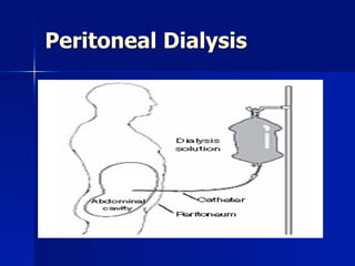
another way to clean the blood- while leaving it in the body\n- put water in peritoneal membrane- has 2 m\n- the waste products will come out through the dialysis\n(dont have to take the blood out of the body)\n- peritoneal cavity = reservoir of dialysis solution\n- need to put catheter in body\n- dialysis will go out with waste products\n
\n
diff kinds of catheters\n\n
when finish dialysis- disconnect, and put catheter in bandage on abdomen/ underwear...\n\n\n
venous clamp-\nhow make out fluid? cant put clamp on venous side of splanchnic circulation- so how take out water?\n- how take the excessive water from the blood\n= osmotic force: needs to be concentrated- put osmotic substance in the water that you are putting into the peritoneum so that it will pull out the water form the blood\n- glucose is the osmotic substance used\n\n
\n
water out in, close: 7-12, then take it out.....\n- can do it alone at home- ambulatory- doesnt need to come in\n- all the time there is fluid - occurs throughout the whole day\n
small machine nowadays- that only have to do it at night\nNIPD: nightly intermittent peritoneal dialysis\n- more small cycles are done- continuous small cycle dialysis\n\n
\n
\n
\n
\n
\n
3 kinds of pores in peritoneum\n- aquaporin (1)\n- small pores- btw the cells\n glycocalyx- lets molecules to pass through small pore (medium molecules)\n- bigger pore- bigger molecules can pass through\n\nso a lot of materials can be cleared- many sizes\n\n\n\n-the protein can pass through- so peritoneal dialysis- lose protein: hypoalbuminemic\n\n
\n
\n- height and weight differs\n\n\n
- peritoneum moves- some molecules go in and out\n\n\n
distribution in pop of how the peritoneum transports the fluids\n- most ppl: peritoneum is average transporter\n- some high transporters\n- some low transporter\n
we need to know if the peritoneum is an active or lazy one- wich determines how the dialysis will work\n- PET: in oreder to assess the peritoneum quality\n- help determine the PD scheme- how long the dialysis needs to be in the stomach (if high transporter- doesnt need to be in for very long)\n
osmotic power lost quickly if high transporter- so, blood will be more osmotic than the fluid, and then it will be absorption of water, rather than clearance of the blood\n- need to change the dialysis on time- or high transporter pt will gain weight\n\n- depend on how transporters work in individual pt\n
\n
\n
\n
\n
after 4 hours- need to change\n\n- if glucose more concentrated - take more time to lose\n\n\n
but glucose not so kind to the body\npolyglucose molecules- not as small as glucose- cant pass- so there is osmosis at all times- so can stay in the abdomen for a long time- wont lose its osmosis\n\n\nIcodextrin is a starch-derived high molecular weight (MW) glucose polymer that is structurally similar to glycogen.1 It consists of oligosaccharide polymers of D-glucopyranose linked by >90% &#x3B1;1-4 and <10% &#x3B1;1-6 glucosidic bonds. \nIcodextrin differs from glycogen in that it has a lower percentage of alpha1-6 linkages, and is thus not as highly branched.2,3 The number of linked glucose molecules ranges anywhere from 4 to >300 with a number-average MW between 5,000 and 6,500 Daltons. The weight-average MW of icodextrin, the most common and accurate way of reporting average MW for large polymers, is between 13,000 and 19,000 Daltons.1,2 \nAs a high molecular weight glucose polymer, icodextrin does not readily diffuse across the peritoneal membrane. Rather, it is slowly removed from the peritoneal cavity via lymphatic absorption. As a result, icodextrin is able to maintain osmotic forces and sustain ultrafiltration over longer dwell periods compared with conventional dextrose-based peritoneal dialysis solutions.\n\n\n
\n
polyglucose- doesnt lose its osmotic force!\n
can cause peritoneal membrane abnormalities\n- glucose degradation products are bad all the time\n
\n
right: accum of glycose end-products (stained brown) after long term peritoneal dialysis \n(diabetics- proliferation of vessels in the eye- retinopathy- glucose can cause proliferation of the vessels- tx of diabetic nephropathy= laser)\n-hypoxia: VEGF\n- when put glucose: make hyperglycemic environment int he abdomen: some pts that are prone to diabetes, become diabetic due to this dialysis\n- EVGF in peritoneum- more blood vessels are formed (similarly to the retina)\n- peritoneum can then transform from average transporter to high transporter- bc absorb faster bc have more vessels\n
right: with time- more VEGF staining\n
\n
during time- start dialysis\n- ppl transform form average transporter to high transporters\n
high transporters- greater mortality\n
\n77% higher RR for mortality HI vs LA\n
\n
\n
\n
\n
\n
\n
\n
\n
\n
\n
\n
\n
less water pulled out from pt\n
\n
Figure 3. Changes in (a) total body water and (b) extracellular fluid determined from bioelectrical impedance. At each time point, values represent mean &#xB1; SEM change from baseline for patients randomized to icodextrin ( ) or 2.27% glucose ( ). Between-group differences, P < 0.04, P < 0.008. Longitudinal differences from baseline, * P < 0.002, ** P < 0.001. \n
- after 5 yrs dialysis: peritoneum is MUCH THICKER\n- peritoneum can also become fibrotic- more collagen, more VEGF\n
\n
\n
more vessels and more fibrosis in the peritoneum\n
rare complication: \n- strangulate/encapsulate the small bowels- obstruction of the intestine\n(after 7/8 yrs)- switch ppl over to hemodialysis: not more than 7 yrs- bc of danger of complications\n
\n
\n
\n
\n
\n
\n
right: calcification fo peritoneum- intestine- \n\nstrangulation of the bowels \n
\n
\n
\n
very difficult surgery\n
rare- but can happen\n\nLAST SLIDE\n
\n
\n
\n
\n
\n
\n
\n
\n
\n
\n
\n
\n
\n
Category: Peritoneal Membrane Preservation \nThis slide shows the beneficial effects of the newer PD solutions containing alternate buffers and osmotic agents.\nAll three solutions reduce levels of GDPs and AGEs. With Physioneal, it is because glucose is sterilized at a very low pH. With Extraneal and Nutrineal, it is because there is no glucose in either solution.\nAdditional benefits of Physioneal : Physiologic pH and pCO2, reduced lactate levels.\nAdditional benefit of Extraneal: Same osmolarity as plasma.\nAs a result of its unique properties, each solution has been shown to improve membrane and peritoneal immune cell function.\n