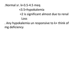
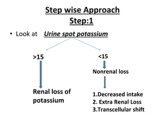
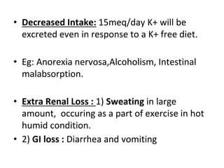
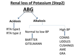
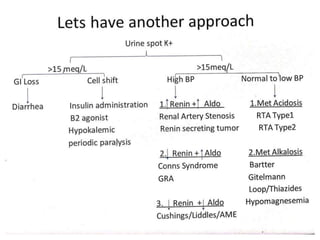
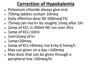
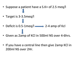
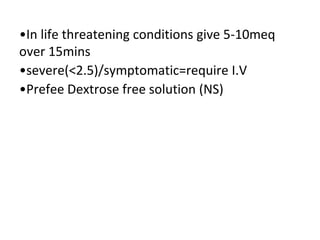
2. PHYSIOLOGY OF POTASSIUM
• Major intracellular cation
. 1 mmol =1meq=40mg of k+
.Total body stores=50meq x BW
=3000meq
=2500meq stores in muscles
.Intracellular K+=120-140meq per lit
. The function of intracellualar k+ is for cell growth
and protein synthesis
. On an average daily 100 meq of k+ is required for
the body
3. .ECF ICF ratio of potassium is important in
maintaining the resting membrane potential
. 90-95% of k+ excreted by kidneys ( chief ion)
.buffering extra cellular pool by a much large
intracellular pool even on infusing 40meq after
1 hr sr. K+can raise only by 0.5meq per lit
.Intracellular accumulation of k+against its
electrochemical gradient made possible by Na+-
k+ ATPase .
.
4. .Digoxin and K+ compete for same binding site
on
Na+k+ATPase , so digoxin inhibits Na+k+ATPase
as a result K+ cannot come inside the cell and
causes hyperkalemia.
.Muscle-2500meq, RBC -250meq,Liver-250meq
Bone -300meq: BODY K+DISTRIBUTION
. K+shifts from blood to the cell (active)by insulin,
B2agonists , alkalosis,alphaantagonists,
Theophylline, barium , chloroquine,
hypokalemic periodic paralysis (ca2+
channelopathy)
5. Hypokalemic periodic paralysis
. It is a genetic thing , AD
. Classic paralysis/ severe muscle weakness
. Recurrent episodes
. PPT by carbohydrate meal / rest /stress
6. .k+ shifts from cell to blood(passive)
By:1) cell destruction ( hemolysis,
rhabdomylosis, rumor lysis)
2)Non selective B blockers
3) alpha agonists
4) acidosis
5) exercise
6) mannitol(water enters from cell to blood ,
which leads to shrinkage of the cell, and K+ moves
out and leads to hyperkalemia)
7. Renal handling of K+
• 90% of K+ is excreted by kidney
• 65-70% of K+ is reabsorbed by PCT(passive transport)
• ThAL reabsorb K+ via Na-K-2Cl symporter
• Principal cells of cortical collecting duct with the help
of aldosteron-reabsorb salt and water and excrete
out K+ and H+
• Alpha intercalated cells of cortical collecting duct has
H+-K+ atpase which excrete H+ and reabsorb K+
8.
9. When to Suspect
• Muscle weakness- Flaccid paralysis with no sensory
involvement.
• Smooth muscle- constipation(paralytic ileus) and Bladder
dysfunction.
• Cardiac-Arrythmias
• Nephrogenic DI
• Rhabdomyolysis
• Respiratory failure
10. ECG changes
• 3-3.5meq/l: Flattening of T waves, Prominent
U waves and prolongation of QT or QU
interval
• <3meq/l: sagging of ST segment
• <2.5meq/l: widening of QRS, Prolongation of
PR interval,risk of arrythmias
11.
12.
13. .Normal sr. k+3.5-4.5 meq
<3.5=hypokalemia
<2 is significant almost due to renal
Loss
. Any hypokalemia un responsive to k+ think of
mg deficiency
14. Step wise Approach
Step:1
• Look at Urine spot potassium
>15
Renal loss of
potassium
<15
Nonrenal loss
1.Decreased intake
2. Extra Renal Loss
3.Transcellular shift
15. • Decreased Intake: 15meq/day K+ will be
excreted even in response to a K+ free diet.
• Eg: Anorexia nervosa,Alcoholism, Intestinal
malabsorption.
• Extra Renal Loss : 1) Sweating in large
amount, occuring as a part of exercise in hot
humid condition.
• 2) GI loss : Diarrhea and vomiting
16. Renal loss of Potassium (Step2)
ABG
Acidosis
RTA type1
RTA type 2
Alkalosis
Normal to low BP
BARTTER
GITELMANN
High BP
CONNS
LIDDLES
CUSHINGS
AME
GRA
17. Renal Potassium Wasting
• There is always a balanced reciprocal relationship
between distal load of Na and circulating
aldosterone which is the most important
mechanism for maintaining normal S.K+.
18. RENAL TUBULAR ACIDOSIS
• Group of disorders having defect in acidification
of urine due to tubular defect.
• Metabolic acidosis with alkaline urine.
• Normal anion gap metabolic acidosis with
high/+ve urine anion gap.
19. TYPES OF RTA
• Type 1 RTA: (Distal RTA): defect in alpha
intercalated cells of cortical collecting duct.
• Defect is in H+ ATPase or H+-K+ ATPase .
• Hence it is an H+ secretion defect.
• It is the most severe form of RTA
• Severe metabolic acidosis with alkaline urine of
pH>5.5
20. • Severe metabolic acidosis cause calcium
resorption from bone.
• Hence Hypercalcemia, hypercalcuria and renal
stones.
• Severe bone changes like short stature can also
occur.
• Severe Metabolic acidosis will decrease the
capacity of PCT to reabsorb salt and water
resulting in severe volume depletion – RAAS
activation - Worsen hypokalemia.
21. Causes
• Type 1 RTA: 1)Idiopathic (most common type)
2)Genetic(AR defect in H+ atpase).
3)Acquired causes: Sjogren syndrome, SLE,
Drugs (Ampho B)
22. Type 2 RTA (Proximal RTA)
• Type 2 RTA: Defect is in proximal convoluted tubule.
• Generalised proximal tubular dysfunction.
• Defect in bicarbonate reabsorption
• Less severe form of RTA.
• Since it is a generalised tubular dysfunction there will
be Glycosuria, Aminoaciduria and Phosphaturia.
(Fanconi syndrome).
23. • Less severe metabolic acidosis-hence less
bone resorption- hence less chance of stones.
• There will be loss of vit D through urine -
hence vit D deficiency/ Rickets/ osteomalacia.
• Less metabolisc acidosis- less volume
depletion – less RAAS activation - less
hypokalemia
24. • M/c cause : Fanconi Syndrome
• Fanconi syndrome can be Genetic or sporadic
or acquired.
• Genetic causes : Cystinosis, Wilsons disease
• Acquired causes: Multiple myeloma,
Hyperparathyroidism, drugs (outdated
tetracycline), Aminoglycoside,
topiramate,cyclofosfamide)
Causes of Type 2 RTA
25. • Type 3 RTA: Carbonic anhydrase 2 gene defect
• Rare form of RTA.
• Type 4RTA: RTA associated with
Hyperkalemia.
• Aldosterone defect: Aldosteron act on eNaC,
cause Na and water reabsorption and H+ & K+
excretion.
• Defect in this mechanism will cause metabolic
acidosis with hyperkalemia.
26. BARTTER SYNDROME
• Severe form of Inherited channelopathy
• AR mode of inheritance.
• Defect lies in ThAL : Inhibitory defect of Na-K-2Cl
channel.
• Simulating an aldosterone exess state.
• Presenting as Hypokalemia ,Alkalosis and high urine
chloride.
27. • Same channel is present in the ear for maintaining
endolymph concentration.
• Hence many have associated SNHL.
• Chronic use of loop diuretic can mimic Bartter.
• Other association is a paracellular pathway defect
where there is hypercalcuria and
hypermagnesuria hence can present as stone
disease.
• Serum Ca and Mg are normal because intestinal
absorption of Ca and Mg is normal.
28. GITELMANN SYNDROME
• Less severe form of inherited channelopathy.
• AR mode of inheritance.
• Defect is in DCT : inhibitory defect of Na-Cl symporter.
• Can simulate aldosterone excess state, Hence cause
Hypokalemia, Alkalosis and high urine Chloride.
• Chronic Thiazide use can mimic Gitelmann.
29. • Whenever there is volume depletion body try
to conserve more calcium, hence
Hypercalcemia and hypocalcuria.
• Same channel in intestine is also defective
hence absorption of Mg from intestine is
defective and hence Hypomagnesemia.
30. LIDDLES SYNDROME
• AD mode of inheritance
• Stimulatory defect of eNaC
• Hence called as PSEUDOHYPERALDOSTERONISM
• Chronic steroid usage can mimic Liddle.
• Treatment : Amiloride
31. Hypomagnesemia
• Think of hypomagnesemia when you have persistant
hypokalemia.
• Under normal circumstances intracellular Mg will inhibit
K+ excretion through ROMK channels of distal nephron.
• Low intacellular concentration of Mg
cause Hypokalemia refractory to Rx.
• Once hypomagnesemia gets corrected K+ will come
back to normal
32.
33. ● Potassium chloride always give oral
● 750mg tablets contain 10meq
● Daily effective dose 40-100meq(75)
● 75meq can rise k+ by roughly 1meq after 1hr
● 2amp of KCL in 200ml NS run over 2hrs
● 1amp of KCL=10ml
● 1ml=2meq of k+
● 1amp=20meq
● 2amp of KCL=40meq rise k+by 0.5meq/L
● Max can given on a day =160meq
● Max dose that can be given through a
peripheral line =20meq/hr
Correction of Hypokalemia
34. • Suppose a patient have a S.K+ of 2.5 meq/l
• Target is 3-3.5meq/l
• Deficit is 0.5-1meq/l 2-4 amp of Kcl
• Given as 2amp of KCl in 500ml NS over 4-6hrs.
• If you have a central line then give 2amp KCl in
200ml NS over 2Hr.
35. •In life threatening conditions give 5-10meq
over 15mins
•severe(<2.5)/symptomatic=require I.V
•Prefee Dextrose free solution (NS)