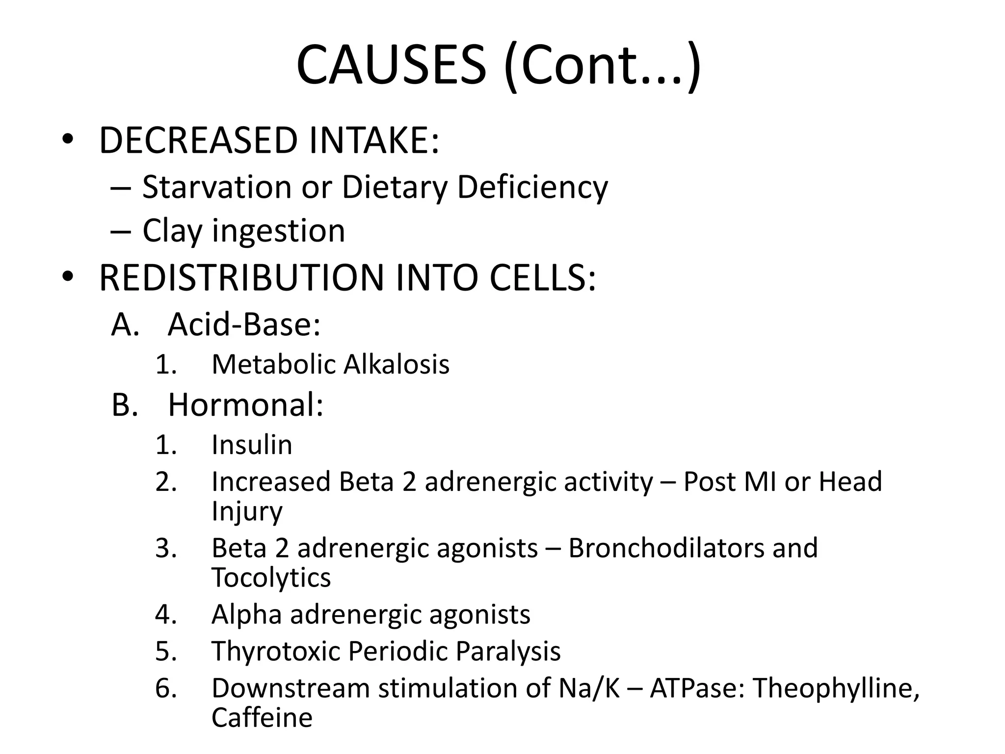
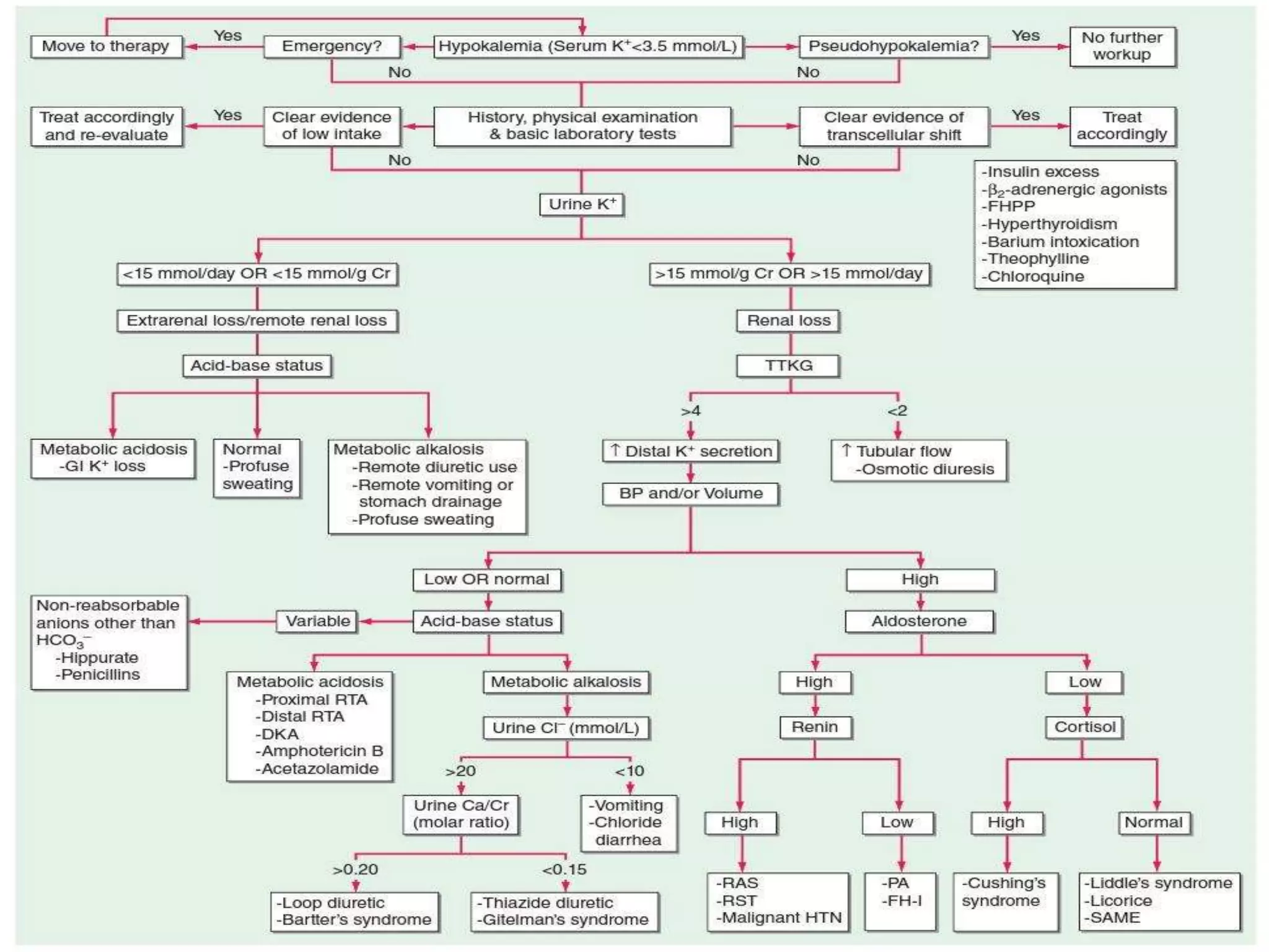
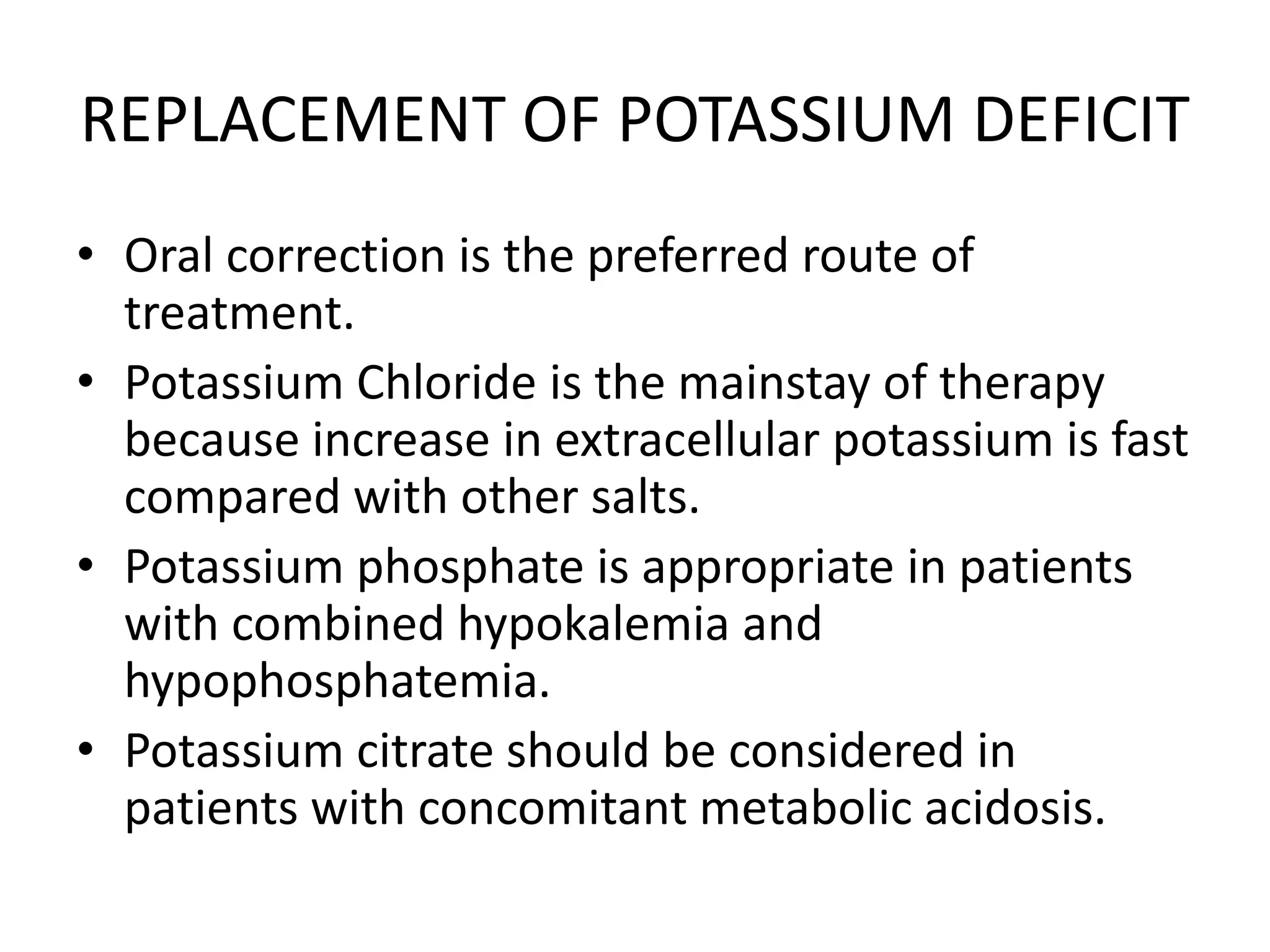
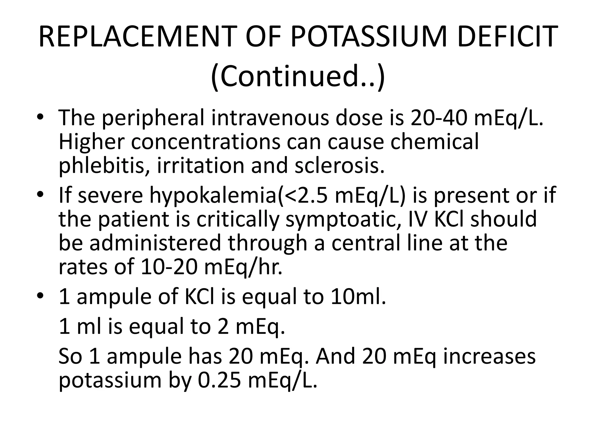
HYPOKALEMIA
Dr. Adhiya.N.S.S
M.D GENERAL MEDICINE
First Year Post Graduate
POTASSIUM
• It is the most abundant cation in the Human
Body.
– Total Body stores are approximately 3000 to 4000
mEq.
• It is a major intracellular cation.
– Intracellular K concentration : 130 -140 mEq/L
– Extracellular K concentration : 4 – 5 mEq/L
• It is found primarily in the muscles (70%), and to
a lesser extent in bone, red blood cells, liver and
skin.
FUNCTIONS OF POTASSIUM
• The normal ratio between extracellular and
intracellular potassium concentration is
important for the maintenance of the resting
membrane potential and neuromuscular
functioning.
• Intracellular Potassium:
– Cell growth and maintenance of cell volume
– DNA and Protein synthesis
– Acid –Base balance
POTASSIUM HOMEOSTASIS
HYPOKALEMIA
• It is an electrolyte disorder characterized by
Serum Potassium concentrations less than 3.5
mEq/L. (Normal Range: 3.5 – 5.0 mEq/L).
• It is present in upto 20% of hospitalized
patients and is associated with a ten-fold
increase in in-hospital mortality.
• It may be asymptomatic or have dangerous
complications such as arrthymias or paralysis.
CLINICAL MANIFESTATIONS OF
HYPOKALEMIA
• CARDIOVASCULAR:
– Increased Blood Pressure.
– Increased sensitivity to Arrhythmias.
– Increased risk of Digitoxicity.
• SKELETAL MUSCLES:
– Weakness, Flaccid muscle paralysis, Rhabdomyolysis.
• SMOOTH MUSCLE:
– Constipation, Ileus and rarely bladder dysfunction.
• ENDOCRINE:
– Worsening of glucose control in diabetics.
• RENAL:
– Polyurea due to Nephrogenic Diabetes Insipidus.
– It may lead to formation of Renal cysts.
– It may precipitate or worsen hepatic
encephalopathy due to increased renal ammonia
production.
– Increased ammonia production might also
contribute to the development of metabolic
alkalosis. Conversely metabolic alkalosis may
increase renal potassium excretion and cause
hypokalemia
– It may also cause Renal Failure as potassium
depletion causes tubulointerstitial fibrosis which is
characterised by vacuolization of PCT.
ECG CHANGES IN HYPOKALEMIA
• Flattening of T waves
• Prominent U waves
• Biphasic T waves (Dip and Rise pattern)
• ST depression
• QT prolongation/ QU prolongation (T-U fusion wave)
• PR prolongation
• Wide QRS
• Ventricular arrhythmias if associated with
hypomagnesaemia
POTASSIUM
LEVELS
ECG CHANGES
3.5 – 4.0 NORMAL
< 3.5 FLATTENING OF T WAVES
PROMINENT U WAVES
PROLONGED QT/ QU INTERVAL
< 3 SAGGING/ DEPRESSION OF ST SEGMENT
< 2.5 QRS WIDENING
PR PROLONGATION
CAUSES OF HYPOKALEMIA
• PSEUDOHYPOKALEMIA:
– After phlebotomy, if the blood is stored for
prolonged periods at room temperature, if large
number of leucocytes are present, they might take
up the potassium present in the serum.
– The most common cause is acute leukemia.
– Rapid separation of plasma and storage at 4
degree celcius is used to confirm this diagnosis.
CAUSES (Cont...)
• DECREASED INTAKE:
– Starvation or Dietary Deficiency
– Clay ingestion
• REDISTRIBUTION INTO CELLS:
A. Acid-Base:
1. Metabolic Alkalosis
B. Hormonal:
1. Insulin
2. Increased Beta 2 adrenergic activity – Post MI or Head
Injury
3. Beta 2 adrenergic agonists – Bronchodilators and
Tocolytics
4. Alpha adrenergic agonists
5. Thyrotoxic Periodic Paralysis
6. Downstream stimulation of Na/K – ATPase: Theophylline,
Caffeine
CAUSES (Cont...)
C. Anabolic state:
1. Vitamin B12 or Folic Acid administration leading to
increased Red Blood Cell production.
2. Granulocyte-Macrophage colony stimulating factor
leading to White Blood Cell production.
3. Total Parenteral Nutrition.
D. Familial Hypokalemic Periodic Paralysis
E. Hypothermia
F. Barium Toxicity: Systemic inhibition of Potassium
leak channels.
CAUSES (Cont...)
• NON-RENAL LOSS:
– Gastrointestinal loss:
• Upper GI loss – Vomiting, Nasogastric aspiration:
Associated with metabolic alkalosis.
• Lower GI loss – Diarrhoea, Laxative abuse, Villous
adenoma, Bowel obstruction/ fistula,
Ureterosigmoidostomy
– Integumentary loss:
• Sweat – In extremes of physical exertion.
CAUSES (Cont...)
• RENAL LOSS:
– Increased Tubular Flow:
• Diuretics
• Osmotic diuresis
• Salt-wasting Nephropathies
– Increased secretion of Potassium:
• Mineralocorticoid excess:
– Primary hyperaldosteronism
– Secondary hyperaldosteronism
– Genetic hyperaldosteronism
– Cushing’s Syndrome
– Bartter’s Syndrome
– Gitelman’s Syndrome
RENAL LOSS: (Cont...)
• Apparent mineralocorticoid excess:
– Genetic deficiency of 11 Beta dehydrogenase 2
– Inhibition of 11 Beta hydrogenase 2 (Carbenoxolone,
licorice)
– Liddle’s syndrome
• Distal delivery of non reabsorbed anions:
– Renal Tubular Acidosis
– Diabetic Ketoacidosis
– Penicillin derivatives
– Toluene Abuse (Glue sniffing)
• Magnesium deficiency
APPROACH TO HYPOKALEMIA
TRANSTUBULAR POTASSIUM
GRADIENT
• TTKG is the ratio of potassium concentration in
the lumen of cortical collecting duct to that in the
peritubular capillaries.
• TTKG = Urine Potassium x Serum osmolality/
Serum Potassium x Urine osmolality
• During hypokalemia, TTKG should fall below 3
indicating an appropriately reduced urinary
excretion of potassium.
• TTKG greater than 4 indicates renal potassium
loss due to increased distal potassium secretion.
TREATMENT OF HYPOKALEMIA
• The goals of therapy are
– To prevent life-threatening or serious consequences
– To replace the potassium deficit
– To correct the underlying cause
• Urgency of therapy depends on severity of
hypokalemia, rate of decline and associated
clinical factors.
• Serum potassium drops by 0.27 mmol for every
100 mmol reduction in body stores.
REPLACEMENT OF POTASSIUM DEFICIT
• Oral correction is the preferred route of
treatment.
• Potassium Chloride is the mainstay of therapy
because increase in extracellular potassium is fast
compared with other salts.
• Potassium phosphate is appropriate in patients
with combined hypokalemia and
hypophosphatemia.
• Potassium citrate should be considered in
patients with concomitant metabolic acidosis.
REPLACEMENT OF POTASSIUM DEFICIT
(Continued..)
• IV administration should be limited to patients
unable to use enteral route or in the setting of
severe complications.
• IV KCl is preferred.
• It should be administered in saline solutions,
rather than dextrose , because the dextrose
induced increase in insulin can acutely
exacerbate hypokalemia.
REPLACEMENT OF POTASSIUM DEFICIT
(Continued..)
• The peripheral intravenous dose is 20-40 mEq/L.
Higher concentrations can cause chemical
phlebitis, irritation and sclerosis.
• If severe hypokalemia(<2.5 mEq/L) is present or if
the patient is critically symptoatic, IV KCl should
be administered through a central line at the
rates of 10-20 mEq/hr.
• 1 ampule of KCl is equal to 10ml.
1 ml is equal to 2 mEq.
So 1 ampule has 20 mEq. And 20 mEq increases
potassium by 0.25 mEq/L.
REFERENCES
• HARRISON’S PRINCIPLES OF INTERNAL
MEDICINE – 20TH EDITION
• Comprehensive Clinical Nephrology by Richard
J. Johnson
• Hypokalemia: a clinical update
Efstratios Kardalas, Stavroula A
Paschou, Panagiotis Anagnostis, Giovanna
Muscogiuri, Gerasimos
Siasos, and Andromachi Vryonidou
THANK YOU

Hypokalemia - Approach and Management

  • 1.
    HYPOKALEMIA Dr. Adhiya.N.S.S M.D GENERALMEDICINE First Year Post Graduate
  • 2.
    POTASSIUM • It isthe most abundant cation in the Human Body. – Total Body stores are approximately 3000 to 4000 mEq. • It is a major intracellular cation. – Intracellular K concentration : 130 -140 mEq/L – Extracellular K concentration : 4 – 5 mEq/L • It is found primarily in the muscles (70%), and to a lesser extent in bone, red blood cells, liver and skin.
  • 3.
    FUNCTIONS OF POTASSIUM •The normal ratio between extracellular and intracellular potassium concentration is important for the maintenance of the resting membrane potential and neuromuscular functioning. • Intracellular Potassium: – Cell growth and maintenance of cell volume – DNA and Protein synthesis – Acid –Base balance
  • 4.
  • 8.
    HYPOKALEMIA • It isan electrolyte disorder characterized by Serum Potassium concentrations less than 3.5 mEq/L. (Normal Range: 3.5 – 5.0 mEq/L). • It is present in upto 20% of hospitalized patients and is associated with a ten-fold increase in in-hospital mortality. • It may be asymptomatic or have dangerous complications such as arrthymias or paralysis.
  • 9.
    CLINICAL MANIFESTATIONS OF HYPOKALEMIA •CARDIOVASCULAR: – Increased Blood Pressure. – Increased sensitivity to Arrhythmias. – Increased risk of Digitoxicity. • SKELETAL MUSCLES: – Weakness, Flaccid muscle paralysis, Rhabdomyolysis. • SMOOTH MUSCLE: – Constipation, Ileus and rarely bladder dysfunction. • ENDOCRINE: – Worsening of glucose control in diabetics.
  • 10.
    • RENAL: – Polyureadue to Nephrogenic Diabetes Insipidus. – It may lead to formation of Renal cysts. – It may precipitate or worsen hepatic encephalopathy due to increased renal ammonia production. – Increased ammonia production might also contribute to the development of metabolic alkalosis. Conversely metabolic alkalosis may increase renal potassium excretion and cause hypokalemia – It may also cause Renal Failure as potassium depletion causes tubulointerstitial fibrosis which is characterised by vacuolization of PCT.
  • 11.
    ECG CHANGES INHYPOKALEMIA • Flattening of T waves • Prominent U waves • Biphasic T waves (Dip and Rise pattern) • ST depression • QT prolongation/ QU prolongation (T-U fusion wave) • PR prolongation • Wide QRS • Ventricular arrhythmias if associated with hypomagnesaemia
  • 12.
    POTASSIUM LEVELS ECG CHANGES 3.5 –4.0 NORMAL < 3.5 FLATTENING OF T WAVES PROMINENT U WAVES PROLONGED QT/ QU INTERVAL < 3 SAGGING/ DEPRESSION OF ST SEGMENT < 2.5 QRS WIDENING PR PROLONGATION
  • 13.
    CAUSES OF HYPOKALEMIA •PSEUDOHYPOKALEMIA: – After phlebotomy, if the blood is stored for prolonged periods at room temperature, if large number of leucocytes are present, they might take up the potassium present in the serum. – The most common cause is acute leukemia. – Rapid separation of plasma and storage at 4 degree celcius is used to confirm this diagnosis.
  • 14.
    CAUSES (Cont...) • DECREASEDINTAKE: – Starvation or Dietary Deficiency – Clay ingestion • REDISTRIBUTION INTO CELLS: A. Acid-Base: 1. Metabolic Alkalosis B. Hormonal: 1. Insulin 2. Increased Beta 2 adrenergic activity – Post MI or Head Injury 3. Beta 2 adrenergic agonists – Bronchodilators and Tocolytics 4. Alpha adrenergic agonists 5. Thyrotoxic Periodic Paralysis 6. Downstream stimulation of Na/K – ATPase: Theophylline, Caffeine
  • 15.
    CAUSES (Cont...) C. Anabolicstate: 1. Vitamin B12 or Folic Acid administration leading to increased Red Blood Cell production. 2. Granulocyte-Macrophage colony stimulating factor leading to White Blood Cell production. 3. Total Parenteral Nutrition. D. Familial Hypokalemic Periodic Paralysis E. Hypothermia F. Barium Toxicity: Systemic inhibition of Potassium leak channels.
  • 16.
    CAUSES (Cont...) • NON-RENALLOSS: – Gastrointestinal loss: • Upper GI loss – Vomiting, Nasogastric aspiration: Associated with metabolic alkalosis. • Lower GI loss – Diarrhoea, Laxative abuse, Villous adenoma, Bowel obstruction/ fistula, Ureterosigmoidostomy – Integumentary loss: • Sweat – In extremes of physical exertion.
  • 17.
    CAUSES (Cont...) • RENALLOSS: – Increased Tubular Flow: • Diuretics • Osmotic diuresis • Salt-wasting Nephropathies – Increased secretion of Potassium: • Mineralocorticoid excess: – Primary hyperaldosteronism – Secondary hyperaldosteronism – Genetic hyperaldosteronism – Cushing’s Syndrome – Bartter’s Syndrome – Gitelman’s Syndrome
  • 18.
    RENAL LOSS: (Cont...) •Apparent mineralocorticoid excess: – Genetic deficiency of 11 Beta dehydrogenase 2 – Inhibition of 11 Beta hydrogenase 2 (Carbenoxolone, licorice) – Liddle’s syndrome • Distal delivery of non reabsorbed anions: – Renal Tubular Acidosis – Diabetic Ketoacidosis – Penicillin derivatives – Toluene Abuse (Glue sniffing) • Magnesium deficiency
  • 19.
  • 21.
    TRANSTUBULAR POTASSIUM GRADIENT • TTKGis the ratio of potassium concentration in the lumen of cortical collecting duct to that in the peritubular capillaries. • TTKG = Urine Potassium x Serum osmolality/ Serum Potassium x Urine osmolality • During hypokalemia, TTKG should fall below 3 indicating an appropriately reduced urinary excretion of potassium. • TTKG greater than 4 indicates renal potassium loss due to increased distal potassium secretion.
  • 22.
    TREATMENT OF HYPOKALEMIA •The goals of therapy are – To prevent life-threatening or serious consequences – To replace the potassium deficit – To correct the underlying cause • Urgency of therapy depends on severity of hypokalemia, rate of decline and associated clinical factors. • Serum potassium drops by 0.27 mmol for every 100 mmol reduction in body stores.
  • 23.
    REPLACEMENT OF POTASSIUMDEFICIT • Oral correction is the preferred route of treatment. • Potassium Chloride is the mainstay of therapy because increase in extracellular potassium is fast compared with other salts. • Potassium phosphate is appropriate in patients with combined hypokalemia and hypophosphatemia. • Potassium citrate should be considered in patients with concomitant metabolic acidosis.
  • 24.
    REPLACEMENT OF POTASSIUMDEFICIT (Continued..) • IV administration should be limited to patients unable to use enteral route or in the setting of severe complications. • IV KCl is preferred. • It should be administered in saline solutions, rather than dextrose , because the dextrose induced increase in insulin can acutely exacerbate hypokalemia.
  • 25.
    REPLACEMENT OF POTASSIUMDEFICIT (Continued..) • The peripheral intravenous dose is 20-40 mEq/L. Higher concentrations can cause chemical phlebitis, irritation and sclerosis. • If severe hypokalemia(<2.5 mEq/L) is present or if the patient is critically symptoatic, IV KCl should be administered through a central line at the rates of 10-20 mEq/hr. • 1 ampule of KCl is equal to 10ml. 1 ml is equal to 2 mEq. So 1 ampule has 20 mEq. And 20 mEq increases potassium by 0.25 mEq/L.
  • 26.
    REFERENCES • HARRISON’S PRINCIPLESOF INTERNAL MEDICINE – 20TH EDITION • Comprehensive Clinical Nephrology by Richard J. Johnson • Hypokalemia: a clinical update Efstratios Kardalas, Stavroula A Paschou, Panagiotis Anagnostis, Giovanna Muscogiuri, Gerasimos Siasos, and Andromachi Vryonidou
  • 27.