Hyperureacmia and gout
•Download as PPT, PDF•
8 likes•1,364 views

This document discusses uric acid and hyperuricemia. It summarizes that uric acid is the final product of purine breakdown in humans. Hyperuricemia can result from increased uric acid production, decreased excretion, or a combination. Complications of long-term hyperuricemia include gouty arthritis, kidney stones, and kidney damage. The diagnosis of gout involves identifying urate crystals in joint fluid. Treatment aims to resolve acute gout attacks and lower uric acid levels to prevent future attacks through medications and lifestyle changes.

Recommended
More Related Content
What's hot
What's hot (20)
Similar to Hyperureacmia and gout
Similar to Hyperureacmia and gout (20)
More from Koppala RVS Chaitanya
More from Koppala RVS Chaitanya (20)
Recently uploaded
Recently uploaded (20)
Hyperureacmia and gout
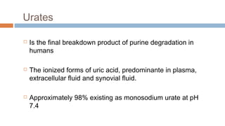
- 1. Urates Is the final breakdown product of purine degradation in humans The ionized forms of uric acid, predominante in plasma, extracellular fluid and synovial fluid. Approximately 98% existing as monosodium urate at pH 7.4
- 2. Plasma is saturated with monosodium urate at a concentration of 6.8 mg/dl. At higer concentrations, plasma is therfore supersaturated, creating the potential for urate crystal precipitation. Urate production varies with the purine content of the diet and the rates of purine biosyntesis, degradation and salvage. 2/3 to ¾ of urate is excreted by kidneys, and most of the remainer is eliminated through the intestines.
- 3. Renal handling Glomerular filtration Tubular reabsorption Secretion Postsecretory reabsorption Serum urate levels vary with age and sex. Children: 3 to 4 mg/dl Adult men: 6 to 6.8 mg/dl
- 4. Uric acid is more soluble in urine than in water. The pH of urine greatly influences its solubility. pH 5 urine is saturated with uric acid at concentrations ranging from 6 to 15 mg/dl. At pH 7 saturation is reached at concentration between 158 and 200mg/dl Defined as a plasma urate concentration > 7.0 mg/dl = ?
- 5. Hyperuricemia Can result from: Increased production of uric acid Decreased excretion of uric acid Combination of the two processes.
- 6. Increased Urate Production Diet provides an exogenous source of purines and, accordingly, contributes to the serum urate in proportion to its purine content. Foods high in nucleic acid: liver, thymus and pancreas, kidney. Restriction intake: reduces: 1 mg/dl Endogenous sources: De novo purine biosynthesis: 11 step
- 7. Decreased Uric Acid Excretion Alterated uric acid excretion could result from decreased glomerular filtration, decreased tubular secretion or enhanced tubular reabsorption. Decreased tubular secretion of urate causes the secondary hyperuricemia of acidosis. Diabetic ketoacidosis, starvation, ethanol intoxication, lactic acidosis, and salicylate intoxication are accompanied by accumulations of organic acids (B-hydroxybutyrate, acetoacetate, lactate or salicylates) that compete with urate for tubular secretion.
- 8. Combined Mechanisms Alcohol intake promotes hyperuricemia: Fast hepatic breakdown of ATP and increases urate production. Can induce hyperlacticacidemia, and inhibition of uric acid secretion. The higher purine content in some alcoholic beverages such as beer may also be a factor.
- 9. Complications of Hyperuricemia The most recognized complication of hyperuricemia is gouty arthritis Nephrolithiasis Urate Nephropathy Uric Acid Nephropathy www.freelivedoctor.com
- 10. Nephrolithiasis The prevalence of nephrolithiasis correlates with the serum and urinary uric acid levels. (Serum urate levels 13 mg/dl & Urinary uric acid excretion > 1100 mg/d) Urate Nephropathy Deposits of monosodium urate crystals surrounded by a giant cell inflammatory reaction in the medullary intrerstitium Uric acid nephropathy Precipitation in renal tubules and collecting ducts cause obstruction to urine flow.
- 11. An acute attack of gout has a rapid onset, with pain being maximal at 6–24 h of onset. The first attack affects a single joint in the lower limbs in 85– 90% of cases (the first metatarsophalangeal joint (big toe). The next affected are the mid-tarsi, ankles, knees and arms. The affected joint is hot, red and swollen with shiny overlying skin. Presentation and diagnosis
- 12. Crystal-induced arthritides MSU (monosodium urate) CPPD (calcium pyrophosphate dihydrate) HA (calcium hydroxyapatite) Calcium oxalate (CaOx) www.freelivedoctor.com
- 13. Monosodiumurate Gout Affecting middle-aged to elderly men. Women represent only 5 to 17% of all patients. www.freelivedoctor.com
- 14. Monosodiumurate Gout Associated with an Increased uric acid, Hyperuricemia, Episodic acute and chronic arthritis, Deposition of MSU crystals in connective tissue tophi and kidneys. www.freelivedoctor.com
- 15. Acute and chronic arthritis Acute arthritis is the most frequent early clinical manifestation of MSU gout. Usually only one joint is affected initially Polyarticular acute gout is also seen in male hypertensive patients with ethanol abuse as well as in postmenopausal women. www.freelivedoctor.com
- 16. The patient may also have a fever, leucocytosis, raised erythrocyte sedimentation rate (ESR). The attack may also be preceded by prodromal symptoms such as anorexia, nausea orchange in mood. Following resolution of the attack, there may be pruritis and desquamation of the overlying skin on the affected joint.
- 18. Several events may precipitate acute gouty arthritis: Dietary excess Genetics (SLC22A1 2, SLC2 A9 ) Comorbidities (obesity, dyslipidemia, glucose intolerance and hypertension) Renal disease (urate crystals in the interstitium and tubules of the kidney.) Trauma Surgery Excessive alcohol ingestion Medication (Glucocorticoid withdrawal) www.freelivedoctor.com Risk factors
- 19. Laboratory Diagnosis Even the clinical appearance strongly suggests gout. The diagnosis should be confirmed by needle aspiration of acute or chronically inflamed joints or tophaceous deposits. Acute septic arthritis several of the other crystalline – associated arthropathies, and psoriatic arthritis may present with similar clinical features. Effusion appear cloudy due to leukocytes and a large amounts crystals ocassionally produce a thick pasty or chalky joint fluid. www.freelivedoctor.com
- 20. Radiographic Features Cystic changes, well-defined erosions described as punched- out lytic lesion. Soft tissue calcified masses (chronic tophaceous gout) www.freelivedoctor.com
- 21. Monosodium urate crystals form in cartilage and fibrous tissues (protected) Crystals are shed into the joint space or bursa that inflammatory reaction occurs The shedding of crystals can be triggered by a number of factors including direct trauma, dehydration, acidosis or rapid weight loss. Pathogenesis
- 22. There is increased urinary urate excretion with a lowering of serum uric acid which leads to partial dissolution of monosodium urate crystals and subsequent shedding of crystals into the joint space. The shed crystals are phagocytosed by monocytes and macrophages, activating protein-3 (NALP3) inflammasome and triggering the release of interleukin-1(IL-1) and other cytokines, a subsequent infiltration of neutrophils and the symptoms of an acute attack
- 24. Course of disease The course of gout follows a number of stages Initially, the patient may be asymptomatic with a raised serum uric acid level often a second attack occurs within 6–12 months. Affect more than one joint and may spread to the upper limbs. Untreated disease can result in chronic tophaceous gout, with persistent low-grade inflammation in a number of joints resulting in joint damage and deformity.
- 25. Tophi deposition can occur anywhere in the body, but they are commonly seen on the helix of the ear, within and around the toe or finger joints, on the elbow, around the knees or on the Achilles tendons. The skin overlying the tophi may ulcerate and extrude white, chalky material composed of monosodium urate crystals.
- 27. Treatment aims in gout Rapid alleviation of the acute attack Prevention of future attacks Lower serum uric acid levels to below saturation point Reduce risk of co-morbidities, for example, cardiovascular disease Lifestyle modification
- 28. Treatment The management of gout can be split into The rapid resolution of the initial acute attack Long-term measures to prevent future episodes. Gout is often associated with othermedical problems including obesity, hypertension, excessive alcohol and the metabolic syndrome of insulin resistance, hyperinsulinaemia, impaired glucose intolerance and hypertriglyceridaemia.
- 29. This contributes to the increased cardiovascular risk and deterioration of renal function seen in patients with gout. Management is not only directed at alleviating acute attacks and preventing future attacks, but also identifying and treating other co-morbid conditions such as hypertension and hyperlipidaemia. Pharmacological measures should be combined with non- pharmacological measures such as weightloss, changes in diet, increased exercise and reduced alcohol consumption.
- 30. Management of acute attackof gout Management of an acute attack of gout Promptly and safely resolve pain First line: NSAID (use maximum dose) Second line: Colchicine Third line: Corticosteroid (consider first line in mono-articular disease) (±Simple and opiate analgesia if needed, for example, paracetamol, codeine dihydrocodeine) Rest the joint 1–2 days and treat with ice Remove contributing factors Review medication Review lifestyle
- 31. Management of chronic gout The presence of hyperuricaemia is not an indication to commence prophylactic therapy. Some patients may only experience a single episode and a change in lifestyle, diet or concurrent medication may be sufficient to prevent further attacks. Patients who suffer one or more acute attacks within 12 months of the first attack should normally be prescribed prophylactic urate-lowering therapy.
- 32. The aim of prophylactic gout treatment The aim of prophylactic gout treatment is to maintain the serum urate level below the saturation point of monosodium urate (300 μmol/L). If the serum urate is maintained below this level, crystal deposits dissolve and gout is controlled. Prophylactic treatment should not be initiated until an acute attack of gout has completely resolved, Usually 2 to 3 weeks after symptom resolution. Once started, prophylactic treatment should be continued indefinitely even if further acute attacks develop.
- 33. Criteria for starting prophylactic therapy for gout One ormore acute attacks within 12 months of the first attack Tophi present at the first presentation of an acute attack Presence of uric acid stones Need to continue medication associated with raised uric acid levels, for example diuretics Young patients with a family history of renal or cardiac disease
- 34. Classification of prophylactic agents used to lower serum urate Drugs that lower serum uric acid can be classified into three groups according to their pharmacological mode of action Uricostatic agents: Allopurinol, Febuxostat Uricosuric agents: Benzbromarone, Probenecid, Sulphinpyrazone Uricolytic agents: Rasburicase, Polyethylene glycol-uricase Inhibit the xanthine oxidase enzyme Increase excretion of uric acid Uric acid to allantoin (urate oxidase)