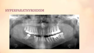
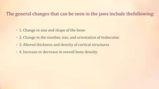
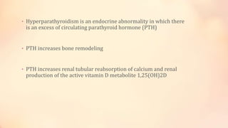
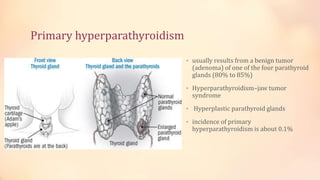
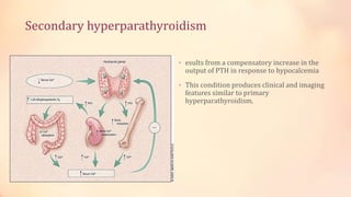
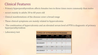
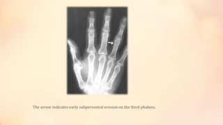
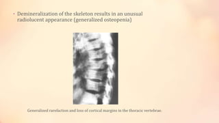
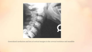
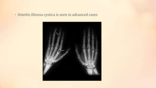
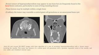
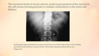
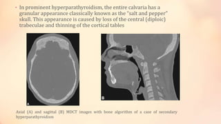
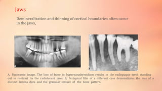
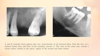
In the name of GOD
Sajad farokhi
HYPERPARATHYROIDISM
The general changes that can be seen in the jaws include thefollowing:
• 1. Change in size and shape of the bone
• 2. Change in the number, size, and orientation of trabeculae
• 3. Altered thickness and density of cortical structures
• 4. Increase or decrease in overall bone density
• Hyperparathyroidism is an endocrine abnormality in which there
is an excess of circulating parathyroid hormone (PTH)
• PTH increases bone remodeling
• PTH increases renal tubular reabsorption of calcium and renal
production of the active vitamin D metabolite 1,25(OH)2D
Primary hyperparathyroidism
• usually results from a benign tumor
(adenoma) of one of the four parathyroid
glands (80% to 85%)
• Hyperparathyroidism–jaw tumor
syndrome
• Hyperplastic parathyroid glands
• incidence of primary
hyperparathyroidism is about 0.1%
Secondary hyperparathyroidism
• esults from a compensatory increase in the
output of PTH in response to hypocalcemia
• This condition produces clinical and imaging
features similar to primary
hyperparathyroidism.
Clinical Features
• Primary hyperparathyroidism affects females two to three times more commonly than males
• occurs mainly in adults 30 to 60 years old
• Clinical manifestations of the disease cover a broad range
• These clinical symptoms are mainly related to hypercalcemia
• The combination of hypercalcemia and an elevated serum level of PTH is diagnostic of primary
hyperparathyroidism
• Laboratory test
Imaging Features
• Only about one in five patients with hyperparathyroidism has observable bone
changes
subtle erosions of bone from the subperiosteal surfaces of the phalanges of the hands
General Features :
The arrow indicates early subperiosteal erosion on the third phalanx.
• Demineralization of the skeleton results in an unusual
radiolucent appearance (generalized osteopenia)
Generalized rarefaction and loss of cortical margins in the thoracic vertebrae.
Generalized rarefaction and loss of cortical margins in the cervical vertebrae and mandible
• Osteitis fibrosa cystica is seen in advanced cases
• In about 10% of cases, brown tumors occur late in the disease
.Brown tumors of hyperparathyroidism may appear in any bone but are frequently found in the
facial bones and jaws, particularly in cases of long-standing disease
.These lesions may be multiple within a single bone
.If solitary, the tumor may resemble a central giant cell granuloma or an aneurysmal bone cyst
Axial (A) and coronal (B) MDCT images with bone algorithm of a case of secondary hyperparathyroidism with a brown tumor
involving the maxilla. This tumor has features of a central giant cell granuloma with a granular expanded cortex of the maxilla and
very subtle and ill-defined internal septa
.The histologic appearance of a brown tumor is identical to giant cell granuloma
• The increased levels of serum calcium result in precipitation of the mineral in
the soft tissues forming punctate or nodular calcifications in the joints and
kidneys
Radiograph depicting bilateral nephrocalcinosis in an adult male patient who initially
presented with features of pancreatitis. Ultimately, hyperparathyroidism was
diagnosed
• In prominent hyperparathyroidism, the entire calvaria has a
granular appearance classically known as the “salt and pepper”
skull. This appearance is caused by loss of the central (diploic)
trabeculae and thinning of the cortical tables
Axial (A) and sagittal (B) MDCT images with bone algorithm of a case of secondary
hyperparathyroidism
Demineralization and thinning of cortical boundaries often occur
in the jaws,
Jaws
A, Panoramic image. The loss of bone in hyperparathyroidism results in the radiopaque teeth standing
out in contrast to the radiolucent jaws. B, Periapical film of a different case demonstrates the loss of a
distinct lamina dura and the granular texture of the bone pattern.
The elevated rate of bone remodeling can cause an abnormal bone
Teeth and Associated Structures
. loss of the lamina dura
A and B, Granular bone pattern that was characteristic in all intraoral films. Note the loss of a
distinct lamina dura and floor of the maxillary antrum. C, This view of the same case reveals a
brown tumor related to the apical region of the second and third molars
Management
After successful surgical removal of the causative parathyroid adenoma, almost all changes revert to normal.
The only exception may be the site of a brown tumor, which often heals with bone that is more sclerotic than
normal
Many people with this disease are being diagnosed earlier, resulting in fewer severe cases
THE END

hyperparathyroidism in oral radiology

  • 1.
  • 2.
  • 3.
  • 4.
    The general changesthat can be seen in the jaws include thefollowing: • 1. Change in size and shape of the bone • 2. Change in the number, size, and orientation of trabeculae • 3. Altered thickness and density of cortical structures • 4. Increase or decrease in overall bone density
  • 5.
    • Hyperparathyroidism isan endocrine abnormality in which there is an excess of circulating parathyroid hormone (PTH) • PTH increases bone remodeling • PTH increases renal tubular reabsorption of calcium and renal production of the active vitamin D metabolite 1,25(OH)2D
  • 6.
    Primary hyperparathyroidism • usuallyresults from a benign tumor (adenoma) of one of the four parathyroid glands (80% to 85%) • Hyperparathyroidism–jaw tumor syndrome • Hyperplastic parathyroid glands • incidence of primary hyperparathyroidism is about 0.1%
  • 7.
    Secondary hyperparathyroidism • esultsfrom a compensatory increase in the output of PTH in response to hypocalcemia • This condition produces clinical and imaging features similar to primary hyperparathyroidism.
  • 8.
    Clinical Features • Primaryhyperparathyroidism affects females two to three times more commonly than males • occurs mainly in adults 30 to 60 years old • Clinical manifestations of the disease cover a broad range • These clinical symptoms are mainly related to hypercalcemia • The combination of hypercalcemia and an elevated serum level of PTH is diagnostic of primary hyperparathyroidism • Laboratory test
  • 9.
    Imaging Features • Onlyabout one in five patients with hyperparathyroidism has observable bone changes subtle erosions of bone from the subperiosteal surfaces of the phalanges of the hands General Features :
  • 10.
    The arrow indicatesearly subperiosteal erosion on the third phalanx.
  • 11.
    • Demineralization ofthe skeleton results in an unusual radiolucent appearance (generalized osteopenia) Generalized rarefaction and loss of cortical margins in the thoracic vertebrae.
  • 12.
    Generalized rarefaction andloss of cortical margins in the cervical vertebrae and mandible
  • 13.
    • Osteitis fibrosacystica is seen in advanced cases
  • 15.
    • In about10% of cases, brown tumors occur late in the disease
  • 16.
    .Brown tumors ofhyperparathyroidism may appear in any bone but are frequently found in the facial bones and jaws, particularly in cases of long-standing disease .These lesions may be multiple within a single bone .If solitary, the tumor may resemble a central giant cell granuloma or an aneurysmal bone cyst Axial (A) and coronal (B) MDCT images with bone algorithm of a case of secondary hyperparathyroidism with a brown tumor involving the maxilla. This tumor has features of a central giant cell granuloma with a granular expanded cortex of the maxilla and very subtle and ill-defined internal septa
  • 17.
    .The histologic appearanceof a brown tumor is identical to giant cell granuloma
  • 18.
    • The increasedlevels of serum calcium result in precipitation of the mineral in the soft tissues forming punctate or nodular calcifications in the joints and kidneys Radiograph depicting bilateral nephrocalcinosis in an adult male patient who initially presented with features of pancreatitis. Ultimately, hyperparathyroidism was diagnosed
  • 19.
    • In prominenthyperparathyroidism, the entire calvaria has a granular appearance classically known as the “salt and pepper” skull. This appearance is caused by loss of the central (diploic) trabeculae and thinning of the cortical tables Axial (A) and sagittal (B) MDCT images with bone algorithm of a case of secondary hyperparathyroidism
  • 20.
    Demineralization and thinningof cortical boundaries often occur in the jaws, Jaws A, Panoramic image. The loss of bone in hyperparathyroidism results in the radiopaque teeth standing out in contrast to the radiolucent jaws. B, Periapical film of a different case demonstrates the loss of a distinct lamina dura and the granular texture of the bone pattern.
  • 21.
    The elevated rateof bone remodeling can cause an abnormal bone Teeth and Associated Structures . loss of the lamina dura
  • 22.
    A and B,Granular bone pattern that was characteristic in all intraoral films. Note the loss of a distinct lamina dura and floor of the maxillary antrum. C, This view of the same case reveals a brown tumor related to the apical region of the second and third molars
  • 23.
    Management After successful surgicalremoval of the causative parathyroid adenoma, almost all changes revert to normal. The only exception may be the site of a brown tumor, which often heals with bone that is more sclerotic than normal Many people with this disease are being diagnosed earlier, resulting in fewer severe cases
  • 24.