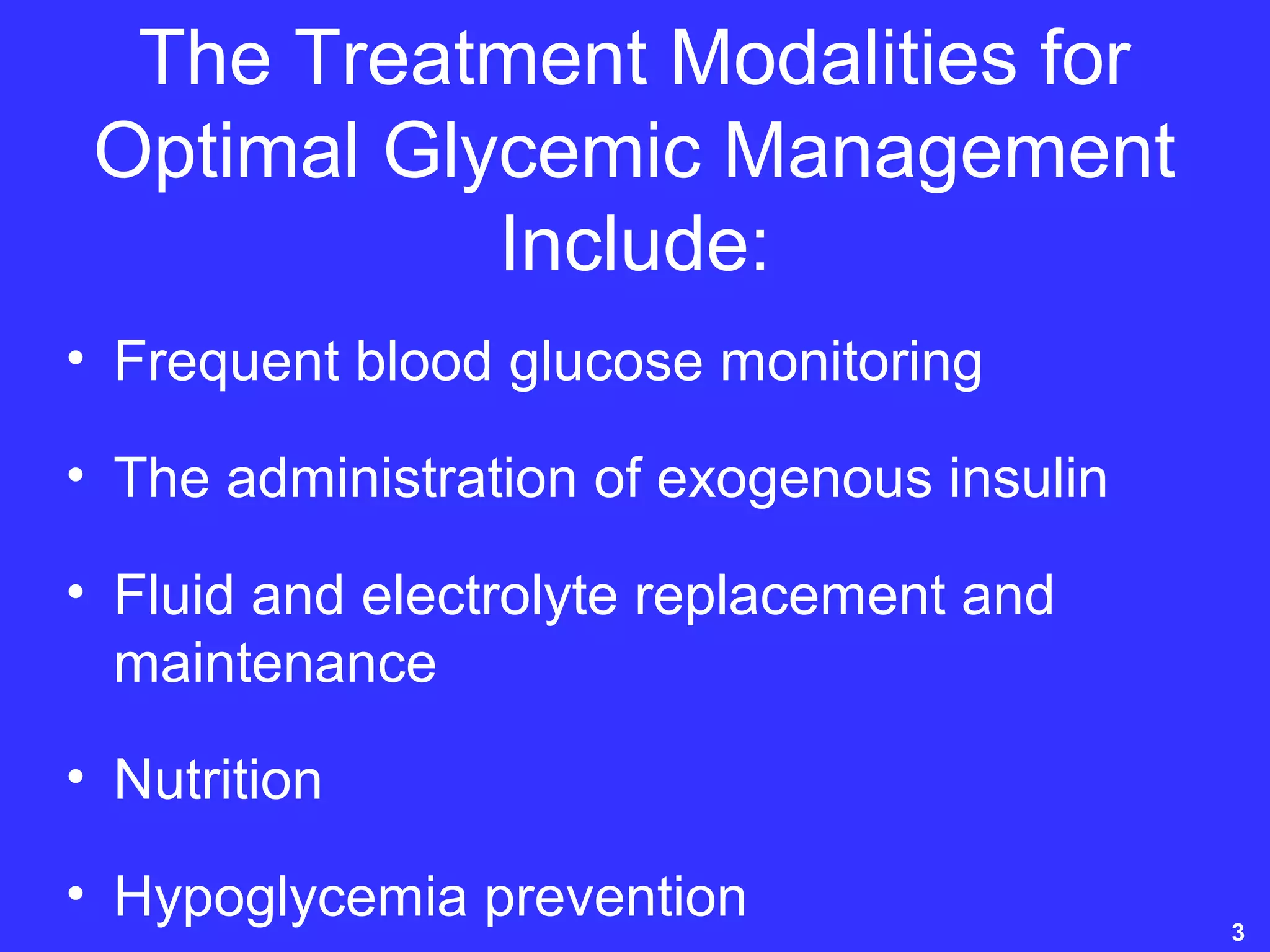
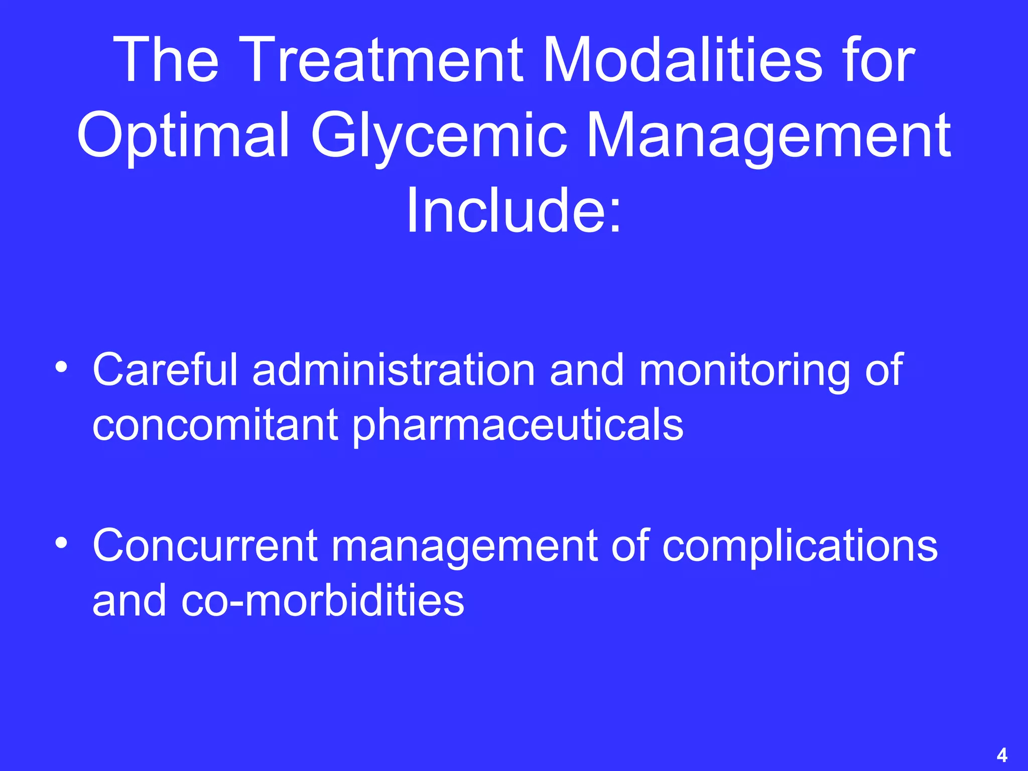
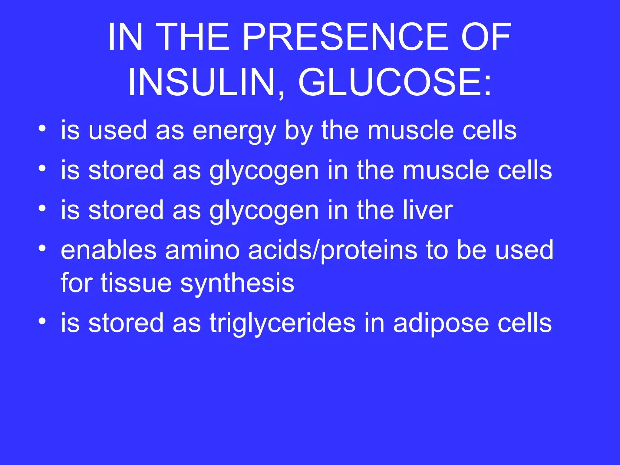
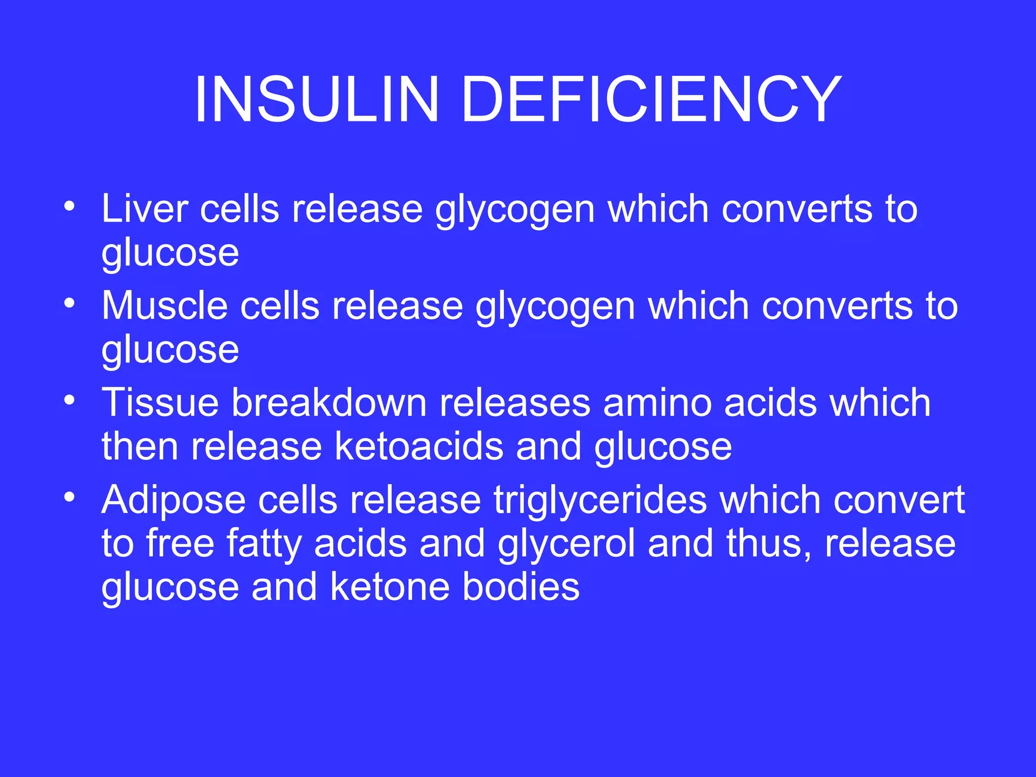
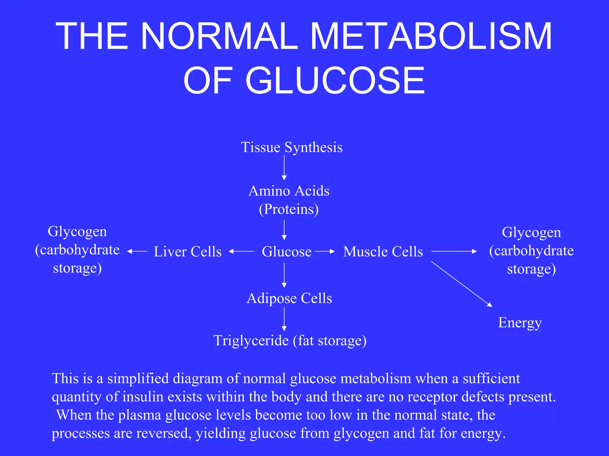
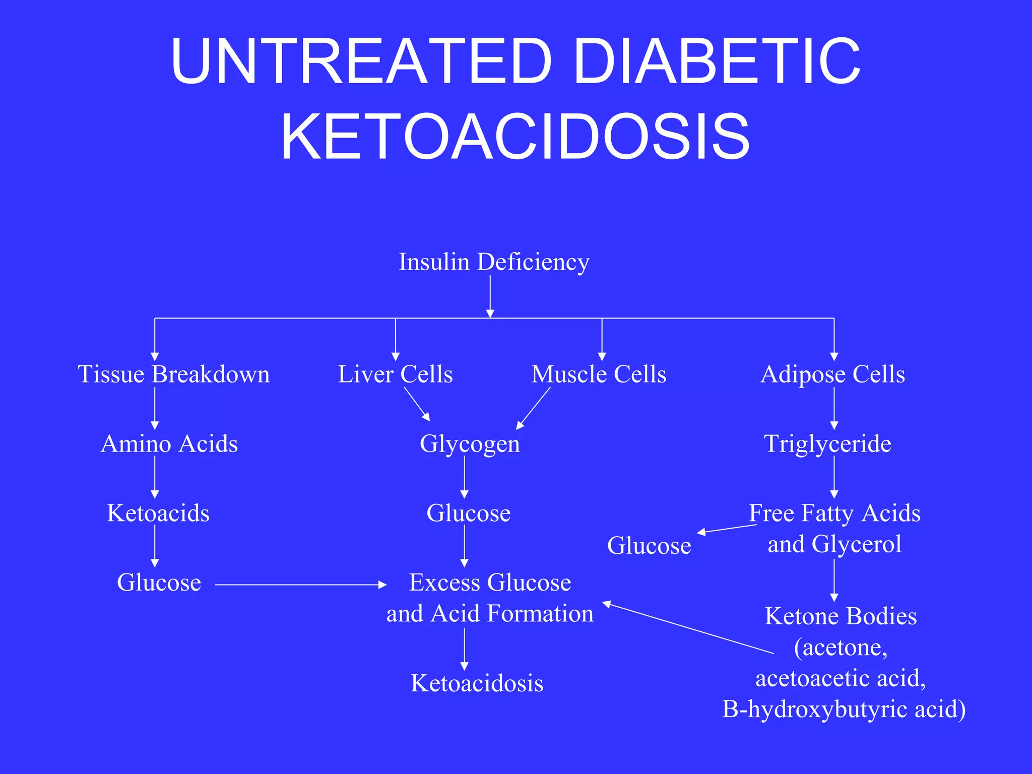
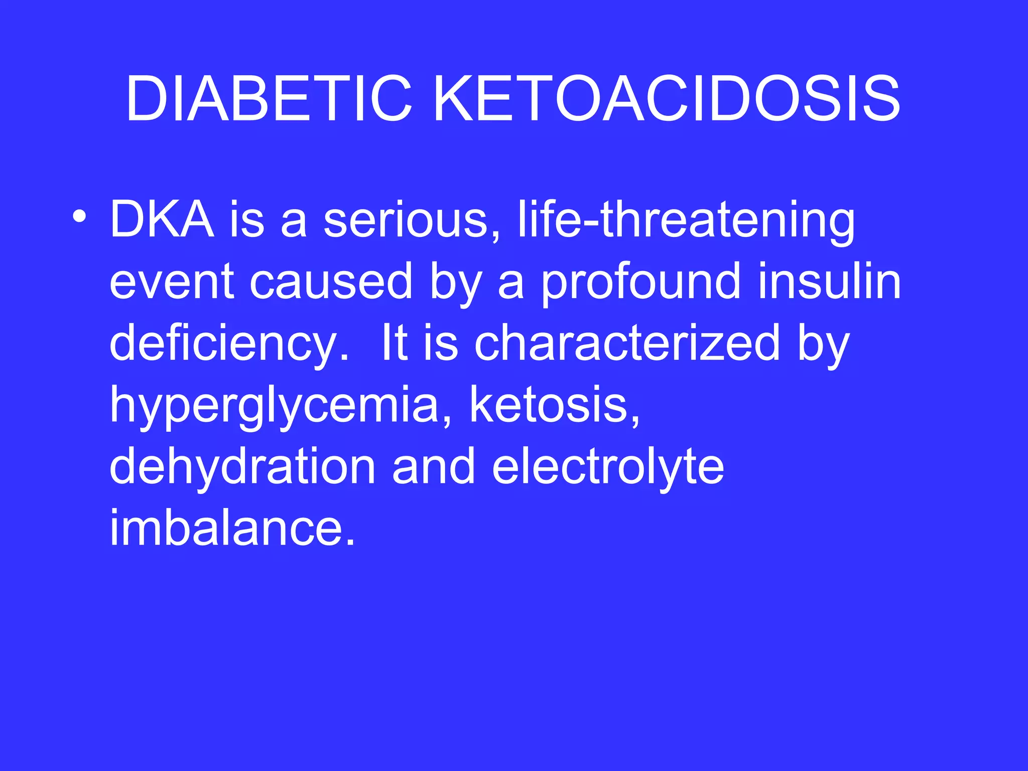
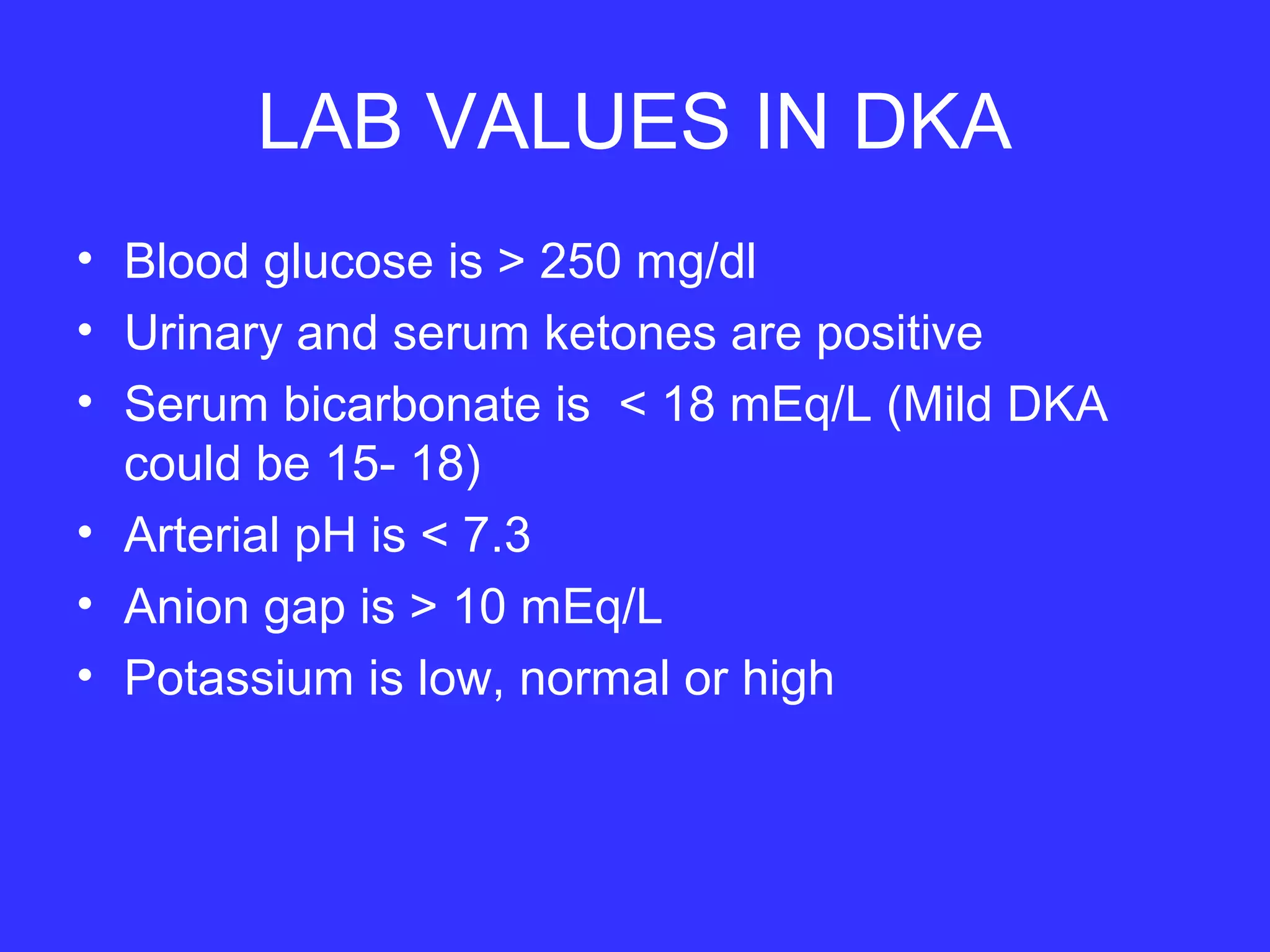
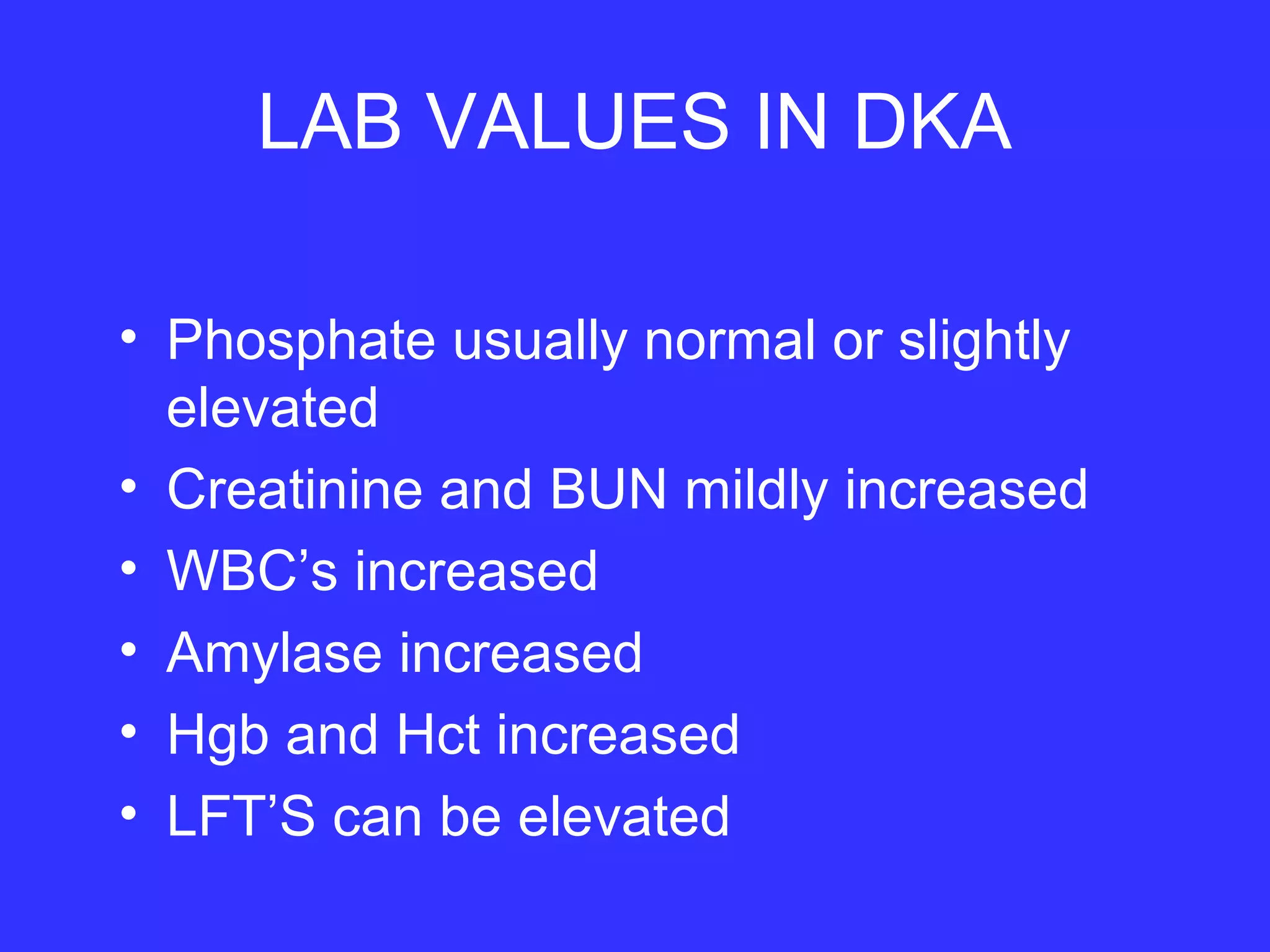
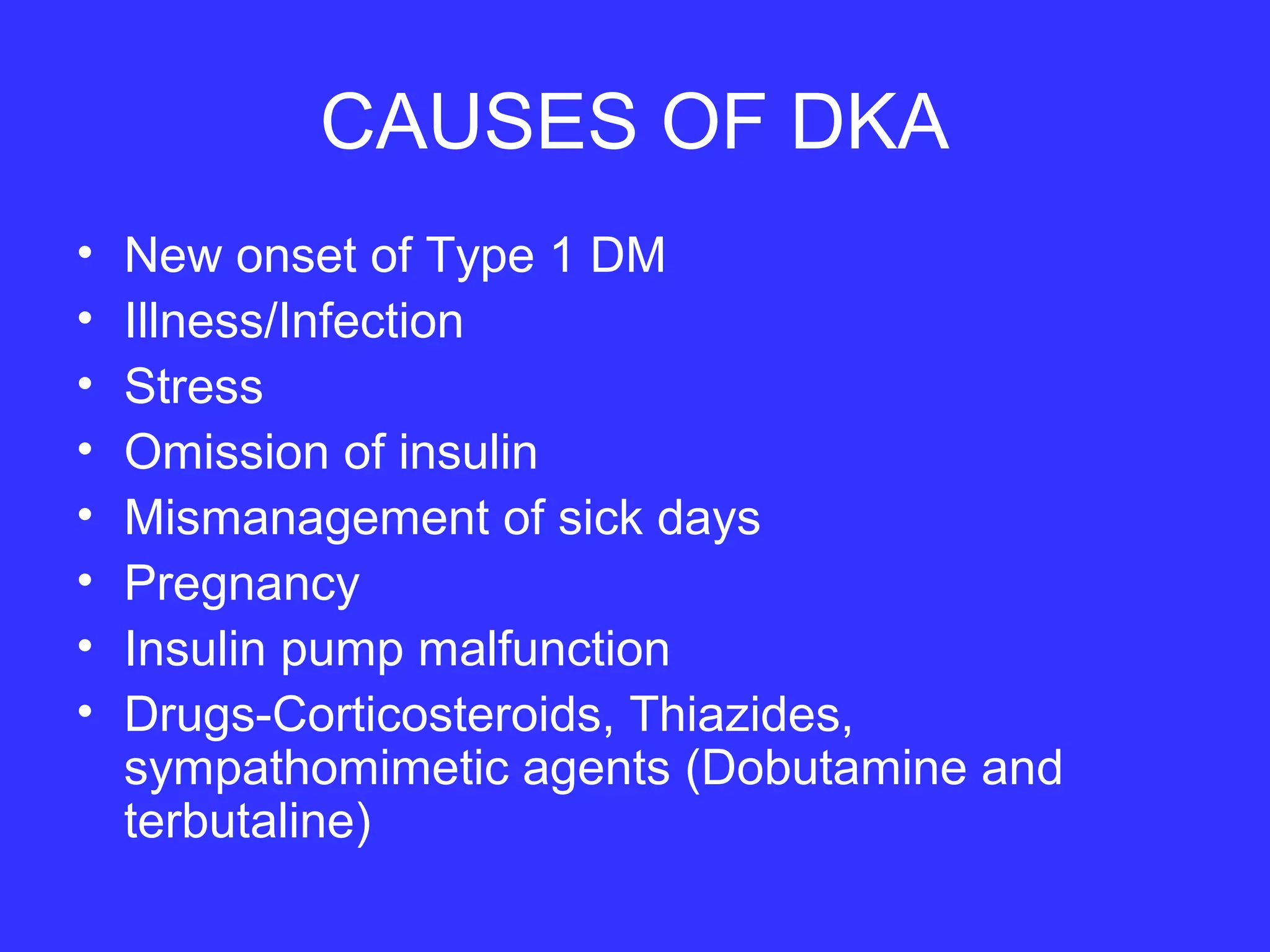
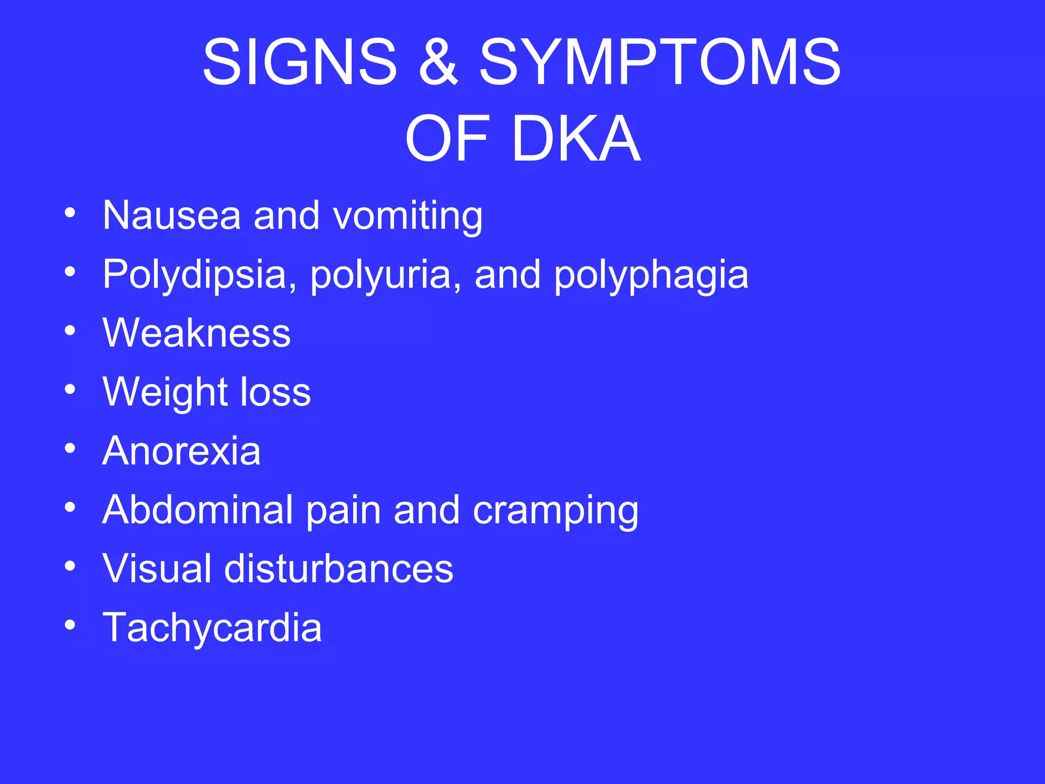
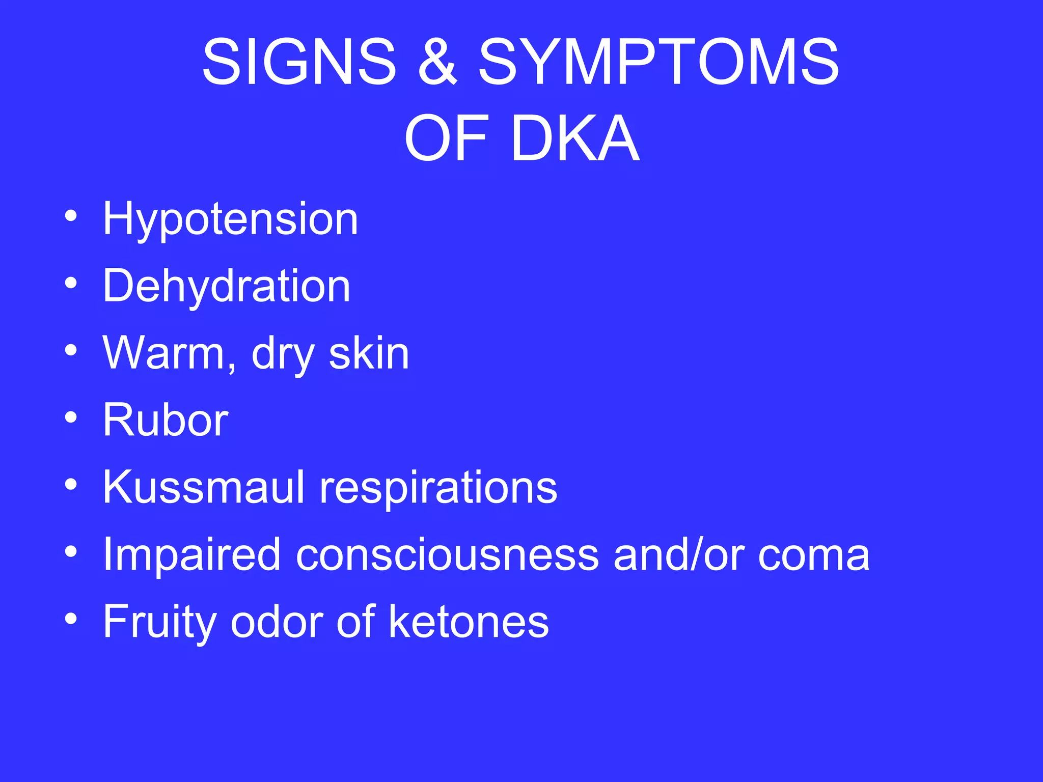
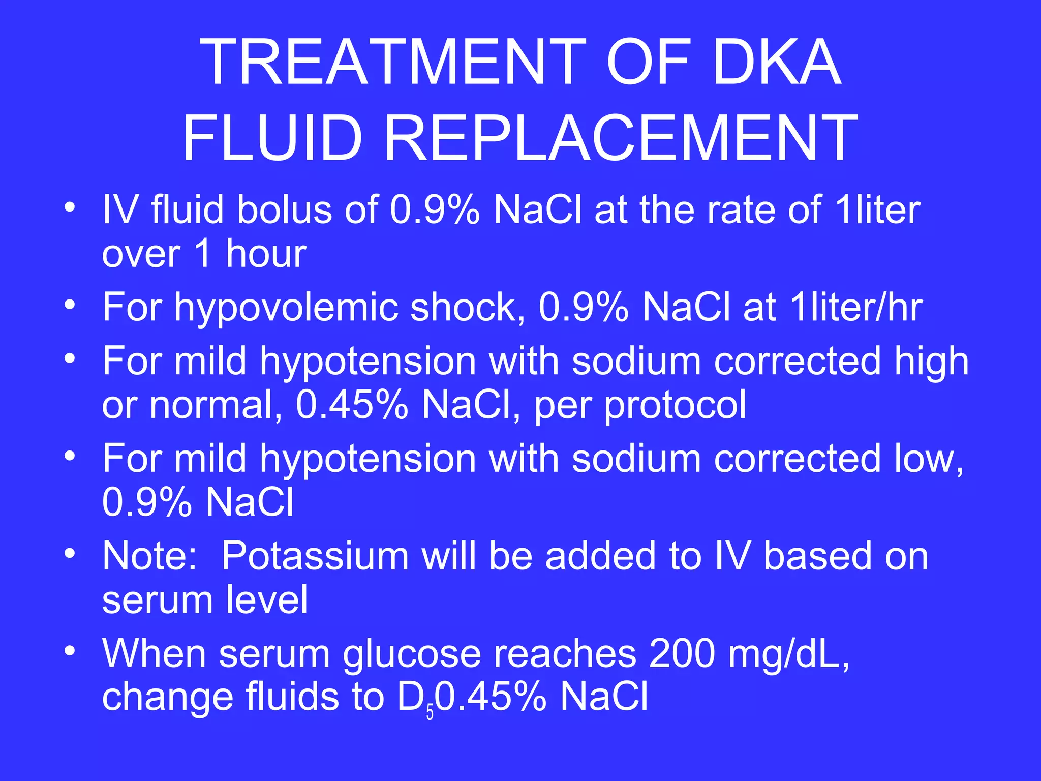
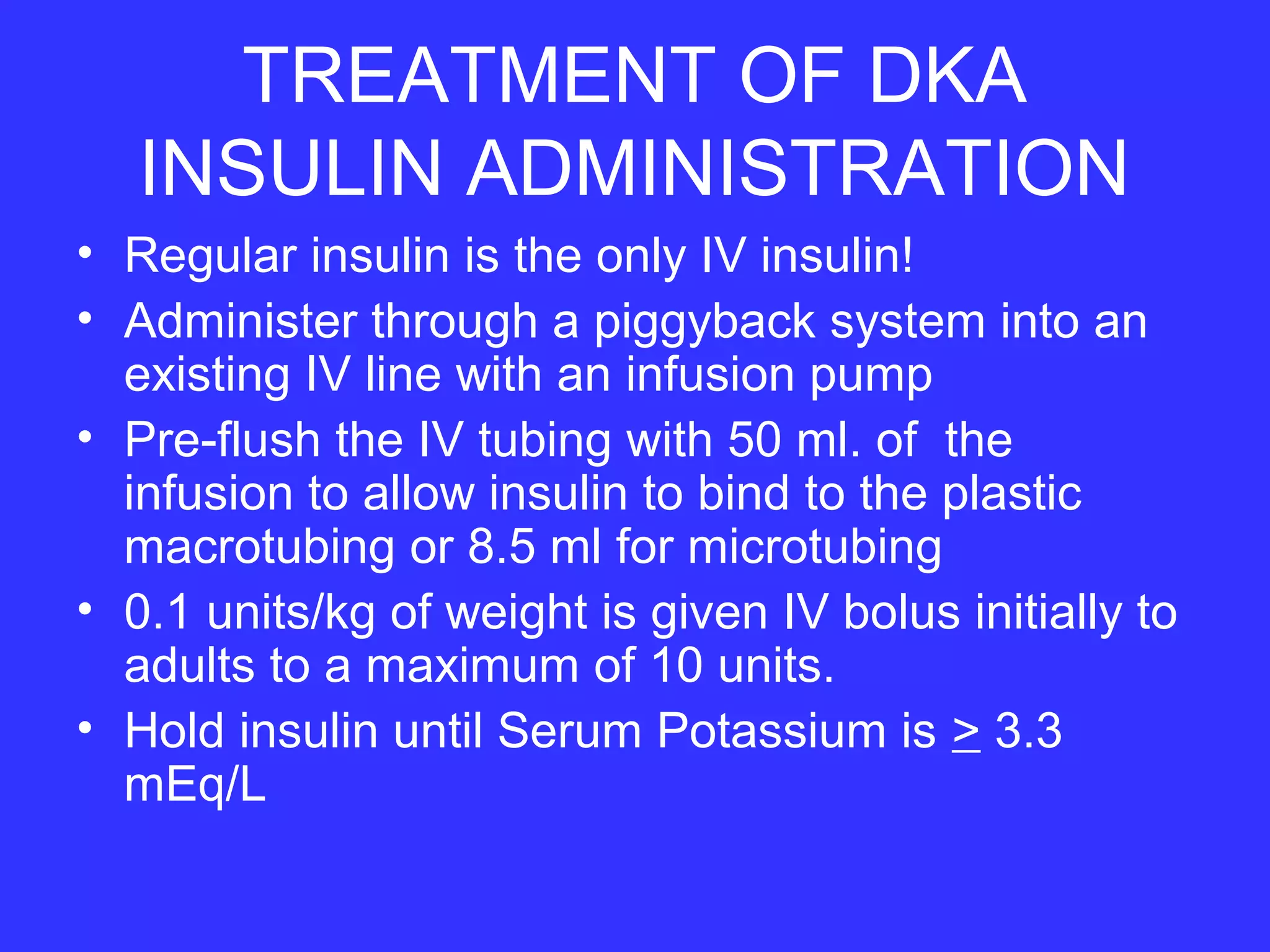
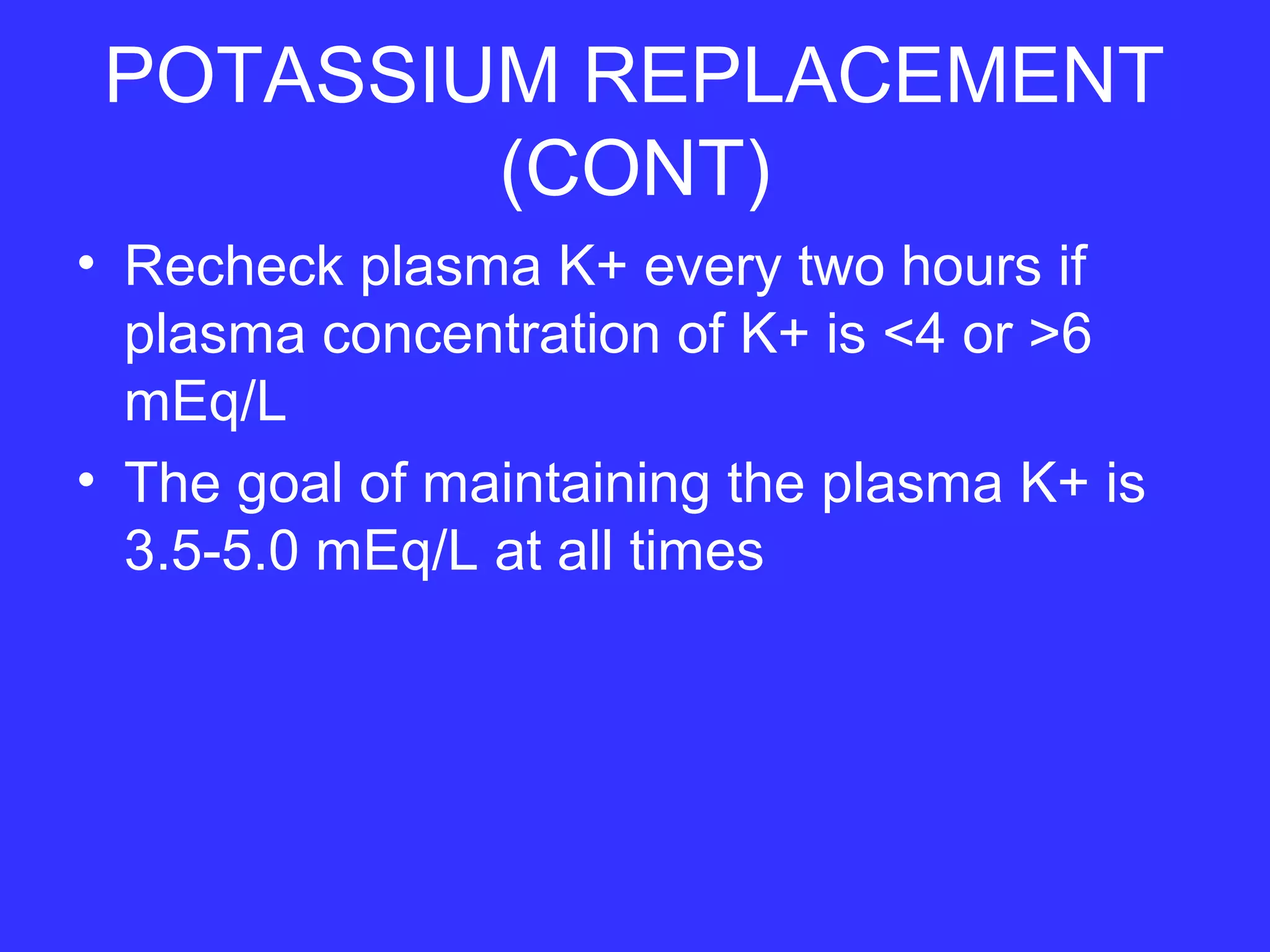
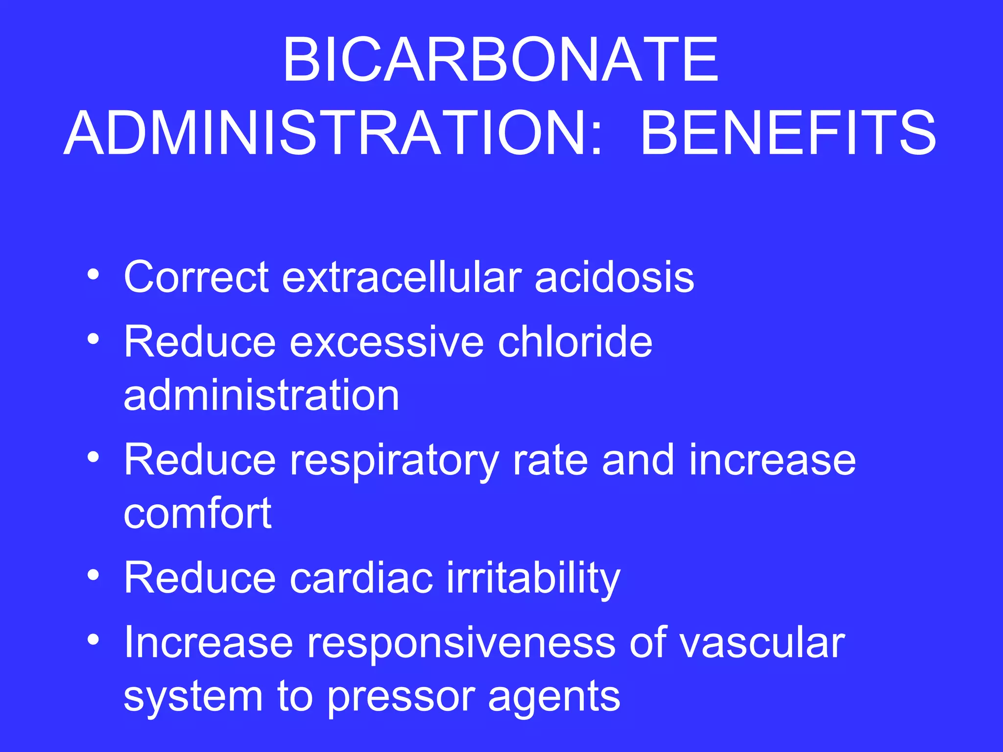
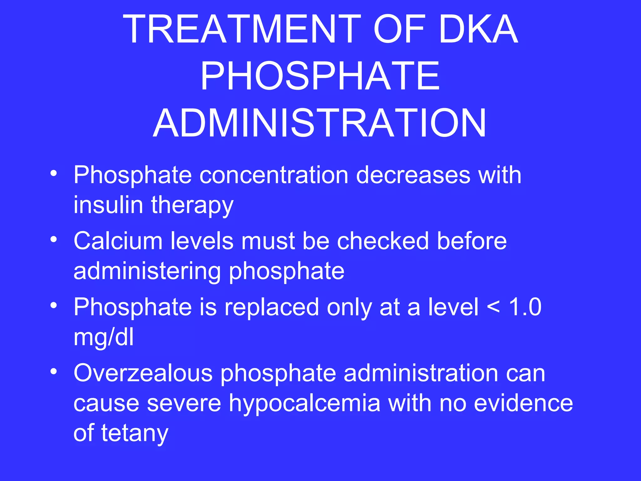
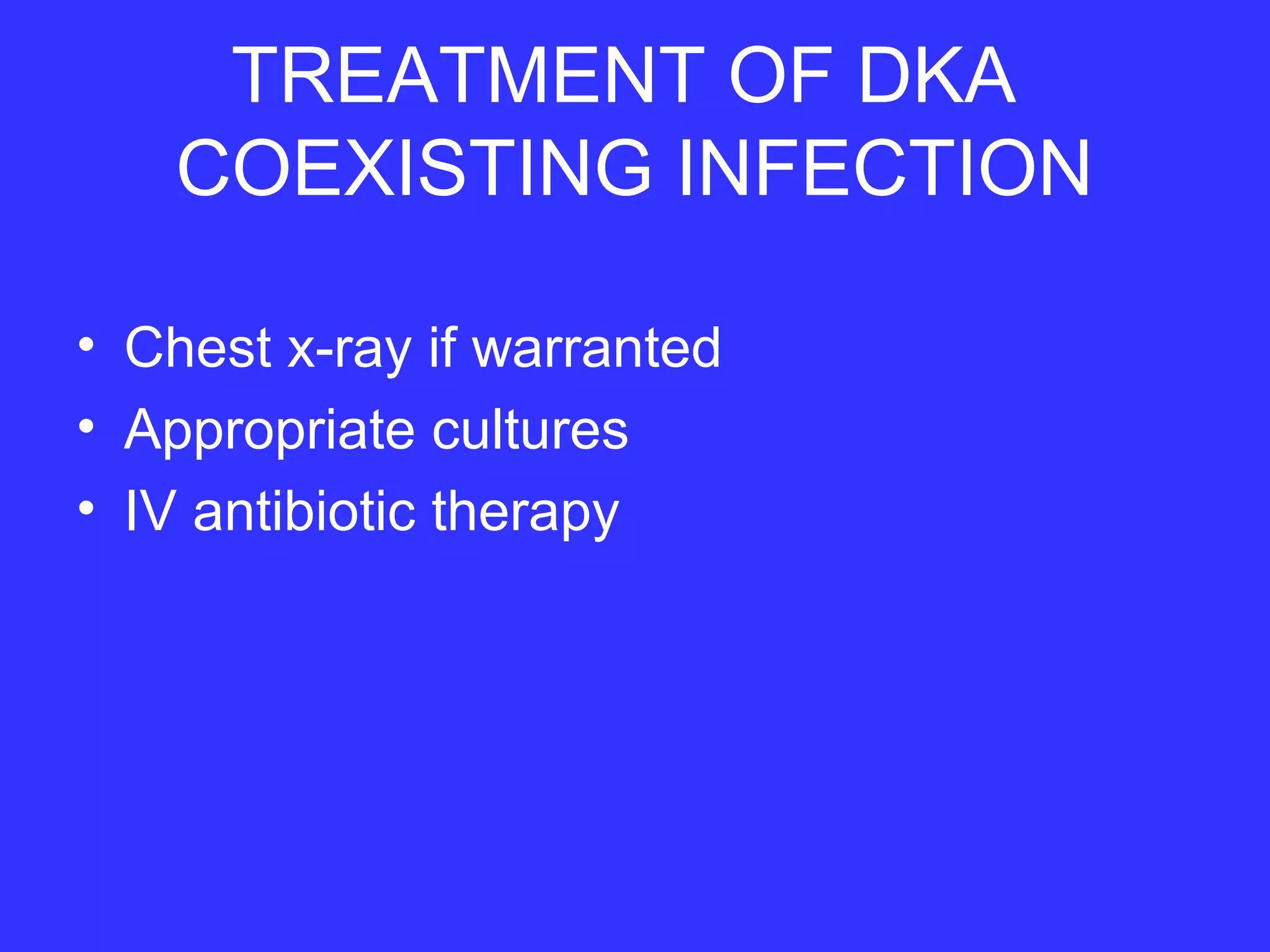
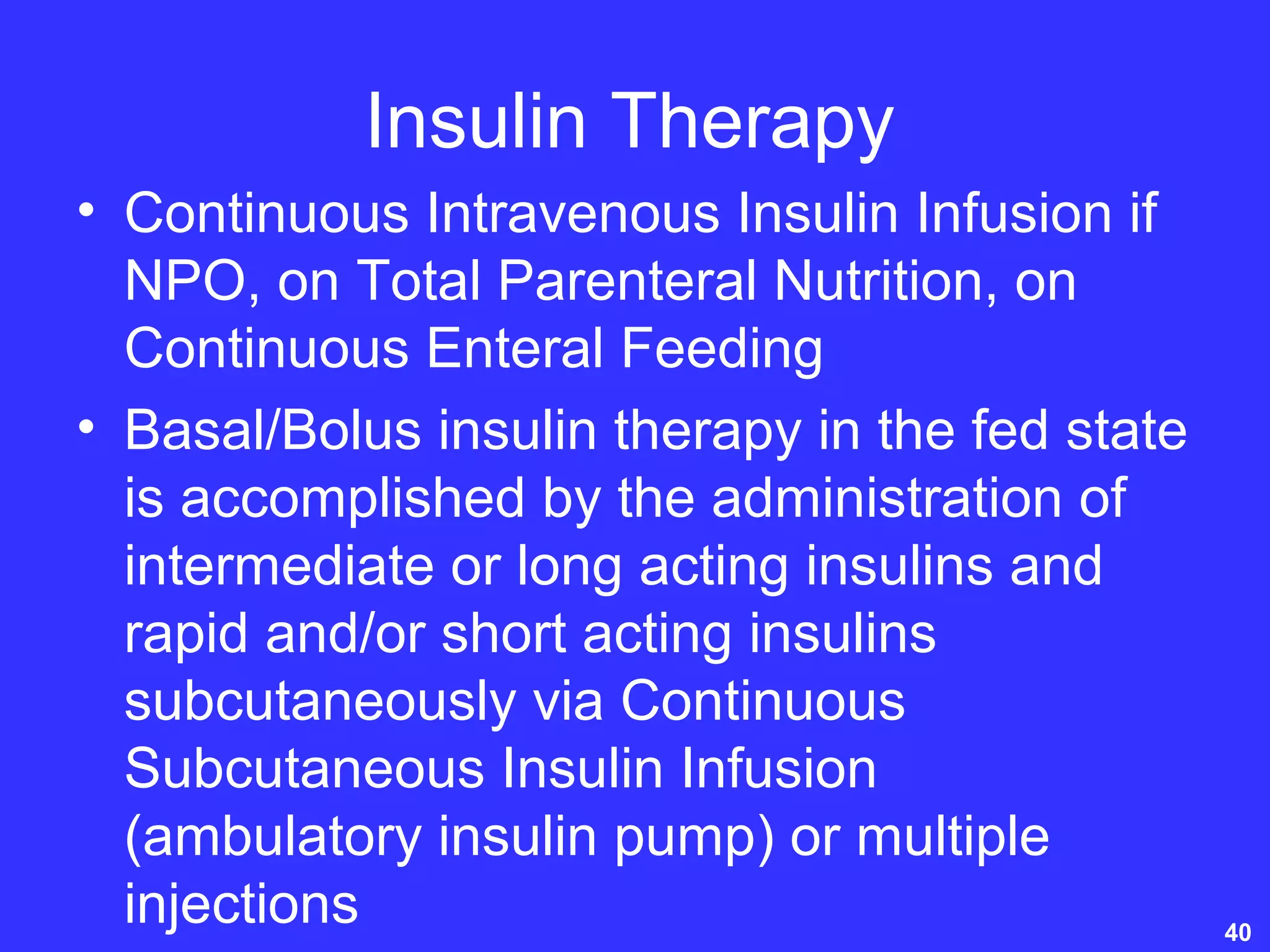
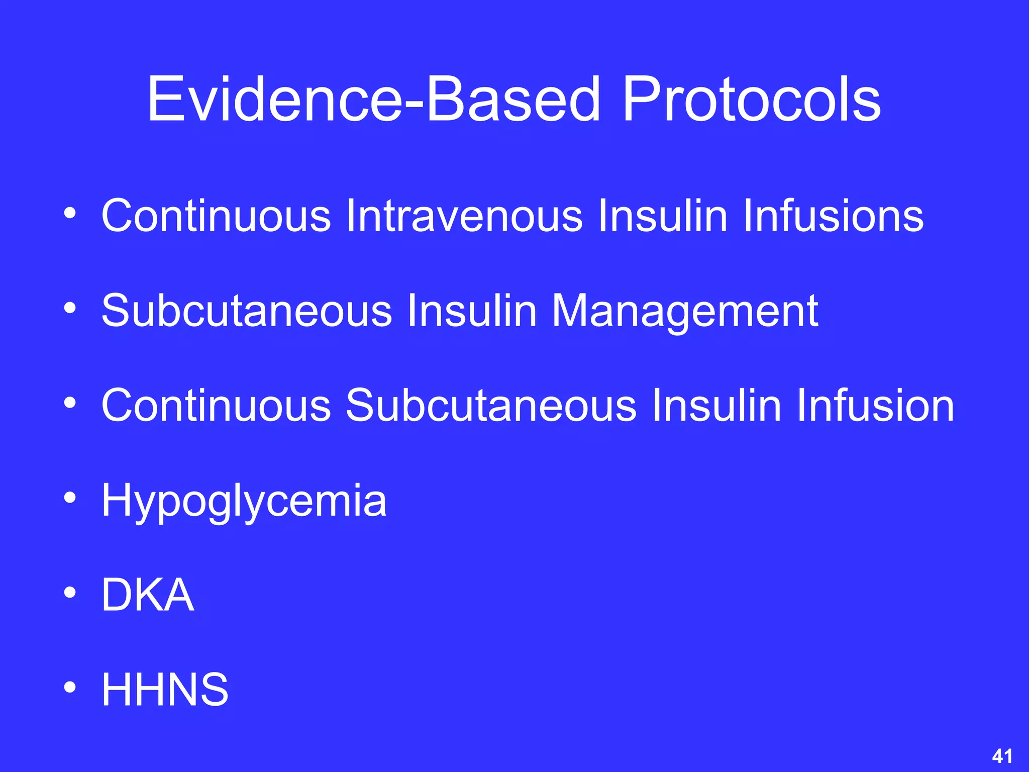
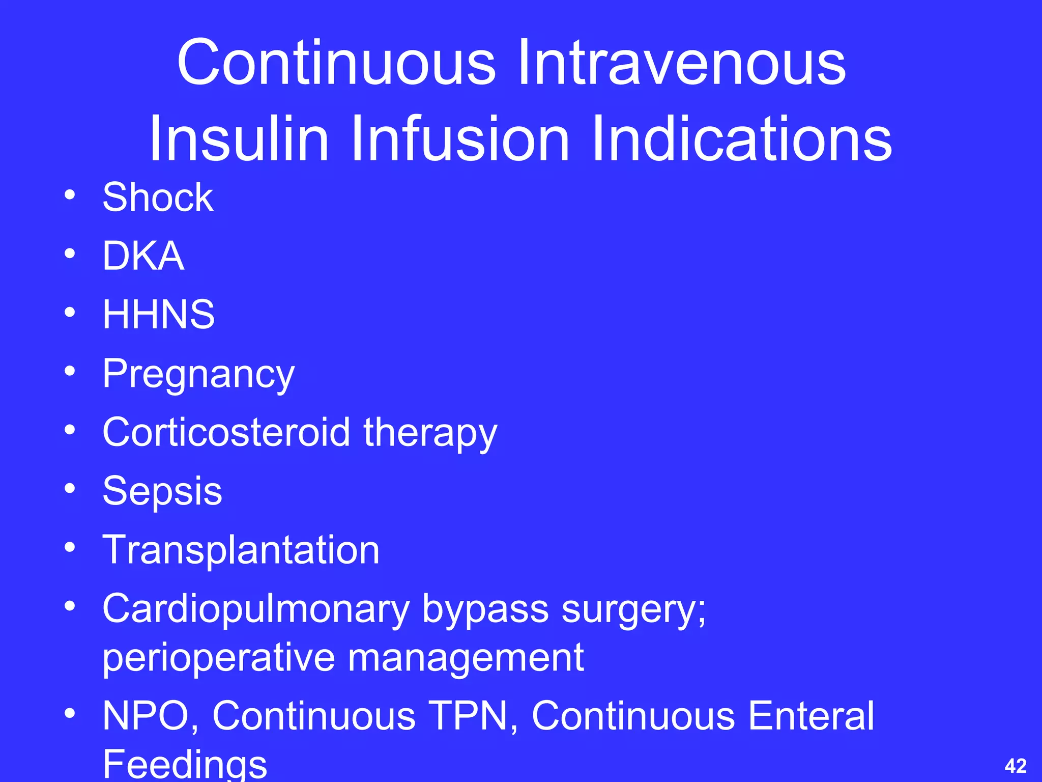
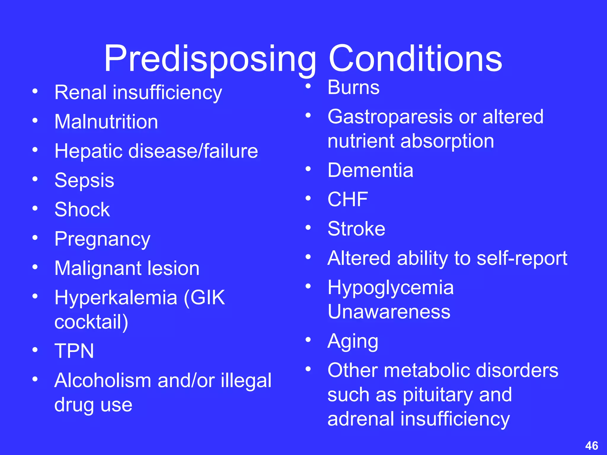
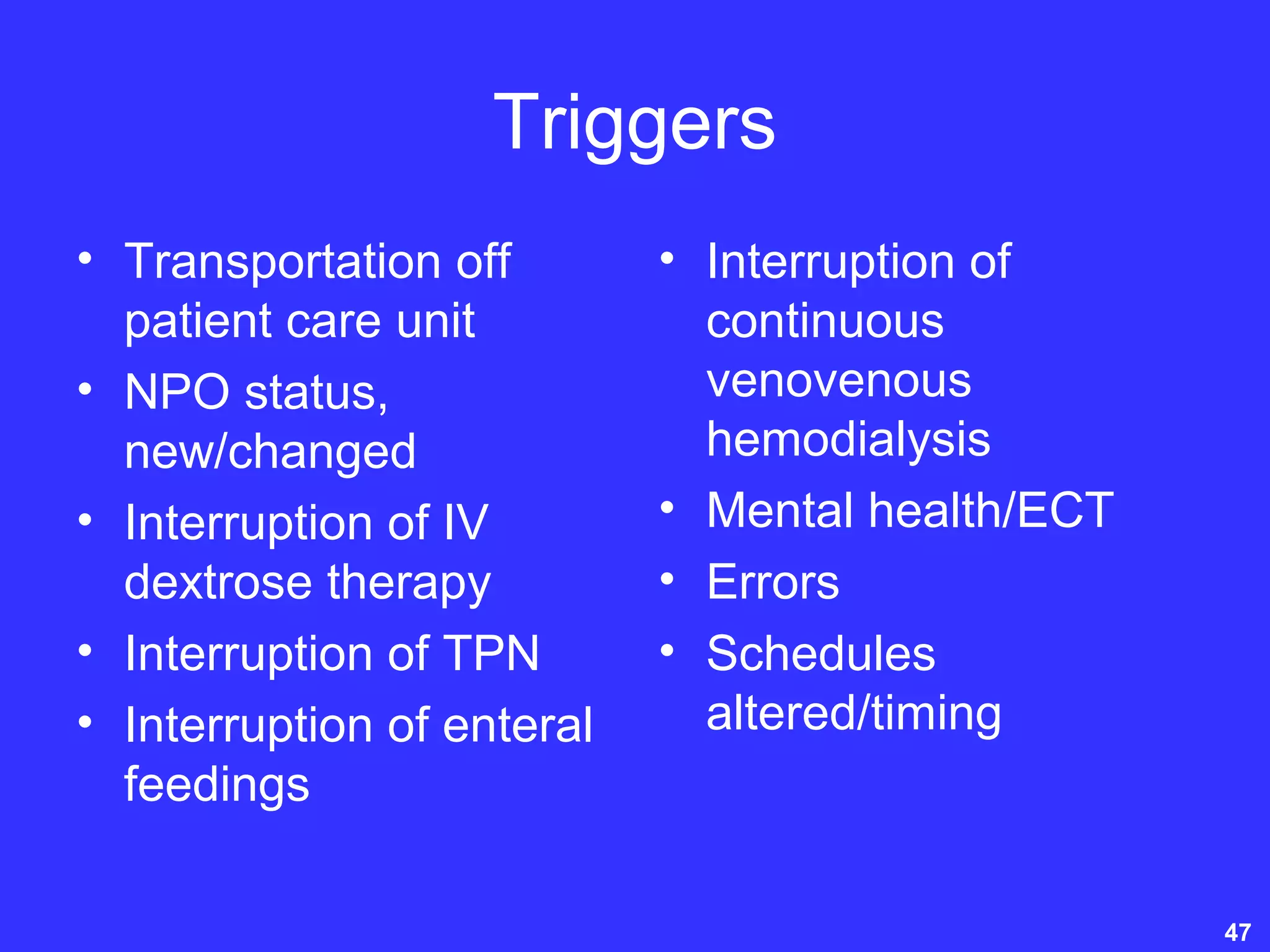
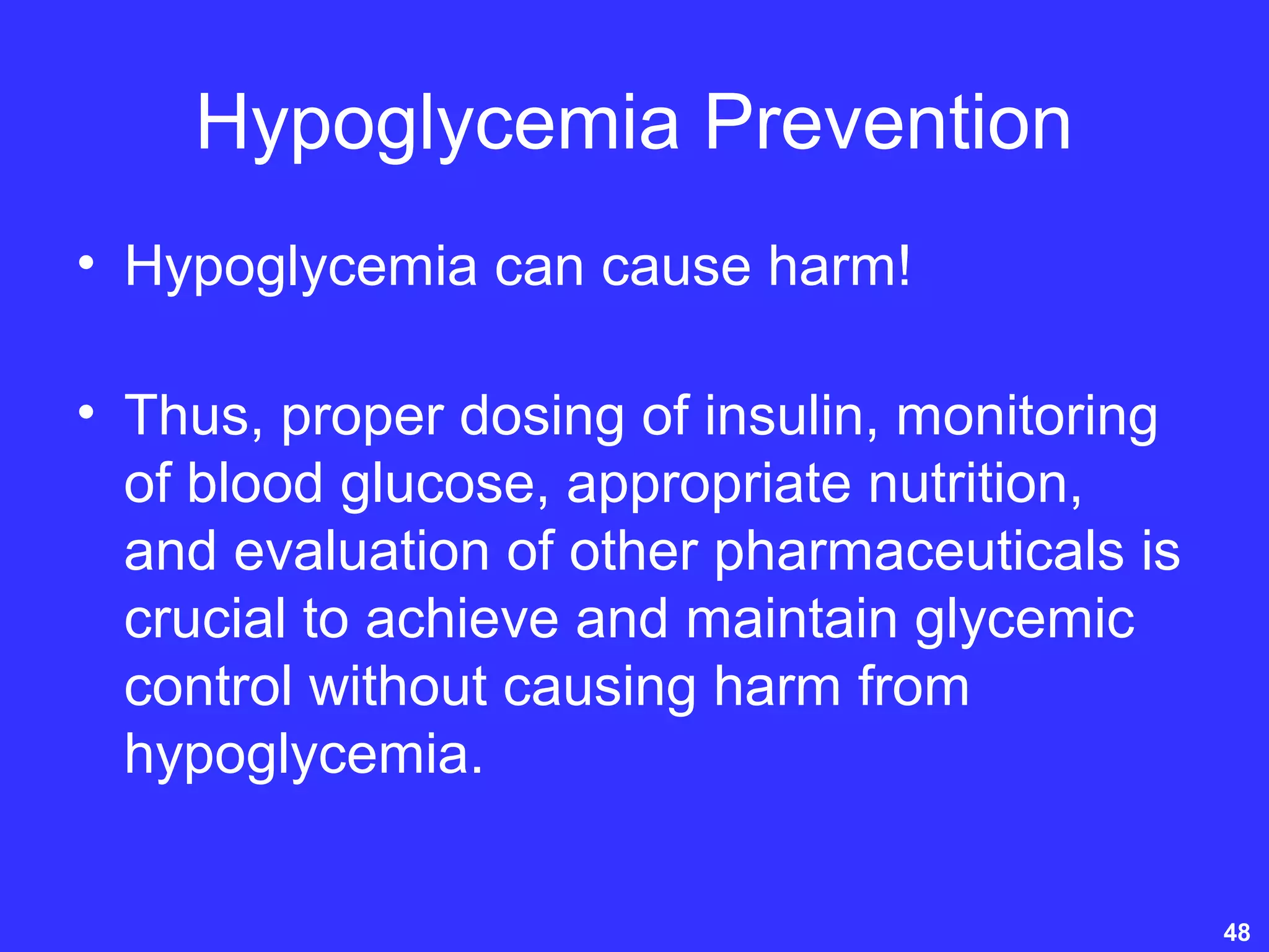
Hyperglycemic Crisis in Acute
Care
Fairview Health Services
2007
Carol Manchester, MSN, APRN, BC-ADM,
CDE
Diabetes Clinical Nurse Specialist
University of Minnesota Medical Center,
Fairview
University of Minnesota Children’s Hospital,
Fairview
GOALS
• Understand the pathophysiology of
Diabetic Ketoacidosis and Hyperosmolar
Hyperglycemic Non-Ketotic Syndrome
• Identify appropriate treatment modalities
for both
• Explain the principles of insulin
administration via intravenous infusion
The Treatment Modalities for
Optimal Glycemic Management
Include:
• Frequent blood glucose monitoring
• The administration of exogenous insulin
• Fluid and electrolyte replacement and
maintenance
• Nutrition
• Hypoglycemia prevention 3
The Treatment Modalities for
Optimal Glycemic Management
Include:
• Careful administration and monitoring of
concomitant pharmaceuticals
• Concurrent management of complications
and co-morbidities
4
IN THE PRESENCE OF
INSULIN, GLUCOSE:
• is used as energy by the muscle cells
• is stored as glycogen in the muscle cells
• is stored as glycogen in the liver
• enables amino acids/proteins to be used
for tissue synthesis
• is stored as triglycerides in adipose cells
INSULIN DEFICIENCY
• Liver cells release glycogen which converts to
glucose
• Muscle cells release glycogen which converts to
glucose
• Tissue breakdown releases amino acids which
then release ketoacids and glucose
• Adipose cells release triglycerides which convert
to free fatty acids and glycerol and thus, release
glucose and ketone bodies
THE NORMAL METABOLISM
OF GLUCOSE
Energy
Adipose Cells
Glucose Muscle Cells
Glycogen
(carbohydrate
storage)
Amino Acids
(Proteins)
Tissue Synthesis
Triglyceride (fat storage)
Liver Cells
Glycogen
(carbohydrate
storage)
This is a simplified diagram of normal glucose metabolism when a sufficient
quantity of insulin exists within the body and there are no receptor defects present.
When the plasma glucose levels become too low in the normal state, the
processes are reversed, yielding glucose from glycogen and fat for energy.
UNTREATED DIABETIC
KETOACIDOSIS
Glucose
Ketoacids
Amino Acids
Tissue Breakdown
Ketoacidosis
Excess Glucose
and Acid Formation
Glucose
Glycogen
Liver Cells Muscle Cells
Glucose
Ketone Bodies
(acetone,
acetoacetic acid,
B-hydroxybutyric acid)
Free Fatty Acids
and Glycerol
Triglyceride
Adipose Cells
Insulin Deficiency
DIABETIC KETOACIDOSIS
• DKA is a serious, life-threatening
event caused by a profound insulin
deficiency. It is characterized by
hyperglycemia, ketosis,
dehydration and electrolyte
imbalance.
LAB VALUES IN DKA
• Blood glucose is > 250 mg/dl
• Urinary and serum ketones are positive
• Serum bicarbonate is < 18 mEq/L (Mild DKA
could be 15- 18)
• Arterial pH is < 7.3
• Anion gap is > 10 mEq/L
• Potassium is low, normal or high
LAB VALUES IN DKA
• Phosphate usually normal or slightly
elevated
• Creatinine and BUN mildly increased
• WBC’s increased
• Amylase increased
• Hgb and Hct increased
• LFT’S can be elevated
CAUSES OF DKA
• New onset of Type 1 DM
• Illness/Infection
• Stress
• Omission of insulin
• Mismanagement of sick days
• Pregnancy
• Insulin pump malfunction
• Drugs-Corticosteroids, Thiazides,
sympathomimetic agents (Dobutamine and
terbutaline)
SIGNS & SYMPTOMS
OF DKA
• Nausea and vomiting
• Polydipsia, polyuria, and polyphagia
• Weakness
• Weight loss
• Anorexia
• Abdominal pain and cramping
• Visual disturbances
• Tachycardia
SIGNS & SYMPTOMS
OF DKA
• Hypotension
• Dehydration
• Warm, dry skin
• Rubor
• Kussmaul respirations
• Impaired consciousness and/or coma
• Fruity odor of ketones
TREATMENT OF DKA
FLUID REPLACEMENT
• IV fluid bolus of 0.9% NaCl at the rate of 1liter
over 1 hour
• For hypovolemic shock, 0.9% NaCl at 1liter/hr
• For mild hypotension with sodium corrected high
or normal, 0.45% NaCl, per protocol
• For mild hypotension with sodium corrected low,
0.9% NaCl
• Note: Potassium will be added to IV based on
serum level
• When serum glucose reaches 200 mg/dL,
change fluids to D50.45% NaCl
TREATMENT OF DKA
INSULIN ADMINISTRATION
• Regular insulin is the only IV insulin!
• Administer through a piggyback system into an
existing IV line with an infusion pump
• Pre-flush the IV tubing with 50 ml. of the
infusion to allow insulin to bind to the plastic
macrotubing or 8.5 ml for microtubing
• 0.1 units/kg of weight is given IV bolus initially to
adults to a maximum of 10 units.
• Hold insulin until Serum Potassium is > 3.3
mEq/L
INSULIN (CONT)
• Step Two will be to initiate the insulin drip
utilizing the Algorithms. Start with Algorithm 1.
• Move to higher algorithm if BG > 200 mg/dL and
BG has not fallen by at least 50 mg/dl within the
previous hour.
• Move to lower algorithm if BG < 150 mg/dl times
2 consecutive readings.
• When blood glucose falls below 200 mg /dl, the
rate is typically decreased and the IV fluid is
changed to a dextrose solution
• Blood glucose should drop 50-70 mg/dl/hr
INSULIN (CONT)
• Hourly blood glucoses are necessary
• Transition from IV insulin to basal/bolus
subcutaneous insulin protocol
• Subcutaneous basal insulin should be
administered 2 hours before discontinuing
the insulin drip
• If the patient will eat, subcutaneous
prandial (rapid-acting insulin) should be
administered during transition.
TREATMENT OF DKA
POTASSIUM REPLACEMENT
• Potassium is replaced based on plasma
K+ concentrations
• Establish urine output to rule out renal
failure
• If hypokalemic, K+ must be given
immediately
• If not hypokalemic, 20-40 mEq/L must be
given within the first 2-4 hours of treatment
POTASSIUM REPLACEMENT
(CONT)
• Administer K+ as K+Cl- or as potassium
phosphate. DO NOT exceed 90 mEq/24
hr. of potassium phosphate because of
danger of hypocalcemia.
• Monitor ECG. Hypokalemia causes a
flattened T and the presence of U waves.
Hyperkalemia causes peaked T waves ,
and if extremely high, a widened QRS
complex.
POTASSIUM REPLACEMENT
(CONT)
• Recheck plasma K+ every two hours if
plasma concentration of K+ is <4 or >6
mEq/L
• The goal of maintaining the plasma K+ is
3.5-5.0 mEq/L at all times
TREATMENT OF DKA
BICARBONATE
REPLACEMENT
• Routine bicarbonate administration is not
recommended if the pH is > 7.0 !
• Sodium bicarbonate enhances
hypokalemia
BICARBONATE
ADMINISTRATION: BENEFITS
• Correct extracellular acidosis
• Reduce excessive chloride
administration
• Reduce respiratory rate and increase
comfort
• Reduce cardiac irritability
• Increase responsiveness of vascular
system to pressor agents
BICARBONATE
ADMINISTRATION:
POTENTIAL HAZARDS
• Rapid reduction in plasma K+
• Na+ overload in elderly persons or
persons at risk for heart failure
• Exacerbate intracellular acidosis
TREATMENT OF DKA
PHOSPHATE
ADMINISTRATION
• Phosphate concentration decreases with
insulin therapy
• Calcium levels must be checked before
administering phosphate
• Phosphate is replaced only at a level < 1.0
mg/dl
• Overzealous phosphate administration can
cause severe hypocalcemia with no evidence
of tetany
TREATMENT OF DKA
COEXISTING INFECTION
• Chest x-ray if warranted
• Appropriate cultures
• IV antibiotic therapy
TREATMENT OF DKA
• Maintain airway
• Consider nasogastric tube if severe
nausea and vomiting
• Observe for signs of cerebral edema,
especially in children
• Auscultate lungs, assessing for heart
failure
• Observe for signs/symptoms of
hypoglycemia
PREVENTION OF DKA
• Provide adequate patient and family
education
• Make sure all items for self-care and
diabetes management are available to the
patient
• Provide follow-up medical care
• Effective communication with health care
provider when ill
HYPEROSMOLAR
HYPERGLYCEMIC NON-
KETOTIC SYNDROME
• HHNS is a syndrome with four primary
features including severe hyperglycemia,
absence of ketosis, profound dehydration
and neurologic manifestations
LAB VALUES IN HHNS
• Usually > 600 mg/dl
• Na+ normal or high
• K+ is high, normal or low
• Serum Bicarbonate is >15mEq/L
• Arterial pH is >7.3
• Serum osmolality is high; >320 mmol/kg
• Minimal ketonuria or ketonemia
CAUSES OF HHNS
• Age; HHNS is more common in elderly
individuals with Types 1 and 2 DM
• Illness such as infections, MI, GI bleeds,
uremia and arterial thrombosis
• Stress
• Massive fluid loss from prolonged osmotic
diuresis
CAUSES OF HHNS
• Hypertonic feedings such as prolonged
parenteral nutrition via IV infusion, high-
protein or gastric tube feedings
• Pharmacologic agents such as thiazides,
propranolol, phenytoin, steroids,
flurosemide and chlorthalidone
SIGNS AND SYMPTOMS
OF HHNS
• Milder gastrointestinal symptoms
• Polydipsia and polyuria
• Weakness
• Anorexia
• Visual disturbances
• Tachycardia
• Hypotension
• Dehydration
SIGNS AND SYMPTOMS
OF HHNS
• Warm, dry skin
• Rubor
• Hyperpnea
• Weight loss
• Decreased mentation
• Focal neurological signs such as
hemisensory deficits, hemiparesis,
aphasia and seizures.
TREATMENT OF HHNS
• The primary goal is rehydration! This is to
restore circulating plasma volume and correct
electrolyte imbalances
• IV fluid bolus of 0.9% NaCl at 1 liter/hour for
initial fluid replacement.
• For hypovolemic shock, 0.9% NaCl at 1 liter/hr
• For mild hypotension with corrected high or
normal sodium, 0.45% NaCl
• For mild hypotension with corrected low sodium,
0.9% NaCl
• When serum glucose reaches 250 mg/dl,
change to D5 with .45% NaCl
• Potassium is added based on serum level
TREATMENT OF HHNS
(CONT)
• 0.1 units/kg of weight is given IV bolus initially to adults
to a maximum of 10 units.
• Hold insulin until Serum Potassium is > 3.3 mEq/L
• Step Two will be to initiate the insulin drip utilizing the
Algorithms. Start with Algorithm 1.
• Move to higher algorithm if BG > 300 mg/dL and BG has
not fallen by at least 50 mg/dl within the previous hour.
• Move to lower algorithm if BG < 250 mg/dl times 2
consecutive readings.
• When blood glucose falls below 250 mg /dl, the rate is
typically decreased and the IV fluid is changed to a
dextrose solution
• Blood glucose should drop 50-70 mg/dl/hr
Treatment of HHNS (cont)
• Glucose hourly until stable while on an
infusion
• Electrolyte levels should be monitored
every 2-4 hours until stable
• Potassium and Phosphate are replaced as
in DKA
• Treat any underlying medical conditions
such as infection, especially urosepsis
and pneumonia
PREVENTION OF HHNS
• Provide patient, family and staff (such as
nursing home) education and follow-up
• Keep fluids within reach or offer fluids
every two hours to hospitalized or nursing
home patients
Insulin Therapy
• Continuous Intravenous Insulin Infusion if
NPO, on Total Parenteral Nutrition, on
Continuous Enteral Feeding
• Basal/Bolus insulin therapy in the fed state
is accomplished by the administration of
intermediate or long acting insulins and
rapid and/or short acting insulins
subcutaneously via Continuous
Subcutaneous Insulin Infusion
(ambulatory insulin pump) or multiple
injections 40
Evidence-Based Protocols
• Continuous Intravenous Insulin Infusions
• Subcutaneous Insulin Management
• Continuous Subcutaneous Insulin Infusion
• Hypoglycemia
• DKA
• HHNS
41
Continuous Intravenous
Insulin Infusion Indications
• Shock
• DKA
• HHNS
• Pregnancy
• Corticosteroid therapy
• Sepsis
• Transplantation
• Cardiopulmonary bypass surgery;
perioperative management
• NPO, Continuous TPN, Continuous Enteral
Feedings 42
Intravenous Insulin
• Fixed-rate insulin infusions
• Individualization of the rate of insulin infusion
• Algorithms based on rate of change in blood
glucose
• Neonatal, Pediatric (< 45 kg), Adult (> 45 kg)
43
Continuous Intravenous Insulin
Infusion Protocol
• Initial IV Bolus-0.1 unit/kg to a maximum
dose of 10 units
• Initiate algorithm 1
• Move to a higher algorithm if BG > 200
mg/dl and BG has not fallen by at least 60
mg/dl within the previous hour.
• Move to a lower algorithm if BG < 80
mg/dl X 2 consecutive readings.
• Transition as with DKA and HHNS.
Classifications of Hypoglycemia
• Severe hypoglycemia; < 45 mg/dL
• Moderate hypoglycemia; 45-59 mg/dL
• Mild hypoglycemia; 60-70 mg/dL
45
Predisposing Conditions
• Renal insufficiency
• Malnutrition
• Hepatic disease/failure
• Sepsis
• Shock
• Pregnancy
• Malignant lesion
• Hyperkalemia (GIK
cocktail)
• TPN
• Alcoholism and/or illegal
drug use
• Burns
• Gastroparesis or altered
nutrient absorption
• Dementia
• CHF
• Stroke
• Altered ability to self-report
• Hypoglycemia
Unawareness
• Aging
• Other metabolic disorders
such as pituitary and
adrenal insufficiency
46
Triggers
• Transportation off
patient care unit
• NPO status,
new/changed
• Interruption of IV
dextrose therapy
• Interruption of TPN
• Interruption of enteral
feedings
• Interruption of
continuous
venovenous
hemodialysis
• Mental health/ECT
• Errors
• Schedules
altered/timing
47
Hypoglycemia Prevention
• Hypoglycemia can cause harm!
• Thus, proper dosing of insulin, monitoring
of blood glucose, appropriate nutrition,
and evaluation of other pharmaceuticals is
crucial to achieve and maintain glycemic
control without causing harm from
hypoglycemia.
48
Concomitant Pharmaceuticals
• Polypharmacy/reconciliation
• Herbals
• Agents that lower blood glucose
– Pharmagrams for fluorquinolones, octreotide
• Agents that raise blood glucose such as
glucocorticoids
– Steroid taper then insulin taper?
– Alternative choices
– Timing of dosages
– Piggyback diluents
49
Comorbid Conditions
• Pneumonia, urinary tract infection,
sinusitus, congestive heart failure,etc.
must be identified and treated.
• Diabetes is usually the comorbid condition
50

Hyperglycemic crisis ppt.

  • 1.
    Hyperglycemic Crisis inAcute Care Fairview Health Services 2007 Carol Manchester, MSN, APRN, BC-ADM, CDE Diabetes Clinical Nurse Specialist University of Minnesota Medical Center, Fairview University of Minnesota Children’s Hospital, Fairview
  • 2.
    GOALS • Understand thepathophysiology of Diabetic Ketoacidosis and Hyperosmolar Hyperglycemic Non-Ketotic Syndrome • Identify appropriate treatment modalities for both • Explain the principles of insulin administration via intravenous infusion
  • 3.
    The Treatment Modalitiesfor Optimal Glycemic Management Include: • Frequent blood glucose monitoring • The administration of exogenous insulin • Fluid and electrolyte replacement and maintenance • Nutrition • Hypoglycemia prevention 3
  • 4.
    The Treatment Modalitiesfor Optimal Glycemic Management Include: • Careful administration and monitoring of concomitant pharmaceuticals • Concurrent management of complications and co-morbidities 4
  • 5.
    IN THE PRESENCEOF INSULIN, GLUCOSE: • is used as energy by the muscle cells • is stored as glycogen in the muscle cells • is stored as glycogen in the liver • enables amino acids/proteins to be used for tissue synthesis • is stored as triglycerides in adipose cells
  • 6.
    INSULIN DEFICIENCY • Livercells release glycogen which converts to glucose • Muscle cells release glycogen which converts to glucose • Tissue breakdown releases amino acids which then release ketoacids and glucose • Adipose cells release triglycerides which convert to free fatty acids and glycerol and thus, release glucose and ketone bodies
  • 7.
    THE NORMAL METABOLISM OFGLUCOSE Energy Adipose Cells Glucose Muscle Cells Glycogen (carbohydrate storage) Amino Acids (Proteins) Tissue Synthesis Triglyceride (fat storage) Liver Cells Glycogen (carbohydrate storage) This is a simplified diagram of normal glucose metabolism when a sufficient quantity of insulin exists within the body and there are no receptor defects present. When the plasma glucose levels become too low in the normal state, the processes are reversed, yielding glucose from glycogen and fat for energy.
  • 8.
    UNTREATED DIABETIC KETOACIDOSIS Glucose Ketoacids Amino Acids TissueBreakdown Ketoacidosis Excess Glucose and Acid Formation Glucose Glycogen Liver Cells Muscle Cells Glucose Ketone Bodies (acetone, acetoacetic acid, B-hydroxybutyric acid) Free Fatty Acids and Glycerol Triglyceride Adipose Cells Insulin Deficiency
  • 9.
    DIABETIC KETOACIDOSIS • DKAis a serious, life-threatening event caused by a profound insulin deficiency. It is characterized by hyperglycemia, ketosis, dehydration and electrolyte imbalance.
  • 10.
    LAB VALUES INDKA • Blood glucose is > 250 mg/dl • Urinary and serum ketones are positive • Serum bicarbonate is < 18 mEq/L (Mild DKA could be 15- 18) • Arterial pH is < 7.3 • Anion gap is > 10 mEq/L • Potassium is low, normal or high
  • 11.
    LAB VALUES INDKA • Phosphate usually normal or slightly elevated • Creatinine and BUN mildly increased • WBC’s increased • Amylase increased • Hgb and Hct increased • LFT’S can be elevated
  • 12.
    CAUSES OF DKA •New onset of Type 1 DM • Illness/Infection • Stress • Omission of insulin • Mismanagement of sick days • Pregnancy • Insulin pump malfunction • Drugs-Corticosteroids, Thiazides, sympathomimetic agents (Dobutamine and terbutaline)
  • 13.
    SIGNS & SYMPTOMS OFDKA • Nausea and vomiting • Polydipsia, polyuria, and polyphagia • Weakness • Weight loss • Anorexia • Abdominal pain and cramping • Visual disturbances • Tachycardia
  • 14.
    SIGNS & SYMPTOMS OFDKA • Hypotension • Dehydration • Warm, dry skin • Rubor • Kussmaul respirations • Impaired consciousness and/or coma • Fruity odor of ketones
  • 15.
    TREATMENT OF DKA FLUIDREPLACEMENT • IV fluid bolus of 0.9% NaCl at the rate of 1liter over 1 hour • For hypovolemic shock, 0.9% NaCl at 1liter/hr • For mild hypotension with sodium corrected high or normal, 0.45% NaCl, per protocol • For mild hypotension with sodium corrected low, 0.9% NaCl • Note: Potassium will be added to IV based on serum level • When serum glucose reaches 200 mg/dL, change fluids to D50.45% NaCl
  • 16.
    TREATMENT OF DKA INSULINADMINISTRATION • Regular insulin is the only IV insulin! • Administer through a piggyback system into an existing IV line with an infusion pump • Pre-flush the IV tubing with 50 ml. of the infusion to allow insulin to bind to the plastic macrotubing or 8.5 ml for microtubing • 0.1 units/kg of weight is given IV bolus initially to adults to a maximum of 10 units. • Hold insulin until Serum Potassium is > 3.3 mEq/L
  • 17.
    INSULIN (CONT) • StepTwo will be to initiate the insulin drip utilizing the Algorithms. Start with Algorithm 1. • Move to higher algorithm if BG > 200 mg/dL and BG has not fallen by at least 50 mg/dl within the previous hour. • Move to lower algorithm if BG < 150 mg/dl times 2 consecutive readings. • When blood glucose falls below 200 mg /dl, the rate is typically decreased and the IV fluid is changed to a dextrose solution • Blood glucose should drop 50-70 mg/dl/hr
  • 18.
    INSULIN (CONT) • Hourlyblood glucoses are necessary • Transition from IV insulin to basal/bolus subcutaneous insulin protocol • Subcutaneous basal insulin should be administered 2 hours before discontinuing the insulin drip • If the patient will eat, subcutaneous prandial (rapid-acting insulin) should be administered during transition.
  • 19.
    TREATMENT OF DKA POTASSIUMREPLACEMENT • Potassium is replaced based on plasma K+ concentrations • Establish urine output to rule out renal failure • If hypokalemic, K+ must be given immediately • If not hypokalemic, 20-40 mEq/L must be given within the first 2-4 hours of treatment
  • 20.
    POTASSIUM REPLACEMENT (CONT) • AdministerK+ as K+Cl- or as potassium phosphate. DO NOT exceed 90 mEq/24 hr. of potassium phosphate because of danger of hypocalcemia. • Monitor ECG. Hypokalemia causes a flattened T and the presence of U waves. Hyperkalemia causes peaked T waves , and if extremely high, a widened QRS complex.
  • 21.
    POTASSIUM REPLACEMENT (CONT) • AdministerK+ as K+Cl- or as potassium phosphate. DO NOT exceed 90 mEq/24 hr. of potassium phosphate because of danger of hypocalcemia. • Monitor ECG. Hypokalemia causes a flattened T and the presence of U waves. Hyperkalemia causes peaked T waves , and if extremely high, a widened QRS complex.
  • 22.
    POTASSIUM REPLACEMENT (CONT) • Recheckplasma K+ every two hours if plasma concentration of K+ is <4 or >6 mEq/L • The goal of maintaining the plasma K+ is 3.5-5.0 mEq/L at all times
  • 23.
    TREATMENT OF DKA BICARBONATE REPLACEMENT •Routine bicarbonate administration is not recommended if the pH is > 7.0 ! • Sodium bicarbonate enhances hypokalemia
  • 24.
    BICARBONATE ADMINISTRATION: BENEFITS • Correctextracellular acidosis • Reduce excessive chloride administration • Reduce respiratory rate and increase comfort • Reduce cardiac irritability • Increase responsiveness of vascular system to pressor agents
  • 25.
    BICARBONATE ADMINISTRATION: POTENTIAL HAZARDS • Rapidreduction in plasma K+ • Na+ overload in elderly persons or persons at risk for heart failure • Exacerbate intracellular acidosis
  • 26.
    TREATMENT OF DKA PHOSPHATE ADMINISTRATION •Phosphate concentration decreases with insulin therapy • Calcium levels must be checked before administering phosphate • Phosphate is replaced only at a level < 1.0 mg/dl • Overzealous phosphate administration can cause severe hypocalcemia with no evidence of tetany
  • 27.
    TREATMENT OF DKA COEXISTINGINFECTION • Chest x-ray if warranted • Appropriate cultures • IV antibiotic therapy
  • 28.
    TREATMENT OF DKA •Maintain airway • Consider nasogastric tube if severe nausea and vomiting • Observe for signs of cerebral edema, especially in children • Auscultate lungs, assessing for heart failure • Observe for signs/symptoms of hypoglycemia
  • 29.
    PREVENTION OF DKA •Provide adequate patient and family education • Make sure all items for self-care and diabetes management are available to the patient • Provide follow-up medical care • Effective communication with health care provider when ill
  • 30.
    HYPEROSMOLAR HYPERGLYCEMIC NON- KETOTIC SYNDROME •HHNS is a syndrome with four primary features including severe hyperglycemia, absence of ketosis, profound dehydration and neurologic manifestations
  • 31.
    LAB VALUES INHHNS • Usually > 600 mg/dl • Na+ normal or high • K+ is high, normal or low • Serum Bicarbonate is >15mEq/L • Arterial pH is >7.3 • Serum osmolality is high; >320 mmol/kg • Minimal ketonuria or ketonemia
  • 32.
    CAUSES OF HHNS •Age; HHNS is more common in elderly individuals with Types 1 and 2 DM • Illness such as infections, MI, GI bleeds, uremia and arterial thrombosis • Stress • Massive fluid loss from prolonged osmotic diuresis
  • 33.
    CAUSES OF HHNS •Hypertonic feedings such as prolonged parenteral nutrition via IV infusion, high- protein or gastric tube feedings • Pharmacologic agents such as thiazides, propranolol, phenytoin, steroids, flurosemide and chlorthalidone
  • 34.
    SIGNS AND SYMPTOMS OFHHNS • Milder gastrointestinal symptoms • Polydipsia and polyuria • Weakness • Anorexia • Visual disturbances • Tachycardia • Hypotension • Dehydration
  • 35.
    SIGNS AND SYMPTOMS OFHHNS • Warm, dry skin • Rubor • Hyperpnea • Weight loss • Decreased mentation • Focal neurological signs such as hemisensory deficits, hemiparesis, aphasia and seizures.
  • 36.
    TREATMENT OF HHNS •The primary goal is rehydration! This is to restore circulating plasma volume and correct electrolyte imbalances • IV fluid bolus of 0.9% NaCl at 1 liter/hour for initial fluid replacement. • For hypovolemic shock, 0.9% NaCl at 1 liter/hr • For mild hypotension with corrected high or normal sodium, 0.45% NaCl • For mild hypotension with corrected low sodium, 0.9% NaCl • When serum glucose reaches 250 mg/dl, change to D5 with .45% NaCl • Potassium is added based on serum level
  • 37.
    TREATMENT OF HHNS (CONT) •0.1 units/kg of weight is given IV bolus initially to adults to a maximum of 10 units. • Hold insulin until Serum Potassium is > 3.3 mEq/L • Step Two will be to initiate the insulin drip utilizing the Algorithms. Start with Algorithm 1. • Move to higher algorithm if BG > 300 mg/dL and BG has not fallen by at least 50 mg/dl within the previous hour. • Move to lower algorithm if BG < 250 mg/dl times 2 consecutive readings. • When blood glucose falls below 250 mg /dl, the rate is typically decreased and the IV fluid is changed to a dextrose solution • Blood glucose should drop 50-70 mg/dl/hr
  • 38.
    Treatment of HHNS(cont) • Glucose hourly until stable while on an infusion • Electrolyte levels should be monitored every 2-4 hours until stable • Potassium and Phosphate are replaced as in DKA • Treat any underlying medical conditions such as infection, especially urosepsis and pneumonia
  • 39.
    PREVENTION OF HHNS •Provide patient, family and staff (such as nursing home) education and follow-up • Keep fluids within reach or offer fluids every two hours to hospitalized or nursing home patients
  • 40.
    Insulin Therapy • ContinuousIntravenous Insulin Infusion if NPO, on Total Parenteral Nutrition, on Continuous Enteral Feeding • Basal/Bolus insulin therapy in the fed state is accomplished by the administration of intermediate or long acting insulins and rapid and/or short acting insulins subcutaneously via Continuous Subcutaneous Insulin Infusion (ambulatory insulin pump) or multiple injections 40
  • 41.
    Evidence-Based Protocols • ContinuousIntravenous Insulin Infusions • Subcutaneous Insulin Management • Continuous Subcutaneous Insulin Infusion • Hypoglycemia • DKA • HHNS 41
  • 42.
    Continuous Intravenous Insulin InfusionIndications • Shock • DKA • HHNS • Pregnancy • Corticosteroid therapy • Sepsis • Transplantation • Cardiopulmonary bypass surgery; perioperative management • NPO, Continuous TPN, Continuous Enteral Feedings 42
  • 43.
    Intravenous Insulin • Fixed-rateinsulin infusions • Individualization of the rate of insulin infusion • Algorithms based on rate of change in blood glucose • Neonatal, Pediatric (< 45 kg), Adult (> 45 kg) 43
  • 44.
    Continuous Intravenous Insulin InfusionProtocol • Initial IV Bolus-0.1 unit/kg to a maximum dose of 10 units • Initiate algorithm 1 • Move to a higher algorithm if BG > 200 mg/dl and BG has not fallen by at least 60 mg/dl within the previous hour. • Move to a lower algorithm if BG < 80 mg/dl X 2 consecutive readings. • Transition as with DKA and HHNS.
  • 45.
    Classifications of Hypoglycemia •Severe hypoglycemia; < 45 mg/dL • Moderate hypoglycemia; 45-59 mg/dL • Mild hypoglycemia; 60-70 mg/dL 45
  • 46.
    Predisposing Conditions • Renalinsufficiency • Malnutrition • Hepatic disease/failure • Sepsis • Shock • Pregnancy • Malignant lesion • Hyperkalemia (GIK cocktail) • TPN • Alcoholism and/or illegal drug use • Burns • Gastroparesis or altered nutrient absorption • Dementia • CHF • Stroke • Altered ability to self-report • Hypoglycemia Unawareness • Aging • Other metabolic disorders such as pituitary and adrenal insufficiency 46
  • 47.
    Triggers • Transportation off patientcare unit • NPO status, new/changed • Interruption of IV dextrose therapy • Interruption of TPN • Interruption of enteral feedings • Interruption of continuous venovenous hemodialysis • Mental health/ECT • Errors • Schedules altered/timing 47
  • 48.
    Hypoglycemia Prevention • Hypoglycemiacan cause harm! • Thus, proper dosing of insulin, monitoring of blood glucose, appropriate nutrition, and evaluation of other pharmaceuticals is crucial to achieve and maintain glycemic control without causing harm from hypoglycemia. 48
  • 49.
    Concomitant Pharmaceuticals • Polypharmacy/reconciliation •Herbals • Agents that lower blood glucose – Pharmagrams for fluorquinolones, octreotide • Agents that raise blood glucose such as glucocorticoids – Steroid taper then insulin taper? – Alternative choices – Timing of dosages – Piggyback diluents 49
  • 50.
    Comorbid Conditions • Pneumonia,urinary tract infection, sinusitus, congestive heart failure,etc. must be identified and treated. • Diabetes is usually the comorbid condition 50