Download to read offline
![Muscular chains:
Muscular chains are groups of muscles that work together or influence each other through
movement patterns. There are three subtypes of muscular chains: synergists, muscle slings, and
myofascial chains. Each type of muscular chain interdepends on both the articular and the
neurological systems.
Synergists: A synergistic muscle works with another muscle (agonist) to produce movement or
stabilization around a joint. Synergists may include secondary movers, stabilizers, or
neutralizers. For example, during shoulder rotation, the rotator cuff is active. However, the
rhomboids, serratus anterior, and trapezius must work as stabilizers of the scapula to ensure a
stable origin for the rotator cuff. Therefore, pseudoweakness of the rotator cuff may be caused by
poor stabilization of the scapula; if the scapula is stabilized manually, the patient demonstrates
normal strength of the rotator cuff. Ususally they work locally.
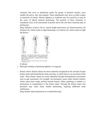
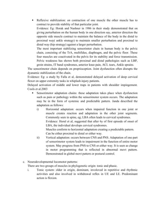
Diag: Scapula, rotator cuff, rhomboid, SA, and trapezius attachment. (Force couple):
Muscle sling: muscle slings are global, providing movement and stabilization across multiple
joints. Muscle slings are thought to facilitate rotation and to transfer forces through the trunk,
particularly from the lower body to the upper body (Vleeming et al. 1995). Muscle slings also
provide stabilization and movement in reciprocal and contralateral movements such locomotion.
Typically, muscle slings are interconnected, as one muscle insertion is connected to the next
muscle's origin via a common keystone structure (see table 3.2). These keystone structures act as
fixation points from which the entire chain of muscles can stabilize.
Lower extremity slings helpful in gait:
Extensor sling Flexor sling
[Type a quote from the document or the
summary of an interesting point. You can position
the text box anywhere in the document. Use the
Text Box Tools tab to change the formatting of the
pull quote text box.]](https://image.slidesharecdn.com/humancontroloflocomotion-200327071244/85/Human-control-of_locomotion-3-320.jpg)
![Only neural control:
1. Introduction to movement, locomotion
2. 3 systems of locomotion control
3. Neural control of locomotion: definition, structure of neuron, muscle and NMJ
4. Interaction of NMJ to provide connection between PNS and Muscle.
5. Neurolgical chain of movt: Protective reflex (crossed extensor and withdrawal reflex),
sensorimotor chain (reflex stabilization[strategies] and sensorimotor adaptation
chain=vertical adaptation[CNS and PNS] and horizontal adaptation[muscular]),
neurodevelopmental locomotion pattern [phasic and tonic chains].](https://image.slidesharecdn.com/humancontroloflocomotion-200327071244/85/Human-control-of_locomotion-7-320.jpg)

The document discusses human locomotion control, defining locomotion and outlining the three systems involved: skeletal, muscular, and neural. It details articular and muscular chains, their roles in biomechanical movement, and how structural positioning influences stability and motion. Additionally, it explores sensorimotor adaptations and neurodevelopmental patterns, emphasizing the importance of coordinated muscle activation for effective gait and posture.