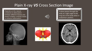
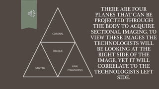
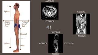
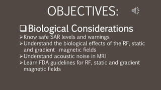
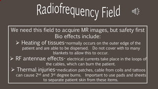
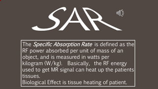
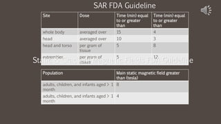
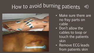
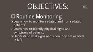
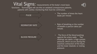
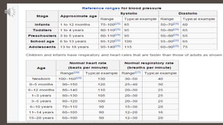
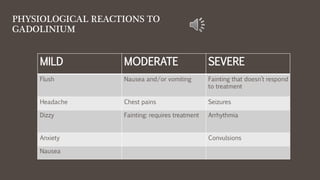
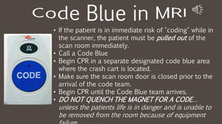
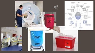
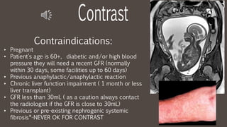
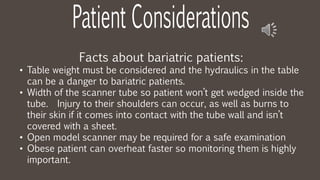
3. OBJECTIVE AND GUIDANCE
ď§ Each presentation will guide you through verbal as well as written information about each weeks lesson.
ď§ This is a self paced review, but it is recommended that you be sure to keep up with the instructors weekly
requirements and due dates.
ď§ We will cover presentations on topics such as:
ď MRI History
ď Safety
ď Anatomy/Pathology
ď Basic Cardiac MRI
ď Physics & Instrumentation
ď MRI Formulas and calculations
ď SNR Trade off parameters
ď Artifacts
7. ď§ Early Greeks discovered
that matter consists of
visible and invisible
particles called atomos
(atoms) which means
âuncut.â
⢠They also studied how
things were attracted or
repelled by invisible forces
now known as static
8. ⢠Mathematics was his specialty and focus,
which is why he was able to come up with
the Fourier Transform.
⢠Ability to create MR images with a theory
on heat.
⢠Mathematically formed a method for
analysis of heat transfer between solid
bodies.
⢠He came up with a way to rapidly process
the phase and frequency signals of the
NMR data and to efficiently utilize the
information for image reconstruction.
⢠Magnetic resonance signal analysis and
image reconstruction used his
mathematical equation by Richard Ernst in
1975 and today in all modern MRI
9.
10. ⢠The father of electricity!
⢠He proved that magnetic
fields traverse through an
electrical coil at 90Âş
angle, which results in a
voltage or current in the
coil.
⢠Further showed it would
need to be pulsed to
keep the magnetic field
11.
12. William Conrad
Rontgen
mistakenly discovered x-rays.
The patient absorbs the x-rays
with some passing through the
patient. When film is placed
behind the patient the rays that
pass through the patient form
the image.
Well, MRI is formed differently,
but this discovery was the basis
for all other imaging
techniques.
13. ⢠Danish physicist
⢠Nobel prize in
1922 for
contributions in
atomic structure
& quantum
theory
14. TIMELINE
1882 1937 1946
1956 1971 1972
Nikola Tesla discovered
the Rotating Magnetic
Field in Budapest,
Hungary.
Professor Isidor I. Rabi observed the quantum
phenomenon dubbed nuclear magnetic resonance
(NMR).He recognized that the atomic nuclei show their
presence by absorbing or emitting radio waves when
exposed to a sufficiently strong magnetic field.
Felix Bloch and Edward Purcell
discover magnetic resonance
phenomenon.
The "Tesla Unit" was proclaimed in
the Rathaus of Munich, Germany by
the International Electro-technical
Commission-Committee of Action.
Raymond Damadian discovered that
hydrogen signal in cancerous tissue
is different from that of healthy tissue
because tumors contain more water.
Damadian applies for a patent for
"Apparatus and Method for Detecting
Cancer in Tissues
15. TIMELINE
1973 1974 1975
1977
Paul Lauterbur, a chemist and an
NMR pioneer produced the first NMR
image. It was of a test tube.
Raymond Damadian receives
his patent
Richard Ernst proposes using phase and
frequency encoding and Fourier transform
for acquisition of MR images..
Raymond Damadian produces MR image of the whole body. Peter Mansfield
improves mathematics behind MRI and develops echo-planar technique, which
allows images to be produces in seconds and later becomes the basis for fast
MR imaging.
16. As early as B.C. our ancestors knew there was a
connection between electricity and magnetism. But it
wasnât until later and through many various attempts
that MRI was perfected.
⢠Michael Faraday asked why magnetism couldnât
produce electricity. Hence came Faradayâs Law,
the means by which MR signal is detected.
Albert Einstein
⢠Stated that mass and energy are one in the same
⢠Unsophisticated equipment hindered his ability to prove it.
Niels Bohr
⢠Nobel Peace Prize in 1922 for his work in atomic physics
Isidor Isaac Rabi
⢠Performed the first NMR experiment
17. NMR or Nuclear
Magnetic Resonance
developed
Nobel Prize was awarded
for discovering and
putting to use atomic
energy for analysis.
A water and paraffin
substance was
magnetically
energized.(RF) Radiofrequency
was used to excite the
atoms, the atoms
responded.
Signals were given
off by the atoms
and recorded into
spectroscopy
images.
Bloch named the
relaxation rates as
T1 and T2
18. After many
advances by
various
individuals the
pieces begin to
develop into
diagnostic MRI
imaging when
two scientist
Felix Bloch and
Edward Purcell
begin their
own research
studies.
Bloch Equation mathematically
states that protons in the nucleus act like
tiny magnets.
Responsible for the term resonance,
which allows energy to efficiently go from
one stable object to another, then making
the receiving object vibrate at the same
frequency as the sender.
19. Dr. Raymond Damadian is the âfather of the
MRIâ (Magnetic Resonance Imaging).
Damadian first got the idea that MRI could be
used to diagnosis cancer in humans while
studying a bacteria with very high potassium
levels. The potassium relaxation times were
much shorter than potassium ions in water
based solutions.
Tired of using a spectrum or graph that
describes how much of an element is present in
the sample material. Damadian modified the
equipment to produce an actual appearance of
the anatomy.
By 1970, he had created a MRI machine that
showed major MR signal differences in normal
24. ⢠DIFFERENTIATE BETWEEN PLAIN FILM
X-RAY AND CROSS SECTIONAL
⢠LIST THE FOUR ANATOMICAL PLANES
⢠LIST THE DIRECTIONAL PLANES
⢠BE ABLE TO DEFINE SECTIONAL
ANATOMY
⢠IDENTIFY STRUCTURES IN THE BODY
USING THE DIRECTIONAL PLANES
OBJECTIVE:
27. ⢠Plain radiographs
show a flat image
to view anatomy
& pathology.
⢠MRI scans use
cross sections to
view anatomy &
pathology.
What is the big
difference?
28. Plain X-ray VS Cross Section Image
D
Depends on the natural
variations in attenuation
between different tissues and
doesnât employ contrast media
to enhance attenuation.
A plane passes through the
body at a right angle to the
long axis of the body when in
anatomical position.
29. Itâs like slicing a loaf of bread. Instead of looking at the
entire loaf at once, individual slices are viewed
separately at different positions. What are the positions
or planes?
How do you
acquire cross
sectional images?
30. Anatomical Position
is achieved when:
⢠Patient is standing erect
⢠Arms are by the side
⢠Faced forward
⢠Feet directed forward slightly
apart
31. THERE ARE FOUR
PLANES THAT CAN BE
PROJECTED THROUGH
THE BODY TO ACQUIRE
SECTIONAL IMAGING. TO
VIEW THESE IMAGES THE
TECHNOLOGISTS WILL
BE LOOKING AT THE
RIGHT SIDE OF THE
IMAGE, YET IT WILL
CORRELATE TO THE
TECHNOLOGISTS LEFT
SIDE.
CORONAL
SAGITTAL
OBLIQUE
AXIAL
(TRANSVERSE)
35. Sagittal
plane
divides the
body into
right and
left
portions.
This is a
vertical
plane
Coronal
plane divides
the body into
front and
back or
anterior and
posterior
portions.
This is a
vertical
plane.
Axial plane
divides the
body into
superior
and inferior
portions.
This is a
horizontal
plane.
Oblique plane
denotes the
direction that a
radiation beam
passes through
the body from a
direction other
than AP*, PA**,
or lateral
projections.
38. DIRECTIONAL PLANES DEFINITION
Superior Above or higher level
Inferior Below or lower level
Anterior Toward the front of the body
Posterior Toward the back of the body
Cranial Direction of the head
Caudal Direction of the feet
Medial Direction of the midsagittal of the body
Lateral Away from the midsagittal plane of the
body
49. OBJECTIVES:
ďąLegal and Ethical Principles
ďExamination Confirmation
ďLegal Issues
ďPatientâs Rights
ďARRT Standards of Ethics
50. ďPatient identifiers (use armband)
1. Patient Name
2. Patient Date of Birth
ďCompare the request to clinical indications
1. Always confirm with the patient the body part (i.e. left or right)
2. Always confirm lab results to confirm kidney function before administering
contrast
3. Make sure the information you get about symptoms from the patient matches with
the order from the doctor as well as the hospital requisition. For example, a
patient states they have left ankle pain and the requisition states MRI right ankle
pain.
51. ďTerminology
1. Intentional Negligence Failure to exercise ordinary care or caution.
2. Malpractice Misconduct in a professional capacity through negligence,
carelessness, lack of skill, or malicious intent.
3. Beneficence To only do good and to prevent evil or harm.
4. Unintentional Negligence Not done on purpose
5. Assault Threatening to cause physical harm.
6. Battery To cause physical harm
7. Spoliation Destroying or altering medical records without legal
authorization or reason
8. Invasion of privacy is a breach of confidentiality
52. ďLegal Doctrines
1. respondeat superior Latin: "let the master answer"; is a legal
doctrine which states that, in many circumstances,
an employer is responsible for the actions of employees
performed within the course of their employment
2. res ipsa loquitur Latin for "the thing speaks for itself" is a
doctrine that infers negligence from the very nature of an
accident or injury, in the absence of direct evidence on how
any defendant behaved
53. ďInformed Consent a process for getting permission before
conducting a healthcare intervention on a person
in possession of all of the facts relevant to the decision.
1. Written
the patient, who signs and keeps a copy of signed consent form
2. Implied is not expressly granted by a person, but rather inferred
from a person's actions and the facts and circumstances of a
particular situation
3. Oral patient has listened to the explanation of indications, risks a
nd benefits of proposed procedure and has given verbal consent
54. ďHIPAA
Is the acronym for the Health Insurance Portability and Accountability
Act that was passed by Congress in 1996. HIPAA does the following:
Provides the ability to transfer and continue health insurance coverage
for millions of American workers and their families when they change or
lose their jobs.
http://www.hhs.gov/ocr/privacy/
55. ďPatient Bill of Rights
Guarantees patients information, fair treatment,
and autonomy over medical decisions, among other rights.
1. Health Care Proxy: because of illness or injury, you are not able to make or communicate your own
decisions. Patient selects another person (called your Health Care Agent) to make health care decisions for
you if you cannot make such decisions yourself, only if your doctor determines that you are unable to make
or communicate your own health care decisions.
2. Privacy: You have the right to talk privately with health care providers and have your health care
information protected
3. Access to information: You also have the right to read and copy your own medical record. You have
the right to ask that your doctor change your record if itâs not correct, relevant, or complete.
56. The purpose standards of ethics in healthcare is to promote high
standards of patient care and allow a basis for restricting or removing
individuals who exhibit behavior that isnât in accordance with these
standards of ethics.
https://www.arrt.org/pdfs/Governing-
Documents/Standards-of-Ethics.pdf
1. CODE OF ETHICS: assist certificate
holders and candidates in
maintaining a high level of ethical
conduct, while giving protection,
safety and comfort to the patients.
2. RULES OF ETHICS: mandatory
standards of minimally
acceptable professional conduct
for all certificate holders and
candidates.
59. OBJECTIVES:
ďąBiological Considerations
ďKnow safe SAR levels and warnings
ďUnderstand the biological effects of the RF, static
and gradient magnetic fields
ďUnderstand acoustic noise in MRI
ďLearn FDA guidelines for RF, static and gradient
magnetic fields
60. We need this field to acquire MR images, but safety first
Bio effects include:
ď Heating of tissues-normally occurs on the outer edge of the
patient and are able to be dispersed. Do not cover with to many
blankets to allow this to occur.
ď RF antennae effects- electrical currents take place in the loops of
the cables, which can burn the patient.
ď Thermal injuries-medication patches, cable from coils and tattoos
can cause 2nd and 3rd degree burns. Important to use pads and sheets
to separate patient skin from these items.
61. The Specific Absorption Rate is defined as the
RF power absorbed per unit of mass of an
object, and is measured in watts per
kilogram (W/kg). Basically, the RF energy
used to get MR signal can heat up the patients
tissues.
Biological Effect is tissue heating of patient.
62. Site Dose Time (min) equal
to or greater
than
Time (min) equal
to or greater
than
whole body averaged over 15 4
head averaged over 10 3
head and torso per gram of
tissue
5 8
extremities per gram of
tissue
5 12
SAR FDA Guideline
Static & Gradient Magnetic Fields FDA Guideline
Population Main static magnetic field greater
than (tesla)
adults, children, and infants aged > 1
month
8
adults, children, and infants aged > 1
month
4
63. FDA GUIDELINES:
⢠the observation of a biological
effect, in and of itself, does not
necessarily suggest the
existence of a biological
hazard.
⢠Biological effects that result
from heating of tissue by RF
energy are often referred to as
"thermal" effects.
⢠the eyes and the testes, are
known to be particularly
vulnerable to heating by RF
energy because of the relative
lack of available blood flow to
dissipate the excessive heat
load
Static field:
studies on alterations in cell growth and morphology, cell
reproduction and teratogenicity, DNA structure and gene
expression, pre- and post-natal reproduction and development,
blood brain barrier permeability, nerve activity, cognitive
function and behavior, cardiovascular dynamics, hematological
indices, temperature regulation, circadian rhythms, immune
responsiveness, neurological processing of visual and auditory
information, and other biological processes.
ď Pulse oximetry monitoring should be used verses ECG
monitoring
ď At field strengths over 2T effects such as headache, fatigue,
hypotension and irritability can occur.
ď Sickle cell red blood cell molecules have magnetic properties
and experience a twisting force during MRI, so many facilities
hesitate to image these patients.
64. Gradient field:
When you change the magnetic field the
voltage within the body in induced. (TVMF)
The results are peripheral nerve stimulation
and magneto-phosphenes.
1. Peripheral Nerve Stimulation: increased
strength, speed and length of gradient
pulses increases the effects. Vision
alterations or alterations in the
biochemistry of cells.
2. Magento-Phosphenes: visual disturbance
or âstars in the eyesâ, possibly due to
stimulation of the retina by the magnetic
field.
ACOUSTIC NOISE:
manifested as loud tapping,
knocking, chirping, squeaking
sounds, or other sounds is
produced when the forces cause
motion or vibration of the
gradient coils as they impact
against their mountings which, in
turn, flex and vibrate.
⢠Passive noise control by
routinely use disposable
earplugs or headphones.
66. The rapid boiling off of the
cryogens that are responsible
for keeping the magnet cool
and as a superconducting
magnet.
67. ⢠Liquid helium is housed in a
cryogent and is a cooling agent.
⢠Liquid helium, normally used
cryogen in MRI, undergoes almost
explosive expansion as it changes
from a liquid to gaseous state.
⢠Oxygen monitor ensures oxygen
levels are safe for
patient/physicians.
⢠Rooms built for superconducting
MRI equipment should be
equipped with pressure relief
mechanisms and an exhaust fan,
plus a required quench pipe.
Blue arrow: tube from scanner for Quench
Green arrow: tube attaches to vent through
ceiling for cryogen/helium to be released
outside
68. ⢠Cryogens are super cooled liquid gases and without this
very important gas the magnetic field is lost.
⢠At room temperature liquid helium boils away very fast so
the MRI machine has to be able to prevent it from leaking
into the air.
⢠The MRI machine contains the helium by having a cryostat
that has a helium condenser that recycles any boil off.
⢠They include helium and sometimes nitrogen.
⢠The concern is helium will over take oxygen in the room.
⢠During a quench the boil off happens and technologist are
trained how to handle such an event.
69. Ultra High Field
Imaging Precautions
⢠Increased SAR because the higher RF power used in higher
field strength.
⢠Implants and devices havenât been tested and approved for
use in higher field strengths.
⢠Clinical experiments are limited at these field strengths.
⢠Protocols have not been fully perfected on humans and/or
animals.
70. OBJECTIVES:
ďąEnvironment
ďKnow why climate control is important
ďKnow what a gauss line is in MRI
ďUnderstand safety zones
ďUnderstand why magnetic and RF shielding is
needed in MRI
71. 5 GAUSS LINES: Reminds you that you are inside
a magnetic field. This is the limit where ferromagnetic items are
strictly prohibited.
⢠Must not
exceed 72
degrees F
⢠Humidity
must not
exceed 60%
Room Temperature & Humidity
Levels
72.
73.
74. These persons must
pass the minimum
safety education to
prove they are safe to
work in Zone III areas.
These persons will have
extensive training and
education when it
comes to thermal
burning and other areas
of MR issues.
75. ⢠Prevents RF noise
from entering scanner
room and causing the
image to be distorted.
⢠Copper is best for this
type of shielding.
⢠Used to block the
magnetic field of the
magnet from interfering
with pacemakers and
other equipment outside
of the scan room.
77. Windows: (image A & B)
consisting of a stainless
steel tube frame, stainless
steel screen frame, two
layers of brass screen, and
two layers of 1/4"
tempered glass. The two
layers of R.F. screen are
offset in order to prevent
the MoirĂŠ effect and also
blackened to achieve
maximum visibility.
Doors: Be careful not to hit
the RF seals (image C & D),
which could rip them off
and degrade the RF
shielding protection for the
(A)
(B)
(C
)
(D)
79. OBJECTIVES:
ďąScreening and Education
ď Understand the need to screen the patient, other healthcare
workers and non patients
ď Distinguish types of topical or surgical procedures, biomedical
devices and implants that are safe, unsafe and conditional
ď Know how to research of a device is safe for MRI
ď Understand medical conditions and how they may be unsafe for
MRI
ď Know the levels of MRI personnel
ď Determine what foreign bodies are appropriate for MRI
80. ⢠Anyone giving information regarding
their health history must be conscious
and coherent. NOTE: Any lack of
memory (i.e. dementia, AMS) or lack of
information about a surgical procedure is
grounds for canceling the patient,
unless a family member can provide a
detailed history.
⢠If you have ANY doubt about a patients
past health history regarding metal in
their body, it is required that the MR
exam be put on hold until the question
can be investigated thoroughly or a
âScreening X-rayâ is done and cleared by
a radiologist.
81.
82.
83. Must fill outâŚ
⢠Patients
⢠Non-
Personnel
⢠Non-Patients
⢠Personnel
(MRI Techs
included)
Who should I have
fill out the MRI
screening form?
84.
85.
86.
87. ⢠Bullet
⢠Shrapnel
⢠Metal in eyes from
welding
⢠Insulin Pumps
⢠Neurostimulators on
spine
⢠BBâs
⢠Pellets
Ferrous: has a presence
of iron and can be
picked up by a magnet.
Heavy
89. ⢠Tattoos must not
be new because
they can heat up.
⢠Place a cool wet
cloth over older
tattoos to help
dissipate the
heat.
⢠Transdermal patches have a
aluminum backing.
⢠Aluminized backings also
serve as electrical conductor.
⢠The patches are not
ferromagnetic, they can result
in burns during an MRI
procedure
90. RENAL FUNCTION patients
with a GFR lower than 30, on
dialysis
PREGNANCY reviewed on a case
by case basis. Suggested that
scanning shouldnât be done on patients
less than 3 months pregnant.
DIABETES requires recent labs
normally within 3 months or and
ISTAT to get the creatinine and
then you must calculate the GFR
RECENT SURGERIES
requires a minimum of 6-8 weeks
before a scan can be done.
BREAST FEEDING mothers
must not use milk for 48 hours
after contrast injection.
91. 1. Abstain from breast-feeding
from the time of contrast
administration for a period of
48 hours.
2. She may wish to use a breast
pump to obtain milk before the
contrast-enhanced study to
feed the infant during the 48
hour period following the
examination
92. Because of the possibility of
burning of the patient because of
heating and artifact it is
recommended
that MRI specific ECG leads be
used.
⢠Body piercings must be
removed to avoid
burning the patient and
causing artifact on the
image.
⢠Jewelry has to be
removed because of
the risk of burning,
artifact and becoming a
projectile.
93. OBJECTIVES:
ďąEquipment Safety
ďLearn the safe place for items such as ECG leads
and coils
ďUnderstand cryogen levels
ďUnderstand the difference of terms such as MRI
safe, conditional and unsafe
ďKnow what to do in the case of a fire and/or
quench
94. ⢠Make sure there are
no fray parts on
cable
⢠Donât allow the
cables to loop or
touch the patients
skin
⢠Remove ECG leads
from patients skin
95. OBJECTIVES:
ďąRoutine Monitoring
ďLearn how to monitor sedated and non sedated
patients
ďLearn how to identify physical signs and
symptoms of patients
ďUnderstand vital signs and when they are needed
in MRI
96. Vital Signs: measurements of the body's most basic
functions. Technologist will use this on sedated/unresponsive patients,
patients with cardiac monitoring that must be continuous.
⢠The number of times the heart
beats per minute
⢠Rate of breathing or the number
of breaths a person takes per
minute.
⢠The force of the blood pushing
against the artery walls. Two
readings are taken: a high systolic
pressure, which occurs during the
maximal contraction of the heart,
and the lower diastolic or resting
pressure.
PULSE
RESPIRATORY RATE
BLOOD PRESSURE
99. Necessary to start:
⢠available personnel and training
⢠pre and post op evaluation by nurses
and doctors
⢠emergency equipment availability
⢠use of additional oxygen
⢠sedative-analgesic agents
⢠discharge instructions
Necessary to Monitor:
⢠patient responsiveness
⢠blood pressure
⢠respiratory rate
⢠heart rate
⢠oxygen saturation
How do I monitor
sedated patients?
101. These patients should
be monitored visually,
with use of the camera
(if available), use the
microphone to
periodically speak with
this type of patient and
use the sound system
to listen for patient
talking.
106. ALLERGIC REACTIONS TO
GADOLINIUM
MILD MODERATE SEVERE
Nasal Congestion Mild Wheezing Anaphylactic Shock
Sneezing Throat tightness,
hoarseness
Labored breathing with
facial swelling
Itchy/Scratchy Throat Facial swelling
Mild hives Hives
107. PHYSIOLOGICAL REACTIONS TO
GADOLINIUM
MILD MODERATE SEVERE
Flush Nausea and/or vomiting Fainting that doesnât respond
to treatment
Headache Chest pains Seizures
Dizzy Fainting: requires treatment Arrhythmia
Anxiety Convulsions
Nausea
108.
109. ⢠If the patient is in immediate risk of "coding" while in
the scanner, the patient must be pulled out of the
scan room immediately.
⢠Call a Code Blue
⢠Begin CPR in a separate designated code blue area
where the crash cart is located.
⢠Make sure the scan room door is closed prior to the
arrival of the code team.
⢠Begin CPR until the Code Blue team arrives.
⢠DO NOT QUENCH THE MAGNET FOR A CODEâŚ
unless the patients life is in danger and is unable to
be removed from the room because of equipment
111. OBJECTIVES:
ďą Safe Patient Transfer, Proper Body Mechanics,
Patients with Medical Equipment
ďIdentify safe and unsafe equipment, such as
stretchers/wheelchairs, that can be used to transfer patients
to MRI table
ďLearn how to safely care for your body while moving patients
ďLearn what medical devices can enter a MRI scanner room
ďUnderstand what implantable devices are allowed in the
scanner.
112.
113.
114.
115.
116.
117. These items pose an
immediate danger!
⢠Pacemakers are NOT to be
scanned. Medtronic &
certain pacemakers can be
scanned under the
guidance of a cardiologist,
radiologist permission, and
Medtronic specialist to turn
pacemaker back on after
scan.
⢠Foreign Bodies like bullets
or shrapnel are unsafe
when near a major blood
vessel or area like an eye,
artery, in the chest or
118. These items are
safe with certain
safety precautions
first.
⢠Stents, titanium
rods and other
surgeries can be
scanned 6-8
weeks post
surgery. This
allows for scar
tissue to form
around the area.
119. ⢠At times you will have
patients who are intubated
yet will need a MRI.
⢠The doctor will at times
intubate a patient on the
MRI scanner table.
⢠The laryngoscope with the
green coloring is MR Safe.
⢠Urinary catheter bags
have a metal clap, which
can not be removed.
⢠It must be hooked onto
the MRI table (the portion
that moves).
120. OBJECTIVES:
ďąCommunication
ďLearn modes and challenges to communicate with
various patients
ďLearn how to educate your patients about the
procedure, during the exam, follow up instructions
121. SPEAKER
HEAD
PHONES
TECH MICROPHONE
TECH
MICROPHONE
HEADPHONES are perfect for
hard of hearing patients.
They can be used for music
to be played for the patient as
well.
TECH MICROPHONES allow
the technologist to speak
directly to the patient.
SPEAKERS transmit the sound
into the room. There are
speakers in the MRI scanner
as well.
123. As a technologist you will need to determine the way
you will communicate with your patients based on their
needs. Some patients require verbal while others you
will need non verbal communication. With non verbal
you may have to touch or use your hands to
communicate instructions. Written communication
will be handy when you need the patient to sign
consents and fill out safety forms.
Modes of communication:
1. Verbal
2. Non Verbal
3. Written
124. As a technologist you will need to determine the way you will meet these challenges.
Always plan prior to bringing your patient back how you will face these communication
challenges.
How will you meet the following challenges?
A. Say your patient speaks Farsi and you need to give breathing instructions for a MRI of
the Abdomen or explain the exam.
B. Because of culture you may have to adjust how you position a patient that is a male if
you are a female technologist or vice versa. Touching a person of the opposite sex
may not be acceptable to the patients culture, but you may need to in order to position
or landmark.
Factors that challenge communication:
1. Language
2. Culture
3. Emotional status
4. Physical makeup of patient
125. 1. Once you bring your patient back to the consult area explain thoroughly from beginning to
end the exam. Let them know what was ordered and why and see if they have anything to
add as to their symptoms. Make sure they know before you place an IV in their arm that it
was ordered with Gadolinium contrast also. Explain the benefits and risks of the contrast as
well. If you have to draw any blood to test their kidney function always explain what you
need to do before drawing the blood.
2. Patients will complain that the last time they had a MRI the technologist walked out and never
talked to them. We understand that MRI is a very long exam already and if you talked to the
patient after each sequence that would double the time. But please communicate with a
patient by updating them on how much longer the exam will be at least at the hallway point.
3. As you help the patient sit up and before they leave always let them know the results will be
sent to the ordering doctor within whatever time frame your facility has for their turn around
time on reports.
4. Always check if the patient is having other exams done at your facility on the same day
before removing an IV or letting them leave. No one likes to be stuck unnecessarily or have
to reschedule.
5. As medical professionals we are use to using medical terminology. Be careful when telling a
patient that you have scanned their abdomen. Sometimes they panic and say âYou were
suppose to scan my liver!â
Educate Your Patient
126. OBJECTIVES:
ďąInfection Control in MRI
ďLearn terminology
ďUnderstand the cycle of infection
ďLearn how to practice standard precautions and
additional precautions for transmissions based
infections
ďUnderstand how to safely clean MRI equipment
127. When the whole
body responds
with inflammation
to an infection
usually
originating in the
lungs, abdomen
or/and urinary
tract.
Septic shock &
hemorrhagic
rash
128. Hospital Acquired Infection:
also known as nosocomial
infection â is an infection
whose development is favored
by a hospital environment,
such as one acquired by a
patient during a hospital visit
or one developing among
hospital staff.
Fomite is any object or substance
capable of carrying infectious
organisms, such as germs or
parasites, and hence transferring
them from one individual to
another. Skin cells, hair, clothing,
and bedding are common hospital
sources of contamination.
Vectors: Creatures such as
fleas, mites, ticks, rats,
snails, and dogsâcalled
vectorsâcan also transmit
disease. The most common
vector for human infection
is the mosquito.
Vehicles: contaminated
food, water, blood, or
other vehicles may
spread pathogens. Ex. E.
Coli
135. Cleaning and Proper Disposal of
Contaminated Items
ďUse the proper sanitizing supplies to
wipe down tables after each patient
ďBleach wipes should be used for C Diff
and all other diseases that require it.
ďClean each coil after using it on
patients
ďLinens MUST be changed after each
patients
ďRegularly wipe down the door handles
and magnet buttons
ďAny soiled and/or blooded lined
should disposed of in a biohazard bag
141. ⢠Gadoliniums job is to
shorten T1 relaxation and
cause lesions to be bright
on T1 weighted imaging.
It shortens T2 relaxation
times causing dark areas
on T2 weighted imaging.
Effects on image:
⢠Shorten relaxation times
in atoms of body tissues
142. ⢠Precession means molecular
tumbling occurs because of the
magnetic field.
⢠Results are fluctuation of the
magnetic field. At certain times
during the tumbling the molecule
will impose, have no net effect or
add to the main magnetic field.
Introducing gadolinium will slow
down the tumbling, which
reduces the relaxation times.
⢠Water tumbles very fast
and to allow for
lesion/tumors that are
water filled to enhance
after gadolinium
injected the water
molecule must be
slowed down.
⢠T1 relaxation time is
decreased in water.
144. ⢠Magnetic susceptibility is the ability of the external magnetic field to
affect the nucleus of an atom and magnetize it.
145. ⢠Magnetic susceptibility is the ability of the external magnetic field to
affect the nucleus of an atom and magnetize it.
146. If you had two wheels and one spun fast it would take
longer for it to come to a stop or relax. Verses the slow
spinning wheel would take a short time to stop spinning or
relax. Water spins faster than the Larmor frequency
resulting in a long T1 & T2 relaxations.
How to reduce the speed of water?
Gadolinium has 7 unpaired electrons, which are much
stronger than hydrogen protons. Resulting in increased
signal of water in T1 weighted images . Image
enhancement is based on the effective tissue not the
particular agent itself.
Factors affecting tumbling are viscosity and temperature
of solution.
148. Dose
Calculation:
http://www.kidney.o
rg/professionals/KD
OQI/gfr_calculator.c
fm
Click on the link below
.1mmol/kg (0.2ml/kg)
of body weight
1lb = 2.2kg
EXAMPLE:
100lbs /2.2 = 45kg x
.2ml/kg
= 9ml
200lbs/2.2 = 91kg x
.2ml/kg
= 18.2ml
*20ml max. dose*
ď Calculate dose by weight
ď Document dose including type of agent used
ď A physician must be present during the injection of
all contrast agents
149. Administration Routes
Facts:
Gadolinium is
excreted
through the
kidneys within
120 minutes
after
administration.
⢠Intraarticular: in the
joint space
⢠Intravenous: into the
vein
⢠Oral: Taken approximately 45
minutes prior to the MR scan.
Patients are to ingest 1L of
VoLumen as well as water prior to
examination. Glucagon used to slow
bowel movement.
150. ⢠Gadolinium is heavy metal and in it pure state it is toxic. Binding with
chelates reduces the chances for toxicity and helps the body excrete
the gadolinium.
151. ⢠Gadolinium (Gd)
doesnât break a
blood brain barrier
that is intact.
When there is a
collapse in the
blood brain barrier
the gadolinium is
able to enhance
lesions.
153. Contraindications:
⢠Pregnant
⢠Patientâs age is 60+, diabetic and/or high blood
pressure they will need a recent GFR (normally
within 30 days, some facilities up to 60 days)
⢠Previous anaphylactic/anaphylactic reaction
⢠Chronic liver function impairment ( 1 month or less
liver transplant)
⢠GFR less than 30mL ( as a caution always contact
the radiologist if the GFR is close to 30mL)
⢠Previous or pre-existing nephrogenic systemic
fibrosis*-NEVER OK FOR CONTRAST
154. ⢠Have an indirect effect by altering the relaxation rates.
⢠Extrinsic factors are affected by contrast media such
as TR, TE, TI, flip angle.
⢠Because tissues may have the same intensity as its
surrounding tissues contrast media is used to cause
contrast enhancement.
⢠Improves the technologist ability to see something
specific in the anatomy.
⢠Signal to Noise (SNR) increases
155. ⢠Micro adenoma in the pituitary enhances fast verses a normal
pituitary enhancement. Rapid contrast enhancement sequences are
needed.
⢠Always acquire post sequences in a minimum of two planes since
lesions enhance at different times.
⢠(1) Metastatic disease, (2) acoustic neuromas and (3) meningioma's
require contrast injection. 21 3
156. Neonate bundle device
Movies and sound are provided
Ear muffs for
neonates
instead of ear
plugs are used
Specialty head coil with immobilization device for pediatric
Sedation or General
157. Facts about pediatric patients:
⢠Normal structures in a childâs body are smaller than in adults.
⢠Pediatrics are made up of 75% of water vs the average adult, which requires
the technologist to use a higher TE for T2 contrast.
⢠At different stages in a child's development anatomy may be normal but at
another time may be abnormal.
158. Facts about geriatric patients:
⢠Alzheimer's is more than being forgetful. Patients loss the ability to reason,
imagine and learn.
⢠Weight of brain and blood and oxygen to their brain decreases.
⢠Increase of ventricle size is normal.
NORMAL DEMENTIA
159. Facts about bariatric patients:
⢠Table weight must be considered and the hydraulics in the table
can be a danger to bariatric patients.
⢠Width of the scanner tube so patient wonât get wedged inside the
tube. Injury to their shoulders can occur, as well as burns to
their skin if it comes into contact with the tube wall and isnât
covered with a sheet.
⢠Open model scanner may be required for a safe examination
⢠Obese patient can overheat faster so monitoring them is highly
important.
160. Bariatric patients may be too wide to fit in the
scanner. Consider if they can bring their
arms above their head if this is possible and
safe. The main issue with a head scan and
these patients is making sure their shoulders
can fit in the scanner and that they are not
exceeding the table weight limit.
Be careful with protecting their skin from
burns and the use of tape as it may pull their
fragile skin off.
Anatomy may be different from adult brain
anatomy as a patient ages.
Editor's Notes
NOTE:
To change the image on this slide, select the picture and delete it. Then click the Pictures icon in the placeholder to insert your own image.
Faraday video
40-50% of MRI make use of contrast so it is extremely important to assess each patients potential risk.
*NSF is a sclerosing disorder that can affect extremities and organs such as lungs, myocardium and occurs 4 weeks after gadolinium administration. It is highly discouraged to administer contrast in patients with a GFR less than 30 even when dialysis is done 12 hours or more post contrast administration.
Pediatric anatomy is different from geriatric and adult anatomy as they havenât fully developed their brain at this age.