MRI & CT basics/ oral surgery courses
•
10 likes•1,425 views

The Indian Dental Academy is the Leader in continuing dental education , training dentists in all aspects of dentistry and offering a wide range of dental certified courses in different formats.for more details please visit www.indiandentalacademy.com

Recommended
More Related Content
What's hot
What's hot (20)
Viewers also liked
Viewers also liked (20)
Similar to MRI & CT basics/ oral surgery courses
Similar to MRI & CT basics/ oral surgery courses (20)
More from Indian dental academy
More from Indian dental academy (20)
Recently uploaded
Recently uploaded (20)
MRI & CT basics/ oral surgery courses
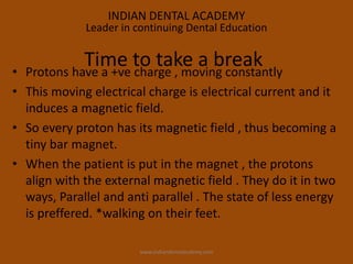
- 1. • Protons have a +ve charge , moving constantly • This moving electrical charge is electrical current and it induces a magnetic field. • So every proton has its magnetic field , thus becoming a tiny bar magnet. • When the patient is put in the magnet , the protons align with the external magnetic field . They do it in two ways, Parallel and anti parallel . The state of less energy is preffered. *walking on their feet. Time to take a break INDIAN DENTAL ACADEMY Leader in continuing Dental Education www.indiandentalacdemy.com
- 2. when the patient is in the MR magnet , his own magnetic field is longitudinal to external magnetic field of MR machine magnet. Because it is longitudinal it cannot be measured. www.indiandentalacdemy.com
- 7. • Protons are like little magnets. • In an external magnetic field they align parallel or anti parallel. • The low energy state(parallel) is preferred , so a few more protons align this way. • The protons perform a motion that resembles the wobbling of a spinning top , that was hit . This is called precession. As there are more protons aligned parallel to the external field there is net longitudinal magnetization. So time to review www.indiandentalacdemy.com
- 8. • A radio frequency pulse that has the same frequency as the precising protons , can cause resonance , ie transfer energy to protons . This results in more protons being anti parallel leading to decrease in longitudinal magnetization. • The RF pulse results in a new magnetic vector , the transversal magnetization. • When the RF pulse is switched off ……. There is an increase in longitudinal magnetization…… Long . relaxation ( T 1 ) And the transversal magnetization decreases … Transversal relaxation (T 2) www.indiandentalacdemy.com
- 9. Long . relaxation Transversal relaxation T1 T2 www.indiandentalacdemy.com
- 10. Implants cardiac pacemakers; defibrillators. CNS aneurysm clips Ocular foreign body e.g. metal shavings. Absolute contraindications to MRI Relative contraindications Lead wires or similar wires. Non-ferromagnetic stapedial implants, Cochlear implants. Claustrophobia. Pregnancy www.indiandentalacdemy.com
- 11. MRI Technique T1 and T2 images Fat saturated images (STIR) Diffusion imaging www.indiandentalacdemy.com
- 16. Diffusion MRI Aims to analyze human brain through diffusion of water molecules Measures the mobility of water within tissues and, may function as a marker for both tissue cellularity. www.indiandentalacdemy.com
- 17. MRI techniques are currently being used in dentistry For evaluation of temporomandibular joint diseases Examination of salivary glands, maxillary sinuses, masseter muscles, & facial skeleton Detection of early bone changes in tumors, fractures, inflammatory conditions and hematoma. Evaluation of bone before applying dental implants www.indiandentalacdemy.com
- 18. Enamel and dentine appeared black due to a lack of unbound protons. Dental pulp chamber,, appeared white or grey on T1 weighted and STIR imaging. Cortical bone was seen as a black zone and high signal internal fatty marrow Moderate signal from external soft tissues T1 weighted scans. Slices taken at 3 mm intervals www.indiandentalacdemy.com
- 19. The two buccal roots (B) of the upper first molar The inferior dental (ID) canal A The ID canal containing the ID nerve is seen as two parallel grey lines running in the ramus and body of the mandible, Between the two lines the neurovascular bundle and surrounding tissue and fat are seen as higher signal (whiter). crowns and roots of premolar and molar teeth are clearly seen www.indiandentalacdemy.com
- 20. Root apices of first and second molars in close proximity to the floor of maxillary antrum. unerupted upper third molar (wisdom tooth) (G) White pulp chambers (F) The root apex of the lower third molar (H) is in close proximity to the inferior dental canal (A). Extensions of the ID neurovascular bundle contents are seen as thin grey lines passing into the pulp chambers of the mandibular teeth via their respective apical foramina (R)www.indiandentalacdemy.com
- 21. ID canal is seen to connect to the mental foramen (C) and through this the mental branches of the ID nerve innervate the lower lip and skin of the chin. The cortex of the mylohyoid ridge (J) (into which the mylohyoid muscle is inserted) is seen as a black line. Maxillary antrum there are two large mucous retention cysts. www.indiandentalacdemy.com
- 22. The crowns of the unerupted upper wisdom teeth (G) small black circles of the apices of the second molars just visible anterior to them. The root apices of teeth are seen as black circles, and pulp chamber (F) seen centrally as a white dot ID nerve (N) enters inferior dental canal via the mandibular foramen. T1 weighted image. www.indiandentalacdemy.com
- 23. Coronal T1 weighted images through the canine and incisor teeth showing nasopalatine suture (S) and pulp chambers (F) www.indiandentalacdemy.com
- 24. The unerupted lower wisdom teeth (H) their crowns angled lingually and roots angled buccally There is high signal from their pulp chambers and a band of high signal immediately surrounding the crown This band or “halo” enveloping the crown is an eruption follicle (Q) developed from the remnants of cells left over from the formation of the enamel and crown of the tooth, and usually disappears on eruption of the tooth into the mouth. Coronal T1 scans www.indiandentalacdemy.com
- 25. shows pulp chambers (F) of the teeth to have high signal due to a relatively high water content. High signal around the un erupted wisdom teeth due to high water content in the dental follicles (P), formed during development of the tooth crown. STIR …… www.indiandentalacdemy.com
- 26. Right Warthin duct …… There are segmental dilatations and stenosis consistent with chronic inflammatory change of duct. www.indiandentalacdemy.com
- 27. STRENGTH ……….. No iodinated contrast • No radiation • Dental artifact is not that big a problem • Soft tissue contrast • Perineural extent of disease • Medullary cavity invovlement WEAKNESS ……… Time to acquire images (20-45 min) • Small bore/claustrophobia • Motion www.indiandentalacdemy.com
- 28. A fish that keeps its mouth shut, Never gets caught. www.indiandentalacdemy.com
- 29. A conventional X-ray image is basically a shadow . Shadows give you an incomplete picture of an object's shape. www.indiandentalacdemy.com
- 30. The X-ray beam moves all around the patient, scanning from hundreds of different angles. The computer takes all this information and puts together a 3-D image of the body. In the CT machine patient lies down on a platform, which slowly moves through the hole in the machine. www.indiandentalacdemy.com
- 31. Dental CT programs , use 1-mm transverse images of the jaw. The transverse images are scanned parallel to the alveolar ridge. Transverse image shows where the cursor is deposited (curved arrows) for the program to produce a curved line (straight arrow) that defines the location for reformatting image in Perpendicular numbered lines (arrowheads) www.indiandentalacdemy.com
- 32. Cross-sectional views show relation of the periapical radiolucency to the buccal and lingual cortex Panoramic image. Transverse image www.indiandentalacdemy.com
- 33. Investigation of jaw pathology including cysts, tumours and fibro- osseous lesions. Investigation of the paranasal sinuses.bony components of the TMJ. Pre- and post-implant assessment . Orthodontic assessment, both dental development and skeletal base relationship. Assessment of wisdom teeth, in particular their relationship to the inferior dental canal Indications www.indiandentalacdemy.com
- 34. CBCT ……………… have 2 major differences . First, CBCT uses a low-energy fixed anode tube, similar to that used in dental panoramic x-ray machines. Second, CBCT machines rotate around the patient only once, capturing the data using a cone-shaped x-ray beam. These changes allow for a less expensive, smaller machine that exposes the patient to approximately 20% of the radiation of a helical CT . www.indiandentalacdemy.com
- 35. All CBCT scanners use the same technology. The difference is in detector , either an amorphous silicon flat-panel detector or a combination of an image intensifier and a charge- coupled device (CCD) camera. www.indiandentalacdemy.com
- 36. TMJ Surface modeRadiographic mode. Cross sectional view www.indiandentalacdemy.com
- 37. Advantages in Dental Imaging Dose: Panoramic: 6-20 µSv CBCT: 20-70 µSv CT 314 µSv Lower dose than helical CT Compact design Superior images to Panoramic Low cost Low heat load www.indiandentalacdemy.com
- 38. Implant Dentistry Advantages of CBCT ….. Alveolus in 3 dimension Precise measurements before surgery. Measurements of bone height, width, nerve position, Objective measures of bone quality. Traditional panoramic radiography ……25% magnification www.indiandentalacdemy.com
- 39. Dental implant planning. Mandibular nerve marked www.indiandentalacdemy.com
- 40. Oral and Maxillofacial Pathology CBCT is replacing conventional CT as it has higher resolution, lower radiation dose, and lower cost. Three dimensional imaging of cysts and tumors of the maxillofacial region can give the surgeon the vital information necessary for planning surgery. www.indiandentalacdemy.com
- 41. cross-sectional view of maxilla and mandible. CBCT images of a Mandibular cyst Anterior view surface mode Lingual view www.indiandentalacdemy.com
- 42. Gold standard for imaging the intra-articular components of the TMJ . Panoramic radiographs provides 2 dimensional image, low sensitivity in evaluating changes in the condyle, poor reliability low accuracy in evaluating the temporal components of the joint. CBCT The resulting images are of high diagnostic quality. With significantly reduced radiation dose and low cost. Temporomandibular Joint Disorders www.indiandentalacdemy.com
- 43. TMJSurface modeRadiographic mode Cross sectional view www.indiandentalacdemy.com
- 44. Craniofacial Surgery Treatment planning for patients with cleft lip and palate . Young age of patients ……. radiation exposure. CBCT allow better evaluation of dental age, arch segment positioning, and cleft size .Better prediction in terms of the morphology of the defect, as well as the volume of graft material necessary for repair www.indiandentalacdemy.com
- 45. Occlusal view of maxilla in surface mode. & radiographic mode. CBCT images of cleft palate Anterior view of maxilla in surface mode & radiographic mode. www.indiandentalacdemy.com
- 46. Lateral cephalography has been the standard modality for diagnosing skeletal and dental deformities, as well as for use in surgical prediction and treatment planning. Orthognathic Surgery Clinicians have long evaluated the usefulness of 3- dimensional imaging in orthodontics and orthognathic Surgery . www.indiandentalacdemy.com
- 47. Identification, treatment planning, and evaluation of potential complications of impacted teeth are greatly improved by adding the third dimension through CBCT. Impacted Teeth www.indiandentalacdemy.com
- 48. CBCT in impacted supra numerary tooth Anterior view of maxilla in radiographic mode. surface mode Occlusal view in radiographic modewww.indiandentalacdemy.com
- 49. Shortcomings • Metal artifacts ? • Worse low contrast detectability • Long scan times = motion artifacts • Slightly Inferior quality to conventional CT Periodontal ligament spaces easily recognizable in the dental CT but not satisfactory in the CBCT CBCT www.indiandentalacdemy.com
- 50. • CBCT offers less dose than conventional CT • CBCT offers superior images and diagnosis than panoramic • More practical than a conventional CT Advantages www.indiandentalacdemy.com
- 52. Slow and steady wins the race. THE STORY DOESN’T END HEREwww.indiandentalacdemy.com
- 53. The hare was disappointed at losing and he did some soul-searching. If he had not taken things for granted, there's no way the tortoise could have beaten him. So he challenged the tortoise to another race. The tortoise agreed. Fast and consistent will always beat the slow and steady. It's good to be slow and steady; but it's better to be fast and reliable. THE STORY DOESN’T END HERE He realized that he'd lost the race only because he had been over confident , careless and lax www.indiandentalacdemy.com
- 54. THE STORY STILL HASN’T ENDED The tortoise did some thinking this time . He thought for a while, and then challenged the hare to another race, but on a slightly different route. change the playing field to suit your core competency. www.indiandentalacdemy.com
- 55. Teamwork They reached the finishing line together. They both felt a greater sense of satisfaction than they'd felt earlier. www.indiandentalacdemy.com