Hypersensitivity reactions
•Download as PPTX, PDF•
3 likes•326 views

This document discusses hypersensitivity reactions, or allergic reactions, caused by an exaggerated immune response. It describes the four main types of hypersensitivity reactions: type 1 involves IgE antibodies and mast cell degranulation, type 2 involves IgG/IgM antibodies attacking self cells, type 3 involves immune complex deposition and complement activation, and type 4 is a delayed T cell mediated response. Tissue injury in hypersensitivity reactions can be caused by the release of inflammatory molecules, phagocytosis, complement activation, and cytokines/enzymes. Each type is then discussed in more detail with examples provided.

Recommended
More Related Content
What's hot
What's hot (20)
Similar to Hypersensitivity reactions
Similar to Hypersensitivity reactions (20)
More from PHARMA IQ EDUCATION
More from PHARMA IQ EDUCATION (20)
Recently uploaded
Recently uploaded (20)
Hypersensitivity reactions
- 1. HYPERSENSITIVITY REACTIONS Priyansha Singh NIPER Guwahati
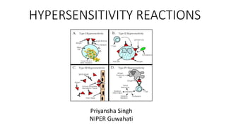
- 2. What is a Hypersensitivity reaction ? • Exaggerated immunologic responses in response to an antigen or allergen • Resulting in tissue injury/cytotoxicity & other pathological changes in blood vessels or other tissues • Our immune system identifies between foreign/ non self & self-molecules & the failure to identify the difference causes hypersensitivity as the immune cells release inflammatory mediators • There are 4 types of Hypersensitivity Type-1: Antibody IgE = Mast cells = release of histamine/PG/LT; quick onset after exposure Type-2: Cytotoxic= IgG/IgM mediated= Attack antigen present on self cells Type-3: Immune complex deposition = complement Type-4: Delayed/ CD4 T (helper) & rarely CD8 T cell mediated
- 3. How tissue injury can be caused ? 1. Release of vasoactive substances like leukotrienes, histamine, prostaglandins etc. 2. Phagocytosis 3. Activation of inflammatory & cytolytic components of complement system (mostly seen in SLE) 4. Release of cytokines, proteolytic enzymes & other inflammatory mediators which destroys the overall cellular structure
- 4. Hypersensitivity reaction Type 1 • Completely due to antigen/allergen/immunogen • Allergens interact with our body cells and the antibodies fight and bind against that specific antigen • Mainly IgE mediated. • IgE recognizes soluble antigens ----> which triggers MAST CELLS to DEGRANULATE to release HISTAMINE hence causing allergic symptoms
- 5. Hypersensitivity reaction Type 2 • By IgG and rarely IgM • Recognize & bind to the cellular self antigens or tissue specific antigens (present on host cells) cytotoxicity/tissue damage. • Includes the recognition and binding to the self-antigen (autoimmunity) • This type of hypersensitivity reaction occurs by 3 mechanisms 1. Antibody dependent cell mediated cytotoxicity 2. Antibody & complement mediated destruction (e.g.- Autoimmune hemolytic anemia, mismatched transfusion of blood cells) 3. Target cell dysfunction (e.g.- Graves disease, Myesthenia gravis, pernicious anemia)
- 6. b) Opsonization Antibody & complement mediated destruction Here the target cells express antigens with which antibody binds to which activates the complement system which activates & cleaves several complement proteins specifically the complement protein C1 causing activation & cleavage of C3 to C3a & C3b which leads to the formation of membrane attack complex of C9 proteins which attack the target cell by lysis. Antibodies & the complement proteins C3b attach to the target cell and act as Opsonins- they attract macrophages to phagocytose the target cell C3b Target cell
- 7. Antibody dependent cell mediated cytotoxicity ADCC can occur when certain medications are involved and they are called HAPTENS which bind to certain tissue and when Ab binds to these tissue cells that can activate complement proteins C5a & C3a They attract WBCs to the site & carry out ADCC E.g.- Medication induced hemolytic anemia, transplant rejection Target cells destroyed by toxic granules It involves antibodies, that bind to antigen on target cells which get recognized by immune cells like Macrophages, Natural killer cells, Eosinophils & Neutrophiles using Fc receptors. Once bound to antibody these cells release cytotoxic substances or granules onto the target cells Cell Death
- 8. Target cell dysfunction lets understand it using an example of Graves disease Hyperthyroidism In Grave’s disease, Follicular cells of thyroid glands are involved & they have TSH receptors. Normal- TSH binds to TSH-R & promotes the production of Thyroid hormone T3 & T4. But in Grave’s disease, there is a presence of auto-antibodies- Thyroid Stimulating Immunoglobulins. Bind to TSH receptors & over activate them as they are not subjected to –ve feedback
- 9. Hypersensitivity Reaction Type 3 When blood soluble antigen (medication, venom, vaccine) enters blood circulation IgM/IgG detect & bind to them forming antigen antibody/ immune complex Attach to endothelium of blood vessel wall (systemic) or other tissues like synovial of joint/ glomerulus of kidney/ epithelial lining of alveoli in lungs (localized) Activation of complement cascade C5a C3b Activation of complement cascade starting with C1 which leads to activation of proteins C5a & C3b Release of chemotactic factors recruitment of neutrophils Neutrophils utilize Fc receptors & complement receptors to bind to Antibody and C3b which deposits on tissue Upon deposition, neutrophils release cytotoxic granules containing ROS causing cell/ tissue damage
- 11. Hypersensitivity reaction Type 4- cell mediated 1. Aka Delayed type HS reaction- cell mediated 2. 2 phases- Sensitization Effector 3. Requires Incubation time to develop for 1-2 weeks then symptoms are shown but on repeated exposure with the same antigen the response takes lesser window period. 4. Sensitization- Antigen on engulfment with macrophage/ APC showcase the fragment of that antigen to the rest of the immune system cells by Major Histocompatibility Complex-II (MHC II) to alert immune cells. Th cells sense the presence of that antigen by T cell receptor & get differentiated into more Th cells 5. Effector- Active Th cells activate other macrophages cytotoxicity/phagocytosis 6. E.g.- TB Test, Poison Ivy
- 12. Binds at proteins in skin & alters them Recognized by immune system as foreign due to alterations Recognized & presented by APC and presented to naïve Th cells APCs are antigen presenting cells where antigens/ altered proteins are expressed by MHC-II and it gets paired up with Th cells and APC then releases Il-12 & Il-6 Differentiation of Th cells into type 1 Th cell (Th1) & Th 17 cells Release of Ifn-g & Il-17 which activates macrophage Activated Macrophage Release of Il-1 & Tnf-a Expression of receptor on endothelial cell to recruit more leukocytes to endothelium Perform phagocytosis & release ROS & lysozymes VASCULAR DERMATITIS Ivy poison (Urushiol) Lets understand it using an example of Poison Ivy