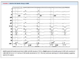
This document discusses His bundle pacing (HBP), which aims to engage the normal conduction system through the His-Purkinje network. HBP may provide physiological stimulation compared to right ventricular pacing. The document covers anatomy and physiology of the His bundle, techniques for selective vs. nonselective HBP, indications including AV node ablation and cardiac resynchronization, long-term outcomes, and challenges such as high capture thresholds and lead revisions. While HBP shows promise, further studies are needed to validate its long-term safety, reliability and impact on arrhythmias.
- Left bundle branch pacing (LBBP) was attempted in 87 patients needing a pacemaker, successfully implanting the lead in the left bundle branch in 80.5% of patients.
- LBBP resulted in a shorter paced QRS duration and left ventricular activation time compared to right ventricular septal pacing. It also had a lower pacing threshold and higher R-wave amplitude.
- The study demonstrated the feasibility of LBBP with potential advantages over traditional right ventricular and His bundle pacing, including more physiological ventricular activation. However, larger long-term studies are still needed to understand the full benefits of LBBP.
This document discusses techniques for localizing the site of origin of ventricular tachycardia based on electrocardiogram characteristics. It describes that right ventricular outflow tract tachycardias typically present with left bundle branch block morphology while left ventricular sites may present with either right or left bundle branch block depending on exit site. Specific leads are discussed that can provide clues about anterior vs posterior, septal vs free wall origin within the outflow tracts. Other areas like fascicles, papillary muscles and mitral/tricuspid annuli are also summarized.
New Heart Failure modalities: HIS Bundle Pacing & Cardiac Contractility Modul...NAJEEB ULLAH SOFI
His bundle pacing activates the ventricles via the native His-Purkinje system, resulting in true physiological pacing, and, therefore, is a promising alternate site for pacing in bradycardia and traditional CRT indications in cases where it can overcome left bundle branch block
CCM signals do not elicit a new contraction; rather, they influence the biology of the failing myocardium
Fascicular ventricular tachycardia is a type of ventricular tachycardia that originates in the Purkinje fibers near the fascicles of the left bundle branch. It typically presents in young males as palpitations or dizziness. The electrocardiogram shows a narrow QRS complex tachycardia with right bundle branch block morphology that is sensitive to verapamil. The reentrant circuit involves abnormal Purkinje fibers as the slow pathway and the left posterior fascicle as the fast pathway. Radiofrequency ablation targeting Purkinje potentials in the left ventricular septum is effective for treatment.
This document describes equipment, catheters, and basic intervals used in electrophysiology (EP) studies. It discusses radiographic tables, EP equipment like cardiac stimulators and mapping/ablation catheters. Patient preparation includes fasting, IV access, monitoring equipment. EP catheters come in different sizes and have electrodes for recording electrical activity. Basic intervals measured include P wave to atrial interval, atrial-His bundle interval, His-ventricular interval, and sinus node recovery time. Drive train stimulation with single, double, or triple extra stimuli is used. The document continues with further discussions of EP protocols, arrhythmias, ablation, and pre-excitation pathways.
This document discusses localization of accessory pathways using electrocardiography. It describes that accessory pathways can be located in eight anatomical positions along the tricuspid and mitral valve annuli. Several algorithms are proposed to determine the location based on delta wave polarity and amplitude in various leads. The most accurate is the Arruda approach, which uses step-wise analysis of delta wave characteristics in leads I, II, aVL, aVF and V1 to identify the specific accessory pathway location with 90% sensitivity and 99% specificity. Characteristic ECG patterns are presented that help localize right anteroseptal, right posteroseptal, left lateral and right free wall accessory pathways.
This document discusses various electrocardiogram (ECG) criteria that can be used to differentiate ventricular tachycardia (VT) from supraventricular tachycardia (SVT) with aberrant conduction. It reviews criteria proposed in several studies from 1965 to 2020, including Sandler's criteria on QRS morphology, Marriott's chest lead concordance, Wellens' criteria on QRS duration and morphology, and the Brugada algorithm - the first multistep algorithm for differentiation. More recent automated methods using computerized ECG interpretation may also help clinicians establish the likelihood of VT.
This document discusses trans-septal puncture, which involves puncturing the septum between the right and left atria to access the left side of the heart. It outlines the evolving indications for trans-septal puncture including interventions for mitral valve disease, closure of defects, left atrial procedures, and arrhythmia ablation. The key steps are reviewed - having the proper anatomical landmarks, hardware including sheaths and needles, and imaging guidance. Complications are discussed and how to successfully perform the puncture is summarized as being familiar with the anatomy, hardware, and vigilance for potential complications.
The document discusses techniques for transseptal puncture (TP). It provides a brief history of septal puncture dating back to the 1950s. It describes the embryology and anatomy of the interatrial septum. The common landmarks and techniques used for fluoroscopy-guided TP are described, including Inoue's angiographic and Hung's modified fluoroscopic methods. Indications for TP include percutaneous mitral commissurotomy and electrophysiology studies. The basic steps of the TP procedure and potential complications are summarized.
Conduction system pacing as resynchronizationSergio Pinski
This document discusses various cardiac resynchronization therapies including biventricular pacing, His bundle pacing, and left bundle branch pacing. It notes that while biventricular pacing achieves imperfect resynchronization in about 20-30% of patients, His bundle pacing has been shown to fully correct left bundle branch block in some cases. Left bundle branch pacing is also discussed as an emerging therapy. The document reviews several studies comparing different resynchronization approaches and outlines criteria for assessing left bundle branch capture during pacing. It concludes by proposing a framework for selecting among resynchronization therapies based on the underlying conduction abnormality.
This document provides guidance on pre-procedural preparation and tips for CRT implantation. It discusses evaluating patients before implantation through imaging, labs, and clinical assessments. Key steps in implantation include accessing veins, placing the RV lead first usually in the septum, cannulating the coronary sinus, performing venograms to select target veins, and placing the LV lead aiming for the lateral or posterior wall. Positioning the leads to maximize electrical delay and hemodynamics is important.
This document discusses various echocardiographic scoring systems used to assess mitral valve anatomy and predict outcomes of percutaneous balloon mitral valvuloplasty (PBMV). The Wilkins score and Commissural Calcification score are described in detail. The Wilkins score grades leaflet thickening, mobility, calcification and subvalvular involvement on a scale of 4-16. A score ≤8 indicates favorable anatomy for PBMV. The Commissural Calcification score quantifies calcification at each commissure. Other discussed scores include the Cormier score, RT-3DE score, Chen score, Reid score and Nobuyoshi score. Limitations of the scoring systems and ideas for an ideal future scoring
The document discusses the history and technique of transseptal puncture (TSP). It describes how TSP provides direct access to the left atrium and has become a routine skill for electrophysiologists performing procedures like atrial fibrillation ablation. The technique involves using a Brockenbrough needle and Mullins sheath inserted via the femoral or jugular vein to puncture the interatrial septum, usually at the fossa ovalis. Landmarks, equipment, steps of the procedure, challenges, and complications are reviewed in detail. The summary emphasizes the importance and increasing use of TSP as well as reviews key aspects of the technique and potential complications.
1) The document discusses various cases involving cardiac resynchronization therapy (CRT) implantation and troubleshooting.
2) Techniques described include accessing the coronary sinus using guidewires when dissection occurs and using a steerable catheter to engage alternative cardiac veins.
3) Optimizing outcomes involves addressing issues like phrenic nerve stimulation, adjusting atrioventricular delays, and replacing unstable leads over time.
This document provides an overview of electrophysiologic studies, including the setup, equipment, procedures, and interpretation. It discusses the basics of the EP lab, including mapping, recording, and ablation systems. It describes patient preparation and catheter placement. It explains concepts such as sinus node recovery time, AH and HV intervals, pacing protocols, and maneuvers to diagnose common arrhythmias like AVNRT and AVRT. Specific examples are given of electrograms during sinus rhythm, pacing, and induced arrhythmias. Ablation of AVNRT is discussed as an example of using these techniques.
1. Complete chordal preservation during mitral valve surgery aims to preserve left ventricular function and geometry by maintaining the continuity between the mitral annulus and left ventricular wall.
2. Early studies found benefits to chordal preservation such as improved cardiac output, exercise capacity, and survival rates.
3. Various surgical techniques have been developed to allow for implantation of an adequate sized prosthesis while preventing complications like left ventricular outflow tract obstruction.
This document discusses the limitations and techniques for assessing right ventricular (RV) function using echocardiography. It is difficult to accurately evaluate RV volume, delineate borders, and image the entire RV using echocardiography due to its complex crescent shape. However, the document recommends using RV fractional area change, tricuspid annular plane systolic excursion, tissue Doppler S' velocity, and Tei index to quantitatively assess RV systolic function as they are reproducible methods. RV dimensions, wall thickness, and outflow tract size can also provide information on RV size and function. Assessment of RV diastolic function includes parameters like E/A ratio, E/E' ratio, and deceleration time.
1. Contrast agents enhance ultrasound images by increasing the backscatter of blood allowing visualization of small vessels. They oscillate non-linearly when exposed to ultrasound, emitting harmonic frequencies not seen in tissue.
2. Harmonic imaging exploits non-linear backscatter to identify the contrast agent signal and suppress tissue echoes, enabling visualization of microvascular flow.
3. Care must be taken in injecting contrast agents to avoid destroying bubbles, and infusion may provide a longer enhancement window for some studies compared to a bolus injection.
In-Stent Restenosis occurs when a blockage redevelops inside a stent placed in a coronary artery. The rate of restenosis is lower with drug-eluting stents (DES) compared to bare-metal stents or balloon angioplasty alone. For treatment of in-stent restenosis, repeat stenting with DES has shown success, though DES for prior DES restenosis remains controversial due to the risk of developing multiple layers of stent struts. Drug-coated balloons are an alternative to repeat stenting and have been shown to be effective for both bare-metal and drug-eluting stent in-stent restenosis. Ongoing randomized clinical trials are further evaluating
1. The document describes the MitraClip procedure for percutaneous mitral valve repair.
2. The MitraClip procedure involves inserting a clip via the femoral vein to grasp and repair the mitral valve leaflets in a technique similar to the surgical Alfieri repair.
3. The key steps involve transseptal puncture, advancing the clip delivery system into the left atrium, positioning the clip below the mitral valve, grasping the leaflets with the clip, and releasing the clip to create a double orifice mitral valve.
- The document discusses the Fontan procedure for univentricular heart defects. It covers the evolution of the Fontan concept from the original atriopulmonary connection to lateral tunnel and extracardiac conduit techniques. It also discusses indications for Fontan, complications such as arrhythmias and ventricular dysfunction, and strategies to optimize outcomes like fenestration.
This document provides an overview of segmental analysis for congenital heart disease. It discusses the key segments that are analyzed which include thoraco-abdominal situs, pulmonary situs, atrial situs, ventricular situs and looping, connections between segments (venous, atrioventricular, ventriculoarterial), and abnormalities that can occur in each segment. The document emphasizes evaluating each segment in a systematic, sequential manner to identify abnormalities.
Today, in addition to measurement of left ventricular ejection fraction, the simple 12-lead surface ECG remains the only evidence-based means of identifying patients who may obtain the substantial benefits of CRT
The document provides an overview of right ventricular assessment using echocardiography. It discusses normal RV anatomy, segmental nomenclature, and coronary supply. Key metrics for evaluating RV size, wall thickness, function, and pressures are outlined. Normal values and technical aspects of measuring RV dimensions, area/fractional area change, tricuspid annular plane systolic excursion, myocardial velocity, and diastolic function are summarized. Hemodynamic assessment of pulmonary pressures is also reviewed.
This document provides information about percutaneous transvenous mitral commissurotomy (PTMC), a procedure used to treat mitral stenosis. It discusses the stages and severity of mitral stenosis, indications and contraindications for PTMC, assessment of valve morphology, the PTMC procedure technique, instruments used, balloon size selection, post-procedure evaluation, complications, follow-up care, and long-term prognosis. PTMC is performed to improve the opening of a stenosed mitral valve by splitting the fused commissures using a balloon catheter, and is an important therapeutic option for treating symptomatic mitral stenosis.
Evaluation and management of Pacemaker malfunctionPRAVEEN GUPTA
The document discusses the evaluation and management of pacemaker malfunctions. It describes how to differentiate between various types of single chamber pacemaker malfunctions including pacing stimuli present with failure to capture, pacing stimuli present with failure to sense, and pacing stimuli absent. Common causes of these malfunctions are then outlined such as lead dislodgment, insulation defects, threshold increases, and undersensing. The document stresses the importance of obtaining baseline pacemaker data during initial programming and follow-up to properly diagnose malfunctions.
His Resynchronization VersusBiventricular Pacing inPatients With Heart Fail...Shadab Ahmad
This study tested the ability of HBP to deliver resynchronization and then compared the electromechanical effects of His resynchronization against conventional BVP, using high-precision hemodynamic assessment and noninvasive epicardial ventricular activation mapping
His Bundle Pacing Versus Cardiac Resynchronization
This document discusses His bundle pacing as an alternative to cardiac resynchronization therapy (CRT) for treating heart failure. Right ventricular pacing can cause left ventricular dyssynchrony, while CRT has limited success due to non-physiological biventricular pacing. His bundle pacing aims to restore normal conduction by capturing the native conduction system. Studies show His bundle pacing improves left ventricular function and reduces dimensions more than CRT. A randomized trial found His bundle pacing had superior electrical resynchronization compared to CRT. However, potential issues with His bundle pacing include lead dislodgement and increased capture thresholds over time. More research is still needed but His bundle pacing
Arrhythmia-induced cardiomyopathy (AIC) refers to left ventricular dysfunction caused by tachyarrhythmias or frequent ectopy. There are two types - type 1 is solely due to the arrhythmia, while type 2 involves an arrhythmia exacerbating an underlying cardiomyopathy. Successful treatment of the arrhythmia via catheter ablation or cardioversion can reverse the left ventricular dysfunction in type 1 AIC. Aggressive treatment with catheter ablation is recommended to eliminate the arrhythmia whenever possible in order to prevent or treat AIC.
Device therapy for heart failure monitoring and managementDIPAK PATADE
The document discusses device therapy for heart failure management and monitoring. It notes that heart failure results in high mortality and morbidity and places a significant financial burden on healthcare systems. Implantable devices like implantable cardioverter defibrillators (ICDs) and cardiac resynchronization therapy (CRT) play an important role in managing advanced heart failure by treating arrhythmias, improving morbidity and mortality, and increasing quality of life. Several landmark randomized controlled trials provided evidence of the benefits of CRT in reducing mortality and improving outcomes for heart failure patients.
Device therapy for heart failure monitoring and managementDIPAK PATADE
1) Device therapy such as ICDs and CRT play an important role in managing advanced heart failure by treating life-threatening arrhythmias and improving morbidity and survival.
2) Clinical trials such as MUSTIC, MIRACLE, and CARE-HF demonstrated that CRT improves symptoms, exercise capacity, quality of life and reduces mortality in patients with heart failure and ventricular dyssynchrony.
3) The COMPANION trial showed that CRT with or without an ICD provided greater benefits than medical therapy alone in reducing the risk of all-cause mortality and hospitalizations.
1) Hydrocephalus is a condition where excess cerebrospinal fluid accumulates in the brain's ventricles, increasing intracranial pressure. It can be treated with shunt surgery but complications are common.
2) Understanding cerebrospinal fluid dynamics is important for diagnosing hydrocephalus and predicting outcomes from shunt surgery. Cerebrospinal fluid flow and pulsatility are assessed using techniques like infusion tests and MRI.
3) Shunt selection is challenging in normal pressure hydrocephalus given variability in intracranial pressure and cerebrospinal fluid dynamics between patients. Adjustable valves allow non-invasive management of complications.
1. The document discusses supraventricular tachycardia (SVT), which refers to tachycardias originating above the ventricles, with a rate over 100 bpm. Common types include atrioventricular nodal reentrant tachycardia (AVNRT) and atrioventricular reentrant tachycardia (AVRT) involving an accessory pathway.
2. AVNRT and AVRT-AP are most commonly treated by catheter ablation, which uses radiofrequency energy to destroy the abnormal tissue causing the reentry circuits. Success rates for ablation of AVNRT and AVRT-AP are over 90% and the procedures have become increasingly safer and more effective.
Left ventriculography provides information about global and segmental left ventricular function, mitral valve regurgitation, ventricular septal defects, and structural abnormalities. It requires a pigtail or multipurpose catheter connected to a power injector. Contrast is injected into the left ventricle to outline the chamber and assess wall motion. Potential complications include arrhythmias, embolism, and endocardial staining. The procedure allows evaluation of conditions like myocardial infarction, hypertrophic cardiomyopathy, and left ventricular aneurysms.
This document discusses the use of ultrasound in critically ill patients. It aims to explain how ultrasound can guide management of hemodynamically unstable patients by rapidly evaluating for reversible causes of shock. The RUSH (Rapid Ultrasound in Shock) protocol is described, which involves using ultrasound to examine the heart (the pump), assess intravascular volume status (the tank), and check for issues with blood vessels (the pipes). Common pathologies that can be identified include cardiac tamponade, pulmonary embolism, hemorrhage, aortic dissection, and thrombosis. Examples of abdominal ultrasound findings in critical illnesses such as gangrenous cholecystitis, emphysematous cholecystitis, liver abscess
This document provides guidance on managing patients who do not respond to cardiac resynchronization therapy (CRT). It discusses that while CRT has been shown to reduce heart failure hospitalizations and mortality, 30-35% of patients do not adequately respond. The document then reviews factors that determine CRT response, including electrical and mechanical dyssynchrony. It also discusses different criteria used to define response and notes lack of agreement. Potential reasons for non-response are explored, including lead placement and individual patient factors. The document concludes by recommending approaches for evaluating and managing CRT non-responders.
This document discusses non-pharmacological management options for atrial fibrillation (AF), including ablation procedures. Pulmonary vein isolation is the cornerstone ablation strategy, as 95% of AF triggers originate from the pulmonary veins. During this procedure, radiofrequency energy is applied to electrically isolate the pulmonary veins from the left atrium. Complete isolation of the pulmonary veins has shown success rates of over 80% for preventing AF recurrence. Other ablation procedures discussed include modifying the atrioventricular node to control ventricular rate during AF and linear ablation techniques. The document also reviews the electrophysiological mechanisms of AF and the rationale behind different ablation strategies.
This document provides an overview of cardiac resynchronization therapy (CRT), including indications, assessment of dyssynchrony, rationale/mechanism, trials, procedures, and programming. It discusses the types and assessment of cardiac dyssynchrony using ECG, echocardiography, MRI, and nuclear imaging. Key trials on CRT are summarized, showing benefits for heart failure patients with reduced ejection fraction and wide QRS duration or echocardiographic evidence of dyssynchrony even in narrow QRS.
Cardiac output can be measured through various invasive and non-invasive methods. The pulmonary artery catheter using thermodilution is still considered the gold standard but is invasive. Minimally invasive methods include lithium dilution, pulse contour analysis devices, esophageal Doppler, and transesophageal echocardiography. Non-invasive options include partial gas rebreathing, thoracic bioimpedance, and Doppler ultrasound. The ideal monitor is accurate, continuous, non-invasive and provides reliable measurements during different physiological states.
Cardiac output can be measured through various invasive and non-invasive methods. The pulmonary artery catheter using thermodilution is still considered the gold standard but is invasive. Minimally invasive methods include lithium dilution, pulse contour analysis devices, esophageal Doppler, and transesophageal echocardiography. Non-invasive methods include partial gas rebreathing, thoracic bioimpedance, and Doppler ultrasound. The ideal monitor is accurate, continuous, non-invasive and provides reliable measurements during different physiological states.
This document provides an overview of cardiac resynchronization therapy (CRT). It discusses how conduction delays can lead to electromechanical dyssynchrony and impair the heart's function. CRT aims to improve this synchrony and thereby improve systolic and diastolic function. The document outlines different types of dyssynchrony and methods to assess it, including echocardiography. Current guidelines recommend CRT for symptomatic heart failure patients with low ejection fraction and wide QRS duration. The implantation procedure involves placing right atrial/ventricular leads and a left ventricular lead via the coronary sinus.
Cardiac resynchronization therapy (CRT) uses electrical pacing of both ventricles to coordinate their contractions and improve heart function in patients with heart failure. It is recommended for patients with left ventricular ejection fraction below 35%, prolonged QRS duration over 150ms, and evidence of electrical or mechanical dyssynchrony. CRT works by pacing both ventricles simultaneously to resynchronize their contractions, improving heart pumping ability and reducing symptoms. About 30% of patients do not respond adequately, often due to factors like lack of sufficient dyssynchrony, lead placement issues, or scar tissue in the ventricles.
Acute myocardial infarction associated with right bundle branch block and cha...YasserMohammedHassan1
Acute myocardial infarction may be associated right bundle branch block.
Accompanied trifascicular heart block had pre-streptokinase left anterior fascicular block
with left axis deviation and post-streptokinase left posterior fascicular block with right axis
deviation.
Recommendations for Cardiac Chamber Quantification by Echocardiography in Adults: An Update from the American Society of Echocardiography and the European Association of Cardiovascular Imaging
Surgeons view on AHA/ACC Coronary revascularisation guidelines .pptxChaitanya Chittimuri
The document summarizes perspectives from surgeons on the 2021 ACC/AHA guidelines for coronary artery revascularization. There are three main areas of concern: 1) Downgrading CABG for treatment of three-vessel CAD, 2) Not recognizing superior long-term benefits of CABG over PCI, and 3) Awarding a high recommendation for radial artery grafts without sufficient evidence. The surgeons argue that studies like ISCHEMIA should not decrease CABG recommendations for multivessel CAD and that earlier studies found CABG superior to PCI. They are also concerned about arbitrarily downgrading CABG and only including one side of the heart team in guidelines development.
Mercurius is named after the roman god mercurius, the god of trade and science. The planet mercurius is named after the same god. Mercurius is sometimes called hydrargyrum, means ‘watery silver’. Its shine and colour are very similar to silver, but mercury is a fluid at room temperatures. The name quick silver is a translation of hydrargyrum, where the word quick describes its tendency to scatter away in all directions.
The droplets have a tendency to conglomerate to one big mass, but on being shaken they fall apart into countless little droplets again. It is used to ignite explosives, like mercury fulminate, the explosive character is one of its general themes.
These lecture slides, by Dr Sidra Arshad, offer a simplified look into the mechanisms involved in the regulation of respiration:
Learning objectives:
1. Describe the organisation of respiratory center
2. Describe the nervous control of inspiration and respiratory rhythm
3. Describe the functions of the dorsal and respiratory groups of neurons
4. Describe the influences of the Pneumotaxic and Apneustic centers
5. Explain the role of Hering-Breur inflation reflex in regulation of inspiration
6. Explain the role of central chemoreceptors in regulation of respiration
7. Explain the role of peripheral chemoreceptors in regulation of respiration
8. Explain the regulation of respiration during exercise
9. Integrate the respiratory regulatory mechanisms
10. Describe the Cheyne-Stokes breathing
Study Resources:
1. Chapter 42, Guyton and Hall Textbook of Medical Physiology, 14th edition
2. Chapter 36, Ganong’s Review of Medical Physiology, 26th edition
3. Chapter 13, Human Physiology by Lauralee Sherwood, 9th edition
Travel Clinic Cardiff: Health Advice for International TravelersNX Healthcare
Travel Clinic Cardiff offers comprehensive travel health services, including vaccinations, travel advice, and preventive care for international travelers. Our expert team ensures you are well-prepared and protected for your journey, providing personalized consultations tailored to your destination. Conveniently located in Cardiff, we help you travel with confidence and peace of mind. Visit us: www.nxhealthcare.co.uk
Cell Therapy Expansion and Challenges in Autoimmune DiseaseHealth Advances
There is increasing confidence that cell therapies will soon play a role in the treatment of autoimmune disorders, but the extent of this impact remains to be seen. Early readouts on autologous CAR-Ts in lupus are encouraging, but manufacturing and cost limitations are likely to restrict access to highly refractory patients. Allogeneic CAR-Ts have the potential to broaden access to earlier lines of treatment due to their inherent cost benefits, however they will need to demonstrate comparable or improved efficacy to established modalities.
In addition to infrastructure and capacity constraints, CAR-Ts face a very different risk-benefit dynamic in autoimmune compared to oncology, highlighting the need for tolerable therapies with low adverse event risk. CAR-NK and Treg-based therapies are also being developed in certain autoimmune disorders and may demonstrate favorable safety profiles. Several novel non-cell therapies such as bispecific antibodies, nanobodies, and RNAi drugs, may also offer future alternative competitive solutions with variable value propositions.
Widespread adoption of cell therapies will not only require strong efficacy and safety data, but also adapted pricing and access strategies. At oncology-based price points, CAR-Ts are unlikely to achieve broad market access in autoimmune disorders, with eligible patient populations that are potentially orders of magnitude greater than the number of currently addressable cancer patients. Developers have made strides towards reducing cell therapy COGS while improving manufacturing efficiency, but payors will inevitably restrict access until more sustainable pricing is achieved.
Despite these headwinds, industry leaders and investors remain confident that cell therapies are poised to address significant unmet need in patients suffering from autoimmune disorders. However, the extent of this impact on the treatment landscape remains to be seen, as the industry rapidly approaches an inflection point.
Travel vaccination in Manchester offers comprehensive immunization services for individuals planning international trips. Expert healthcare providers administer vaccines tailored to your destination, ensuring you stay protected against various diseases. Conveniently located clinics and flexible appointment options make it easy to get the necessary shots before your journey. Stay healthy and travel with confidence by getting vaccinated in Manchester. Visit us: www.nxhealthcare.co.uk
Kosmoderma Academy, a leading institution in the field of dermatology and aesthetics, offers comprehensive courses in cosmetology and trichology. Our specialized courses on PRP (Hair), DR+Growth Factor, GFC, and Qr678 are designed to equip practitioners with advanced skills and knowledge to excel in hair restoration and growth treatments.
2. Introduction
• Despite years of successful pacing therapy,
persistent debate regarding optimal ventricular
pacing sites.
• Initial ventricular-only pacing devices provided
adequate rate support but were not synchronized
to atrial contraction, and led to negative
hemodynamic consequences including an
increased risk of heart failure (HF) and atrial
fibrillation
3. Introduction
Even atrioventricular (AV) synchronized pacing
delivered at the right ventricular (RV) apex,
however, was noted to worsen contractile
function in many patients.
Eventually, the connection between the degree
of right ventricular apical (RVA) pacing and
cardiac dysfunction became well established.
4. • Pursuit of alternate pacing sites has included the
RV septum, the RV outflow tract, and the left
ventricle (LV).
• Although biventricular pacing has unequivocally
improved HF outcomes and reduced mortality in
patients with left bundle branch block (LBBB) and
severe LV systolic dysfunction, its role in patients
with preserved LV systolic function remains
unresolved.
5. An ideal physiological approach to ventricular
stimulation should engage the normal
conduction through the His-Purkinje
conduction system.
• In this seminar we will discuss anatomy,
physiology, and clinical role of permanent HBP.
6. Anatomy of HIS Bundle
• Anatomical continuation of the AV node.
• Provide connection for electrical signals from
the AV node to right and left ventricles
through right and left bundle branches,
respectively.
7. • There are 3 common variations of the His
bundle relative to the membranous part of
the ventricular septum.
8. Type I
• 46.7%
• The His bundle consistently coursed along the
lower border of the membranous part of the
interventricular septum, but was covered with
a thin layer of myocardial fibers spanning from
the muscular part of the septum.
9.
10. TYPE II
• 32.4% cases
• The His bundle was apart from the lower
border of the membranous part of the
interventricular septum and ran within the
interventricular muscle
11.
12. TYPE III
• 21% cases,
• The His bundle was immediately beneath the
endocardium and coursed onto the
membranous part of the interventricular
septum (naked AV bundle)
13.
14.
15. Physiological properties of HIS bundle
• The bulk of the His bundle is comprised of cells
that eventually course into the left bundle
branches (only a small number enter the right
branch).
• The cells that make up the His-Purkinje fibers are
broader and shorter than the usual working
myocardial cells with relatively few myofibrils.
• These cells are elongated and oblong in shape,
and make contact predominantly at their
terminal ends and to a lesser extent across the
lateral margins.
16. • These cells are partitioned intricately by collagen
fibers; in fact, longitudinal division of the His
bundle by collagen makes it unique from a
histological standpoint when compared with the
AV node and the working myocardium.
• The collagen may minimize or even prevent
lateral spread of the propagated impulse, while
the compartmentalized tissue with specialized
intercellular connections would facilitate rapid
longitudinal spread of the propagated impulse.
17. • An implication of these findings is that some
patients with His-Purkinje conduction disease
(HPCD) may have relatively proximal disease,
and that pacing distal to the site of block
might overcome the block and narrow the
QRS.
18.
19.
20. SELECTIVE HBP
During S-HBP, ventricular activation occurs directly and
completely over the HPS and is accompanied by the
following :
• The pacing stimulus to QRS (S-QRS) onset interval is
equal to the native His-QRS onset interval (H-QRS).
• However, in patients with HPCD, the S-QRS interval can
be shorter than the H-QRS intervals, as in patients with
BBB or HV block due to capture of latent fascicular
tissue.
• The local ventricular electrogram on the pacing lead
will be discrete from the pacing artifact.
21. • The paced QRS morphology is the same as the
native QRS morphology. In patients with HPCD,
the paced QRS duration may be narrower than
the native QRS with BBB or the escape rhythm.
• Usually a single capture threshold (His capture) is
observed. However in patients with HPCD, 2
distinct His capture thresholds—with and without
correction of underlying BBB—may be seen .
22.
23. NONSELECTIVE HBP
During NS-HBP, there is culmination of both His
bundle and ventricular capture.
• The S-QRS interval is usually zero, as there is no
isoelectric interval between pacing stimulus and
QRS due to the presence of a pseudo-delta wave
(due to local myocardial capture).
• The local ventricular electrogram is directly
captured by the pacing stimulus and is not seen
as a discrete component.
24. • The paced QRS duration will usually be longer
than the native QRS duration by the H-QRS
interval, and the overall electrical axis of the paced
QRS will be concordant with the electrical axis of
the intrinsic QRS.
• In patients with HPCD, the paced QRS duration
may be narrower than the native QRS due to
correction of underlying BBB.
25. • There will usually be 2 distinct capture thresholds –
right ventricular and His capture.
• The His capture threshold may be lower or higher
than the ventricular capture threshold.
• In patients with HPCD, 3 distinct capture thresholds
may be observed in varying combination (RV capture,
His capture with correction of BBB, and His capture
without correction of BBB).
26.
27. Implantation technique
• Early studies used conventional screw-in leads
utilizing manually shaped stylets targeting the
Hisian region identified by a mapping
electrophysiology catheter.
• Subsequent studies have demonstrated the
improved success rates of HBP using a dedicated
4.1 Fr lead (SelectSecure 3830) with an exposed
screw, delivered through a steerable catheter
(SelectSite C304-L69, Medtronic), or fixed curve
sheath (Medtronic C315His).
28.
29.
30.
31. Indications of HIS Bundle Pacing
• AV node block
• Infra nodal block(Intra or infra His block)
• Cardiac resynchronisation therapy
• HIS bundle pacing in RBBB and heart failure
32. HIS BUNDLE PACING FOR AV NODE
ABLATION
• ACC/AHA/HRS AF practice guidelines
recommend that AV junction ablation with
permanent ventricular pacing is a reasonable
strategy to control heart rate in AF when
pharmacological therapy is inadequate and
rhythm control cannot be achieved (Class IIa,
Level of Evidence: B)
33. AV BLOCK AND HBP
• While the feasibility of permanent HBP in
patients with AV nodal block is expected,
surprisingly high numbers of patients with
infranodal block can be corrected with HBP
• The postulated mechanisms for this recruitment
of distal His and bundle branches in patients with
intra-His block are: longitudinal dissociation in
the His bundle with pacing adjacent or distal to
the site of delay/block
34.
35. HIS BUNDLE PACING FOR CARDIAC
RESYNCHRONIZATION THERAPY
• Despite the development of sophisticated tools to
facilitate implant and intraprocedural strategies ,rates
of nonresponse to CRT remain high—between 30% and
40%.
• In addition, rates of implant failure for CRT range
between 5% and 9%, in part due to high rates of CS
lead dislodgement (3% to 7% reported across major
trials)
• In light of this, alternative strategies to achieve
resynchronization have gained momentum, including
endocardial LV lead pacing, “wireless” LV lead
stimulation, and permanent HBP.
36.
37.
38. Permanent His Bundle Pacing for Cardiac
Resynchronization Therapy in Patients With Heart
Failure and RBBB
39.
40. Long term outcome of HIS Pacing
• Compared with RVA pacing, HBP has been
associated with improved fractional shortening,
dP/dt, LVEF, and myocardial performance index
(Tei index).
• Also, improvement in interventricular
electromechanical delay, intraventricular
dyssynchrony, systolic diastolic electromechanical
delay, LV isovolumetric contraction and relaxation
times, and LV ejection time have been
demonstrated.
41.
42. HBP: CLINICAL CHALLENGES
CAPTURE THRESHOLDS-
His capture thresholds >2 V at 1 ms may be seen in 10%
of patients at implant.
Vijayaraman et al. reported that His capture thresholds
remained relatively stable during 5-year follow-up of
75 patients (1.35 +_0.9 V at implant vs. 1.62+_ 1.00 V
at 0.5 ms; p < 0.05).
An increase in chronic pacing threshold >1 V from
baseline was noted in 9 patients in HBP compared with
6 patients in RVP (12% vs. 6%; p = 0.04)
43. LEAD REVISIONS
• Vijayaraman et al. , acute loss of capture
occurred in 2 of 100 patients with AV block
and HBP.
• Lead revisions were required in 3 additional
patients at 2 to 6 months post-implant due to
progressive increases in capture threshold for
a lead revision rate of 5%.
44. • In a long-term study of 75 patients with HBP,
lead revisions were required in 5 patients
(6.7%), 4 of whom underwent successful lead
replacement at the His bundle region even as
late as 5 years after the initial implant.
45. • Acute increase in HBP threshold or loss of
capture is most likely due to inadequate
fixation of the HBP lead.
• The mechanism for delayed increase in HBP
threshold during longer-term follow-up is less
clear.
• It is likely that due to the anatomical proximity
of the loop of the HBP lead, the tricuspid valve
motion causes slow unhinging of the lead
46. BATTERY DEPLETION
• Recent studies have demonstrated that the
majority of patients undergoing HBP do well
without need for early generator changes .
• In patients undergoing CRT with HBP, capture
thresholds required to correct underlying BBB
are often higher, and early battery depletion
can still be a major obstacle
47. DEVICE FOLLOW-UP
• During follow-up, assessment of His bundle
capture using multilead ECG (preferably 12-
lead) is recommended.
• At 3-month follow-up, the pacing output is
programmed to at least 1 V above the His
capture threshold, as confirmed with
multilead ECG rather than at twice-safety
margin, to conserve battery life.
48. FUTURE DIRECTIONS
• permanent HBP may be an attractive option
for physiological pacing in several groups of
patients, its reliability and long-term
performance are yet to be fully validated in
large prospective studies.
• Particularly relevant are patients with
infranodal, intra-Hisian AV block and BBB,
where long-term safety of HBP has not been
well studied.
49. • In such patients, should a backup RV lead be
placed with HBP?
• What happens to the His bundle when it is
traumatized by the screw on the tip of the
lead in the long term?
• Can a second His Bundle pacing lead be placed
successfully if the earlier lead fails in the long
run?
50. •Beyond implant, what are the implications of extracting
a chronic HBP lead?
•And beyond pacing hemodynamics, what is the impact
of HBP on arrhythmia?
• Does HBP reduce the risk of ventricular
tachyarrhythmias in the presence of myocardial scar?
51. CONCLUSIONS
• HBP, an attractive mode of physiological
pacing with significant promise for future
applications in patients who are traditional
candidates for RV pacing as well as CRT
• Widespread adaptation of this technique is
dependent on the improvement of tools and
further validation of its efficacy in large
randomized clinical trials.