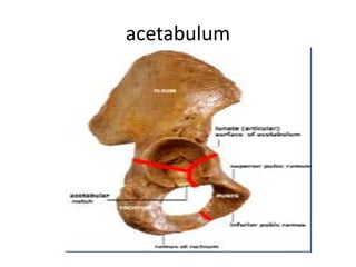
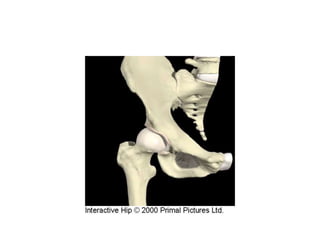
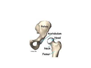
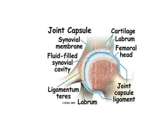
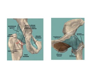
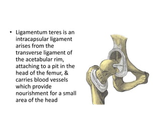
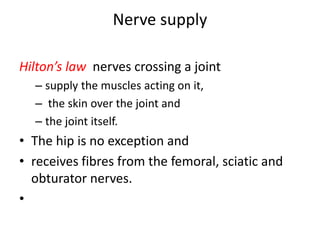
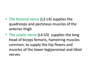
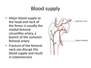
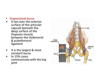
Hip joint anatomy
Dr Md Ruhul Amin
Assistant Registrar
Dept of Medicine
JRRMCH,SYLHET.
Surface anatomy
Hip joint
• Hip joint is a ball-and-socket joint that is
relatively fixed to the body by the pelvic girdle
• Largest and strongest joint of body
• Structures of the hip can be divided into
several categories, These include;
–bones and joints
–capsules and ligaments
–muscles
–nerves
–blood vessels
–bursae
acetabulum
• Acetabulum is deficient inferiorly by acetabular
notch which is bridged with transverse acetabulur
ligament.
• Ligamentum teres passess through acetabular
notch which goes to fovea on the femoral head
• Articular cartilage covers the horse-shoe shaped
articular surface of acetabulum
• The center is free of cartilage-acetabular fossa
which is filled with fibrofatty tissue.
Acetabular labrum:
• fibrocartilage
• Deepens the acetabulum
• Distributes stresses to the joint during joint
loading
• Maintains intra-articular pressure
Femur head
• The femoral head articulates with the cup-
shaped (cotyloid) acetabulum,
• its Centre lying a little below the middle third
of the inguinal ligament.
• The femoral head is covered by articular
cartilage, except over the rough pit where the
ligamentum teres is attached
SYNOVIAL MEMBRANE
• Starting from the femoral articular margin, the
synovial membrane covers
– intracapsular part of the femoral neck,
– internal surface of the capsule
– acetabular labrum,
– ligamentum teres and
– fat in the acetabular fossa.
capsule
• There is a strong dense fibrous capsule
• This is attached proximately to the
acetabulum, the glanoid & the transverse
ligament
• Distally it covers the lateral margin of the
femoral head & most of the neck
• Anteriorly it is attached to the
intertrochanteric line & posteriorly to the neck
just above this
ligament
Capsule is reinforced by
• iliofemoral ligament,-infront
• pubofemoral ligament- inferiorly
• ischiofemoral ligament- behind
• iliofemoral (Y-shaped ligament of Bigelow)—
which arises from the anterior inferior iliac spine,
bifurcates, and is inserted at each end of the
trochanteric line (Fig. 167);
• pubofemoral—arising from the iliopubic junction
to blend with the medial aspect of the capsule;
• ischiofemoral—arising from the ischium to be
inserted into the base of the greater trochanter.
the iliofemoral is by far the strongest and resists
hyperextension
• Ligamentum teres is an
intracapsular ligament
arises from the
transverse ligament of
the acetabular rim,
attaching to a pit in the
head of the femur, &
carries blood vessels
which provide
nourishment for a small
area of the head
Muscle
Muscles causing movement of the Hip
joint
Nerve supply
Hilton’s law nerves crossing a joint
– supply the muscles acting on it,
– the skin over the joint and
– the joint itself.
• The hip is no exception and
• receives fibres from the femoral, sciatic and
obturator nerves.
•
• The femoral nerve (L2-L4) supplies the
quadriceps and pectineus muscles of the
anterior thigh
• The sciatic nerve (L4-S3) supplies the long
head of biceps femoris, hamstring muscles
common, to supply the hip flexors and
muscles of the lower leg(peroneal and tibial
nerves
• superior gluteal nerve (L4,5)- gluteus medius,
minimus and TFL.
• Inferior gluteal nerve (L5, S1) - gluteus maximus.
• The obturator nerve (L2-4)
-anterior division-- hip joint and adductor longus,
brevis, and gracilis,
- posterior division obturator internus and adductor
magnus.
Blood supply
• Major blood supply to
the head and neck of
the femur is usually the
medial femoral
circumflex artery, a
branch of the common
femoral artery
• Fracture of the femoral
neck can disrupt the
blood supply and result
in osteonecrosis
bursae
• Approximately 18 bursae have been identified
• Clinically most important
– Iliopectineal or iliopsoas bursa
– Trochanteric bursa
– Ischiogluteal bursa
• Iliopectineal bursa
• It lies over the anterior
surface of the articular
capsule beneath the
deep surface of the
iliopsoas muscle
between the iliofemoral
& pubofemoral
ligament
• It is the largest & most
constant bursa
• In 15% cases
communicate with the hip
joint
Trochanteric bursa
• Trochanteric bursa
– Situated between the posterolateral surface of the
greater trochanter & the gluteus maximus muscle
– Actually comprises three bursae, the principal
being the gluteus maximus bursae; the bursae of
gluteus medius & gluteus minimus are at sites of
the prospective tendon attachment
• Ischiogluteal bursa
– Over the ischial
tubersity & overlies
the sciatic nerve
– Facilitates gliding of
the gluteus maximus
over the tuberosity
Criteria for Diagnosis of Trochanteric
Bursitis
1.Lateral hip pain
2.Distinct tenderness about the greater trochanter
3.Pain at the extreme of rotation, abduction, or
adduction,especially positive Patrick-FABERE test
4.Pain on hip abduction against resistance
5.Pseudoradiculopathy–pain radiating down the lateral
aspect of the thigh
6.Patrick-FABERE (Flexion, abduction, external rotation,
extension)
Need first 2 criteria plus one of the remaining criteria to
make diagnosis
.
thank you

hipjointanatomy

  • 1.
  • 2.
    Dr Md RuhulAmin Assistant Registrar Dept of Medicine JRRMCH,SYLHET.
  • 3.
  • 4.
    Hip joint • Hipjoint is a ball-and-socket joint that is relatively fixed to the body by the pelvic girdle • Largest and strongest joint of body
  • 5.
    • Structures ofthe hip can be divided into several categories, These include; –bones and joints –capsules and ligaments –muscles –nerves –blood vessels –bursae
  • 6.
  • 9.
    • Acetabulum isdeficient inferiorly by acetabular notch which is bridged with transverse acetabulur ligament. • Ligamentum teres passess through acetabular notch which goes to fovea on the femoral head • Articular cartilage covers the horse-shoe shaped articular surface of acetabulum • The center is free of cartilage-acetabular fossa which is filled with fibrofatty tissue.
  • 10.
    Acetabular labrum: • fibrocartilage •Deepens the acetabulum • Distributes stresses to the joint during joint loading • Maintains intra-articular pressure
  • 11.
    Femur head • Thefemoral head articulates with the cup- shaped (cotyloid) acetabulum, • its Centre lying a little below the middle third of the inguinal ligament. • The femoral head is covered by articular cartilage, except over the rough pit where the ligamentum teres is attached
  • 14.
    SYNOVIAL MEMBRANE • Startingfrom the femoral articular margin, the synovial membrane covers – intracapsular part of the femoral neck, – internal surface of the capsule – acetabular labrum, – ligamentum teres and – fat in the acetabular fossa.
  • 15.
    capsule • There isa strong dense fibrous capsule • This is attached proximately to the acetabulum, the glanoid & the transverse ligament • Distally it covers the lateral margin of the femoral head & most of the neck • Anteriorly it is attached to the intertrochanteric line & posteriorly to the neck just above this
  • 16.
    ligament Capsule is reinforcedby • iliofemoral ligament,-infront • pubofemoral ligament- inferiorly • ischiofemoral ligament- behind
  • 18.
    • iliofemoral (Y-shapedligament of Bigelow)— which arises from the anterior inferior iliac spine, bifurcates, and is inserted at each end of the trochanteric line (Fig. 167); • pubofemoral—arising from the iliopubic junction to blend with the medial aspect of the capsule; • ischiofemoral—arising from the ischium to be inserted into the base of the greater trochanter. the iliofemoral is by far the strongest and resists hyperextension
  • 19.
    • Ligamentum teresis an intracapsular ligament arises from the transverse ligament of the acetabular rim, attaching to a pit in the head of the femur, & carries blood vessels which provide nourishment for a small area of the head
  • 20.
  • 22.
    Muscles causing movementof the Hip joint
  • 25.
    Nerve supply Hilton’s lawnerves crossing a joint – supply the muscles acting on it, – the skin over the joint and – the joint itself. • The hip is no exception and • receives fibres from the femoral, sciatic and obturator nerves. •
  • 26.
    • The femoralnerve (L2-L4) supplies the quadriceps and pectineus muscles of the anterior thigh • The sciatic nerve (L4-S3) supplies the long head of biceps femoris, hamstring muscles common, to supply the hip flexors and muscles of the lower leg(peroneal and tibial nerves
  • 27.
    • superior glutealnerve (L4,5)- gluteus medius, minimus and TFL. • Inferior gluteal nerve (L5, S1) - gluteus maximus. • The obturator nerve (L2-4) -anterior division-- hip joint and adductor longus, brevis, and gracilis, - posterior division obturator internus and adductor magnus.
  • 28.
    Blood supply • Majorblood supply to the head and neck of the femur is usually the medial femoral circumflex artery, a branch of the common femoral artery • Fracture of the femoral neck can disrupt the blood supply and result in osteonecrosis
  • 29.
    bursae • Approximately 18bursae have been identified • Clinically most important – Iliopectineal or iliopsoas bursa – Trochanteric bursa – Ischiogluteal bursa
  • 30.
    • Iliopectineal bursa •It lies over the anterior surface of the articular capsule beneath the deep surface of the iliopsoas muscle between the iliofemoral & pubofemoral ligament • It is the largest & most constant bursa • In 15% cases communicate with the hip joint
  • 31.
  • 32.
    • Trochanteric bursa –Situated between the posterolateral surface of the greater trochanter & the gluteus maximus muscle – Actually comprises three bursae, the principal being the gluteus maximus bursae; the bursae of gluteus medius & gluteus minimus are at sites of the prospective tendon attachment
  • 33.
    • Ischiogluteal bursa –Over the ischial tubersity & overlies the sciatic nerve – Facilitates gliding of the gluteus maximus over the tuberosity
  • 34.
    Criteria for Diagnosisof Trochanteric Bursitis 1.Lateral hip pain 2.Distinct tenderness about the greater trochanter 3.Pain at the extreme of rotation, abduction, or adduction,especially positive Patrick-FABERE test 4.Pain on hip abduction against resistance 5.Pseudoradiculopathy–pain radiating down the lateral aspect of the thigh 6.Patrick-FABERE (Flexion, abduction, external rotation, extension) Need first 2 criteria plus one of the remaining criteria to make diagnosis
  • 37.