This document summarizes various screening methods for evaluating potential anti-atherosclerotic agents. It discusses methods such as inducing atherosclerosis in animal models like rabbits fed a high-cholesterol diet and evaluating the effects of test compounds. It also covers assays measuring effects on lipid metabolism like inhibiting cholesterol biosynthesis and absorption. Specific assays are described in detail, including the procedures, evaluations, and purposes of evaluating agents' abilities to inhibit enzymes involved in cholesterol synthesis.

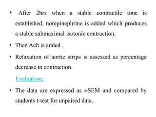
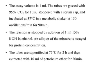
![• After one hour, the rats are sacrificed, the livers
removed and transferred to chilled oxygenated Krebs-
Ringer-bicarbonate buffer (pH 7.4).
• The livers are then chopped into 0.8-mm pieces using
a tissue chopper and are suspended in the same
buffer.
• Aliquots of the suspension are pipetted, in triplicate,
into culture tubes which contain [14C]sodium
octanoate](https://image.slidesharecdn.com/antiarterioscleroticactivity-181201150755/85/screening-methods-for-anti-atherosclerotic-agents-38-320.jpg)
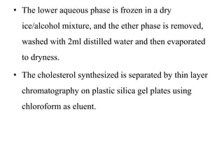
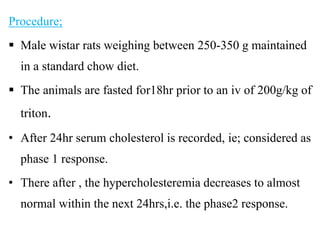
![procedure;
• Male Sprague-Dawley rats weighing 200–225 g are fed
with a diet containing 5.5% peanut oil, 0.5% cholic acid
and 1.5% cholesterol with or without (controls)drugs
for 1 week.
• On the last day, food is removed at 8:00 A.M. and the
isotopes are administered beginning at 2:00 P.M.
[3H]cholesterol is given by oral gavage and
[14C]cholesterol is given is given by tail vein injection.](https://image.slidesharecdn.com/antiarterioscleroticactivity-181201150755/85/screening-methods-for-anti-atherosclerotic-agents-49-320.jpg)
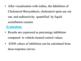
![• The [3H]cholesterol is prepared as an emulsion by
dissolving 125 mg cholesterol in 1625mg olive oil.
The oil phase is suspended by sonication in 25ml of
water containing 156 mg taurocholate(sodium salt).
Each animal receives 1 ml
The rats are allowed to take their food and sacrificed
after by isotop administration](https://image.slidesharecdn.com/antiarterioscleroticactivity-181201150755/85/screening-methods-for-anti-atherosclerotic-agents-50-320.jpg)