SCREENING OF HEPATOPROTECTIVE
DRUGS
PRESENTED BY-
MS.DIPANJALI D KAMTHE
M.PHARM PHARMACOLOGY
STES’ KASHIBAI NAVALE COLLEGE OF PHARMACY KONDHWA(BK),PUNE
1
CONTENT:
 Introduction and function of liver
 Hepatotoxicity and drug causing DILI
 Hepatoprotective agent
 Methods of Screening of hepatoprotectives
2
INTRODUCTION:
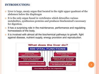
 Liver is large, meaty organ that located in the right upper quadrant of the
abdomen below the diaphragm
 It is the only organ found in vertebrates which detoxifies various
metabolites, synthesizes proteins and produces biochemical's necessary
for digestion.
 It has a surprising role in the maintenance, performance and regulating
homeostasis of the body.
 It is involved with almost all the biochemical pathways to growth, fight
against disease, nutrient supply, energy provision and reproduction.
3
LIVER TOXICITY
 The major cause in India is ethanol and it is suspected that more
than half of the cases of Hepatotoxicity is caused by alcohol.
 Chemicals like carbon tetrachloride CCL4, phosphorous ,
aflatoxins,chlorinated hydrocarbon etc
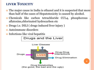
 Drugs i.e. DILI ( drugs induced liver injury )
 Autoimmune disorders
 Infections like viral hepatitis
4
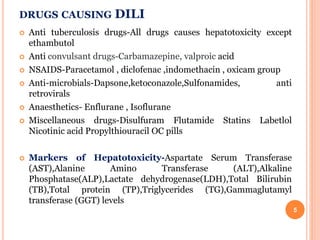
DRUGS CAUSING DILI
 Anti tuberculosis drugs-All drugs causes hepatotoxicity except
ethambutol
 Anti convulsant drugs-Carbamazepine, valproic acid
 NSAIDS-Paracetamol , diclofenac ,indomethacin , oxicam group
 Anti-microbials-Dapsone,ketoconazole,Sulfonamides, anti
retrovirals
 Anaesthetics- Enflurane , Isoflurane
 Miscellaneous drugs-Disulfuram Flutamide Statins Labetlol
Nicotinic acid Propylthiouracil OC pills
 Markers of Hepatotoxicity-Aspartate Serum Transferase
(AST),Alanine Amino Transferase (ALT),Alkaline
Phosphatase(ALP),Lactate dehydrogenase(LDH),Total Bilirubin
(TB),Total protein (TP),Triglycerides (TG),Gammaglutamyl
transferase (GGT) levels
5
LIST OF HEPATOPROTECTIVE AGENTS
 N acetylcysteine
 Penicillamine
 Anti oxidants
 cardiotropin 1
 Herbal medications
e.g. silymarine
 S adenosyl methionine
(SAM)
Vitamins
Melatonin
Glutathion
Beta-carotene
6
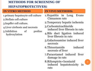
METHODS FOR SCREENING OF
HEPATOPROTECTIVES:
7
1.primary hepatocyte cell culture
2.Stellate cell culture
3.kupffer cell culture.
4.Liver cirrhosis and necrosis
5.Inhibition of proline
hydroxylation
1.Hepatitis in Long Evans
Cinnamon rats
2.Temporary hepatic ischemia
3.Carbontetrachloride
induced liver fibrosis in rats
4.Bile duct ligation induced
liver fibrosis in rats
5.Galactosamine induced liver
necrosis
6.Thiocetamide induced
necrosis of liver
7.Paracetamol induced liver
damage in rats
8.Rifampicin+Isoniazid
induced hepatotoxicity in
rats
IN VITRO METHOD IN VIVO METHODS:
IN VITRO METHODS
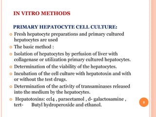
PRIMARY HEPATOCYTE CELL CULTURE:
 Fresh hepatocyte preparations and primary cultured
hepatocytes are used
 The basic method :
 Isolation of hepatocytes by perfusion of liver with
collagenase or utilization primary cultured hepatocytes.
 Determination of the viability of the hepatocytes.
 Incubation of the cell culture with hepatotoxin and with
or without the test drugs.
 Determination of the activity of transaminases released
into the medium by the hepatocytes.
 Hepatotoxins: ccl4 , paracetamol , d- galactosamine ,
tert- Butyl hydroperoxide and ethanol.
8
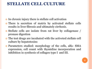
STELLATE CELL CULTURE
 In chronic injury there is stellate cell activation
 There is secretion of matrix by activated stellate cells
results in liver fibrosis and ultimately cirrhosis.
 Stellate cells are isolate from rat liver by collagenase /
pronase digestion
 The test drugs are incubated with the activeted stellate cell
culture by hepatotoxins
 Parameters studied: morphology of the cells, alfa- SMA
expression, cell count with thymidine incorporation and
inhibition in synthesis of collagen type I and III.
9
KUPFFER CELL CULTURE
 Isolation: kupffer cells are isolated from the liver by
perfusion of the liver with pronase followed by
differential centrifugation.
 The isolated cells are maintained in culture where
their phagocytic properties are retained.
 On exposure of the cells phagocytic stimuli like
zymosan particles, there is increased oxygen
consumption and superoxide production.
 The inhibitory effects of various agents or drugs on
kupffer cells are tested in the in vitro cultures
10
LIVER CIRRHOSIS AND NECROSIS
 Excessive formation of connective tissue with collagen over
production reduces hepatic blood flow.
 Collagen is formed as a response to chronic injury.The
collagenous fibers consist of triple helical molecules.Their
formation depends on the presence of hydrogen bonds.
 If the number of hydrogen bonds is reduced,the resulting
collagen can not form the triple helix and is degraded instead of
being deposited in the extracellular matrix.
 The aim of fibrosuppressive compounds is to reduce only the
excessive formation of insoluble collagen.
 Fibrosuppressive effects by inhibition of proline hydroxylation
can be screened with in vitro methods, however, the desired
organ specificity has to be tested in models of liver cirrhosis and
fibrosis in vivo.
11
INHIBITION OF PROLINE HYDROXYLATION
 The thermal stability of the triple helix of collagen is
depend on intramolecular hydrogen bond synthesized by
the enzyme prolyl 4-hydroxylase.
 PROCEDURE
Reaction volume of 1 ml contains Tris buffer pH 7.5,
glutarate, FeSO4,ascorbate,catalase,bovine serum
albumin,dithiotreitol, inhibitors.
After incubation at 37 °C for 30 min, the generated CO2 is
trapped and determined.
12
IN-VIVO METHODS
Hepatitis in long evans cinnamon rat:
Purpose and Rationale:
 The long evans cinnamon rat is useful model to study
genetically transmitted hepatitis and chronic liver
disease
 Due to excessive copper accumulation in the liver of
long evans cinnamon rats,making this animal a
model for Wilson’s disease in human.
 Chelation therapy or feeding copper deficient diet
decreases symptoms in Long Evans Cinnamon rat
and Wilson’s disease.
13
PROCEDURE
Long evans cinnamon rats of age of 5 weeks are
housed in temprature and humidity controlled room.
Group of 6-10 rats are given different diets on a 15%
purified egg protein diet and supplemented with
vitamins or drugs.drugs are supplied via minipumps
intraperitoneally implanted under ether anesthesia.
The occurance of jaundice is esasily observable as the
time when the ears and tails tuen yellow and the
urine becomes bright ending in death of aminal
within about a week.
14
TEMPORARAY HEPATIC ISCHEMIA:
Hepatocellular function is altered by temporary hepatic
ischemia as occuring during surgical management of
acute hepatic trauma and being essential during hepatic
transplantation.
PURPOSE AND RATIONALE:
 Total hepatic ischemia in rats is produced by placing a
ligature around the hepatic artery, portal vein, and the
common bile duct.
 Procedure:
Male albino rats(300–350 g )are fasted for 16 h prior to
the experiment but are allowed water ad libitum.
The rats are anesthetized lightly with ether and the
abdominal cavity is opened through a midline incision. 15
Portal vein as the hepatic artery and the bile duct are
occluded by placing a tourniquet around the vessels.
Blood pressure is measured via a catheter inserted into the
right femoral artery.During the ischemic period, 0.7 ml of
saline is given i.v. at 20-min intervals for volume
replacement.
At the end of the 60-min ischemic period, the tourniquet
around the portal vein, hepatic artery and the bile duct is
removed in order to reestablish blood flow to the liver.
The abdominal incision is then closed and the animals
receive either saline (nontreated) or the drug. 16
ALLYL ALCOHOL INDUCED LIVER NECROSIS IN RATS
 PURPOSE AND RATIONALE
 Administration of allyl alcohol induces liver necrosis in rats
which can be partially prevented by treatment with several drugs
such as antibiotics.
 Procedure:
Female Wistar rats weighing 120–150 g are fasted overnight with
water and libitum.
On following morning test drug is given orally or i.p to 10
rats.After 1hour 0.4ml/kg of 1.25% allyl alcohol solution is given
orally.
Test drug given again on 2nd day and sacrificed on 3rd day,liver is
removed 17
EVALUATION
 The parietal sides of the liver are checked using a
stereomicroscope with 25 times magnification.
 Focal necrosis is observed as white-green or yellowish
hemorrhagic areas clearly separated from unaffected tissue. The
diameter of the necrotic areas is determined using a ocular
micrometer. These values are added for each animal to obtain an
index for necrosis.
 Mean of necrosis index is calculated and compared with Student’s
t-test.
 The protective effect is expressed as percentage decrease of the
necrosis index versus controls
18
CARBON TETRACHLORIDE INDUCED LIVER FIBROSIS
IN RATS
 PURPOSE AND RATIONALE
Chronic administration of tetrachloride to rats induces severe
disturbances of hepatic function together with histologically observable
liver fibrosis.
 PROCEDURE
Groups of 20 female Wistar rats(100–150g)are used. The animals are
treated orally twice a week with 1 mg/kg carbon tetrachloride, dissolved
in olive oil 1 : 1, over a period of 8 weeks.
Animals are fed on chow diet with water ad libitum.Control group
receives only olive oil. Test and standard drugs given twice daily except
on Sunday when only one dose is given.
The animals weighed every week, at the end of 8th week animals are
sacrificed using anesthetic ether .
19
 In serum, parameters determined includes-Total bilirubin,total bile
acids,7S fragment of type IV collagen,procollagen III N-peptide.
 Hydroxyproline is determined in Liver, kidney, aortic wall, and tail
tendons.
 The specimens of the organs are weighed and completely hydrolyzed
in 6 N Hal. Hydroxyproline is measured by HPLC and expressed as
mg/mg wet weight of the organs.
 For histological analysis, 3–5 pieces of the liver weighing about 1 g
are fixed in formalin and 3 – 5 sections of each liver are embedded,
cut and stained with azocarmine aniline blue (AZAN) and evaluated
for the development of fibrosis using a score of 0–IV.
Grade 0: Normal liver histology.
Grade I: Tiny and short septa of connective tissue.
Grade II: Large septa of connective tissue.
Grade III: Nodular transformation of the liver.
Grade IV: Excessive formation and deposition of connective tissue. 20
 EVALUATION
 For detection of significant differences (p < 0.05), the unpaired
t-test is used. For comparison of the scores in the histological
evaluation, chi-square test is used.
 MODIFICATIONS OF THE TEST
 Instead of chronic intoxication with carbon tetrachloride
resulting in liver fibrosis, acute Hepatocellular damage can be
achieved by short term application of carbon tetrachloride.
 The rats are treated daily for 5 days with various oral doses of
the compound under investigation. From day 4 to day 5 the rats
receive 1 mg/kg carbon tetrachloride dissolved in olive oil (1 :
1).
 Blood is withdrawn every day and the aminotransferases and
total bilirubin determined in the serum. 21
BILE DUCT LIGATION INDUCED LIVER FIBROSIS IN RATS
 PURPOSE AND RATIONALE
 Bile duct ligation in rats induces liver fibrosis which can be
evaluated by histological means and by determination of serum
collagen parameters.
 Procedure:
Male Sprague Dawley rats weighing approximately 250 g are
anesthetized with ketamineHCL.Laparatomy is performed under
antiseptic conditions.
Expose the common bile duct, which pursues an almost straight
course of about 3 cm from the hilum of the liver to its opening
into the duodenum. There is no gallbladder, and the duct is
embedded for the greater part of its length in the pancreas,
which opens into it by numerous small ducts.
22
A blunt aneurysm needle is passed under the part of the duct selected,
stripping the pancreas away with care, and the duct is divided between
double ligatures of cotton thread. The peritoneum and the muscle
layers as well as the skin wound are closed with cotton stitches.
The animals receive normal diet and water ad libitum throughout the
experiment. Groups of 5–10 animals receive the test compound in
various doses or vehicle twice daily for 6 weeks.
Then, they are sacrificed and blood is harvested for determination of
bile acids, 7S fragment of type IV collagen, and precollegen III N-
peptide. The liver is used for histological studies and for
hydroxyproline determinations.
Control animals show excessive bile duct proliferation as well as
formation of fibrous septa.
23
GALACTOSAMINE INDUCED LIVER NECROSIS
 Single dose or a few repeated doses of D-galactosamine cause acute
hepatic necrosis in rats .
 Prolonged administration leads to cirrhosis.
 PROCEDURE:
Divided doses of 100 to 400 mg/kg D-galactosamine are injected to rats
i.p. or i.v. during one day.
For induction of liver cirrhosis, male Wistar rats weighing 110–180 g are
injected intraperitoneally three times weekly with 500 mg/kg D-
galactosamine over a period of one to 3 months.
Potential protective substances are administered orally with the food or
by gavages every day.
The rats are sacrificed at various time intervals and the livers obtained by
autopsy.
24
EVALUATION
 The livers are evaluated by light microscopy and immuno-
histology using antibodies against macrophages, lymphocytes
and the extracellular matrix components, e.g., laminin,
fibronectin, desmin, collagen type I, III, and IV.
 The extent of liver cell necrosis and immuno reactivity for
macrophages, lymphocytes and the extracellular matrix
components is graded semiquantitatively on a 0 to 4 scale (0 =
absent, 1+ = trace, 2+ = weak,3+ = moderate, and 4+ = strong).
 Furthermore, serum enzyme activities, such as GOT and GPT
are determined.
25
THIOCETAMIDE INDUCED HEPATOTOXICITY IN RATS:
 Thiocetamide actcs as a hepatocarcinogen and hepatotoxicant
 This action is mediated by formation of S- oxide, which
covalently binds with liver cell macromolecules like protein,
nucleic acid and lipids.
Procedure:
Wistar/Sprague dowel rats of either sex weighing between
180-250 g are used.
They are maintained on a standard chow diet with water and
libitum at 21±2°c room temprature and under 12h dark/12h
light cycle
The food is withdrawn 18h before the experiment but water is
allowed ad libitum. 26
Hepatotoxicity is induced by 200mg/kg body weight of dissolved in
saline and administered orally or 100mg/kg given S.C
 The serum is used for determination of aminotransferases and the liver
is used for histopathological studies
27
PARACETAMOL INDUCED LIVER DAMAGE IN RATS:
 Paracetamol is one of the most commonly and widely used analgesic
anti-pyretic drug used in higher dose lead to hepatic damage.
 It gets metabolised to an active metabolite N-acetyl-p-benzoquinone
imine by the cytocromeP-450 microsomal enzyme system which
results in an oxidative stress producing liver glutathione and glycogen
depletion.
 Procedure:
Wistar rats of either sex weight(150-200g)are used.Paracetamol 2g/kg
body weight is administered orally as a single dose
The animal are given the test drug foe 6 days prior to paracetamol
administration and on the seventh day along with paracetamol
The animal are sacrificed after 24h and the blood/serum is used for
biochemical analysis and the liver for histopathological studies 28
RIFAMPICIN+ISONIAZID INDUCED HEPATOTOXICITY
IN RATS:
 Rifampicin and isoniazid are used together as 1st line anti-T.B drug.
 Procedure:
Wistar/sprague dowley rats of either sex weighing between 150-200 g
are used.
RMP and INH both are given in the dose of 50mg/kg body weight of
each by i.p injection once daily for 15 days.
The test drug were also administered along with RMP+INH
combination daily for 15 days.
At the end of experiment the rats are killed by decapitation and used
for further biochemical analysis and histopathology of the liver.
29
REFERENCES:
 H. Gerhard Vogel Drug Discovery and Evaluation
Pharmacological Assays ,Second Edition .Page no: 936-944
 N.S Parmar, Shiv PrakashʻʻScreening methods in pharmacology.”Page
No:281-286
30