The document summarizes three cardiovascular diagnostic tests:
1) Doppler ultrasound uses sound waves to assess blood flow through arteries and veins, helping doctors evaluate reduced or blocked blood flow. It can detect blood clots that increase risks of stroke or pulmonary embolism.
2) Echocardiography uses ultrasound to create images of the heart, allowing doctors to evaluate the heart valves and detect abnormalities in blood flow or thickened heart walls. It is noninvasive.
3) A Holter monitor continuously records a patient's EKG for 24 hours during daily activities to associate symptoms with heart rhythms like ischemia or reduced blood supply. Electrodes are attached to record the EKG for later analysis.
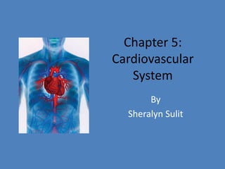
Cardiovascular assessment and diagnostic proceduresANILKUMAR BR
Cardiovascular disease is the leading killer for both men and women among all racial and ethnic groups in the world wide.
According to the Centers for Disease Control (CDC) studies among coronary heart disease (CAD) patients, 90% of patients have had prior exposure to at least one heart disease risk factor that contributed to their disease.
La ecografía Doppler consiste en una técnica especial de ultrasonido que evalúa la circulación a través de los vasos sanguíneos, mediante el registro de la onda del pulso y la determinación de su presión.
step by step presentation on ultrasound evaluation of shoulder and knee joints with illustrations of probe positioning.multiple examples of pathologies also added.
Overview of role of imaging in different intraconal and extraconal pathologies including infective,inflammatory and neoplastic pathologies.Also included is insight into anatomy,trauma,post operative imaging and certain miscellaneous disorders
presentation on ultrasound elastography-introduction ,techniques,physics,application, interpretation and future prospects.sourced from multiple articles.
A moderately frequent illness called congestive heart failure occurs when the heart is unable to pump enough blood to meet the body's demands. It frequently happens as a result of a chronic illness or aging. The body makes an effort to make up for this by boosting blood salt levels and fluid retention.
Swelling, weight gain, and shortness of breath may result from this. Diabetes and high blood pressure are other conditions linked to congestive heart failure. Congestive heart failure, however, is most frequently brought on by coronary artery disease (CAD). This occurs when the arteries that carry blood to the heart start to constrict and narrow.
When calling a doctor is important to question Dr. Sumit shejol Cardiologist from Hrudaysparsh Clinic Suggests that if you recognize the majority of the symptoms of heart failure. Certain signs and symptoms, such as chest pain, acute breathlessness, an irregular heartbeat, extreme weakness, or fainting, demand rapid medical attention. Do not delay in seeking assistance, do not self-diagnose, and do not self-medicate if you feel any of that. Some of these symptoms may also be a sign of heart failure or another serious lung, heart, or cardiovascular disease. Your condition is stabilized as emergency room doctors try to identify the source of your symptoms. Call your doctor right away if you've already been given a heart failure diagnosis and you realize that your symptoms have gotten worse or a new symptom has appeared.
Congestive heart failure is a fatal condition with a high mortality rate. Congestive heart failure has a wide range of risk factors. Smoking, high blood pressure, diabetes, high cholesterol, being obese, and having experienced a heart attack in the past are some of them. It can also be brought on by a hereditary condition like cardiomyopathy. The condition can cause the heart muscle to expand and become excessively thick, which can result in heart failure. Congestive heart failure can be exacerbated by lifestyle choices including smoking, excessive alcohol intake, or tobacco use.
Ultrasonography of Heart or Cardiac ultrasonography or Echocardiogram or ultrasound of the heart is the production of two-dimensional cross-sectional images of intracardiac anatomy by stop-action compound scan pulse-echo ultrasound.
The images show the size and shape of the cardiac chambers in systole and diastole, the appearance of heart valves, and the orientation of the great vessels.
The stop-action display is created by repetitively activating the recording oscilloscope for a selected short segment of each cardiac cycle.
The activating signal is timed by the patient's electrocardiogram.
The asynchronous motion of the scanner accumulates additional echoes with each cycle.
As a non-invasive technique, it is without risk or morbidity.
Echocardiogram or Echocardiography or Heart UltrasoundNishuVerma20
Echocardiogram
Echocardiography,
Heart ultrasound
TOPICS:
Introduction
Indication
Types
Transthoracic Echocardiogram (TTE)
Transesophageal Echocardiogram (TOE)
Stress Echocardiogram
Doppler echocardiogram
Axes in echocardiography
Windows of echo
How to prepare for an echocardiogram
Recovery after an echocardiogram
After an echocardiogram
Results
invasive non invasive procedures.pdf for bsc nursing studentsshanmukhadevi
Chest X-ray:
The chest X-ray is a noninvasive tool used to visualize internal structures, such as the heart, lungs, soft tissues, and bones.
Most chest X-rays are taken while the patient is inhaling so that the lungs are fully expanded.
Several types of chest X-rays can be used to assess heart size, contour, and position; other types reveal cardiac and pericardial calcification as well as physiologic alterations in pulmonary circulation.
2. Doppler Ultrasound A Doppler ultrasound test uses reflected sound waves to assess blood as it flows through a blood vessel. This procedure can help doctors evaluate blood flow through all of the major arteries and veins throughout the legs, arms, and neck. The ultrasound can show if the blood flow has been reduced or blocked through any narrowed arteries. Blood clots can dislodge and travel to any parts of the body causing severe damage or death. A few examples are blood clots to the brain which can lead to stroke, or blood clots in the leg veins that can travel to the lungs and cause pulmonary embolism.
3. The Doppler ultrasound is a handheld instrument that is passed gently over the skin above a blood vessel. The instrument (transducer) will receive and send sound waves that can be heard through the Doppler machine. The sound waves bounce of solid objects like blood cells. The blood cells movement changes the pitch of the sounds waves which is called the Doppler effect. The pitch does not change if there is no blood flow. The computer process the information from the sound waves into graphs or pictures showing the flow of blood through the blood vessels.
4. Echocardiography Echocardiography is a diagnostic test which uses ultrasound waves to make images of the heart chambers, valves, and surrounding structures. It can measure cardiac output and is a sensitive test for inflammation around the heart. Echocardiography can also detect abnormal anatomy or infections of the heart valves. This procedure is used to diagnose certain cardiovascular diseases especially heart disease. Echocardiography can provide the size and shape of the heart, the strength in which the heart can pump, and it can also locate any damage of the surrounding tissues.
5. Doctors use this to evaluate the heart valves, and detect abnormalities of blood flow in the heart valves for example the backward flow of blood through partly closed heart valves. Not only does it notice the presence of coronary artery disease, but it can also detect hypertrophic cardiomyopathyin which the walls of the heart thicken. This procedure is noninvasive and no known risks or side effects. Echocardiography creates an image of the heart using ultra-high frequency sound waves. The examination will last between 15-20 minutes. The transducer is positioned on the chest and acts like a microphone that directs ultra-sound waves into the chest which then bounces back to the transducer. The sound waves are processed into an image of the heart that is displayed on the monitor.
6. Holter Monitor It is a portable ECG monitor that continuously records a patient’s EKG for 24 hours during the patient’s regular activities in order for the physician to associate symptoms if dizziness, palpitations or black outs. The patient’s EKG can also include episodes of chest pain which can suggest ischemia or reduced blood supply to the muscle.
7. EKG electrodes are attached to the chest and thin wires connect the electrodes to a small tape recorder that is secured to the patient’s belt. The recorder is in place for 34 hours as the patient continues his or her daily activities. The patient is to record all activities while the monitor is on along with the time. The Holter monitor has an internal clock for the EKG strips which correlate the heart rhythm with symptoms and complaints. After 24 hours, the Holter monitor is returned for analysis.